

Load-bearing properties of minimal-invasive monolithic lithium disilicate and zirconia occlusal onlays: finite element and theoretical analyses
- PMID: 23683531
- PMCID: PMC3698988
- DOI: 10.1016/j.dental.2013.04.004
Load-bearing properties of minimal-invasive monolithic lithium disilicate and zirconia occlusal onlays: finite element and theoretical analyses
Abstract
Objective: The aim of this study was to test the hypothesis that monolithic lithium disilicate glass-ceramic occlusal onlay can exhibit a load-bearing capacity that approaches monolithic zirconia, due to a smaller elastic modulus mismatch between the lithium disilicate and its supporting tooth structure relative to zirconia.
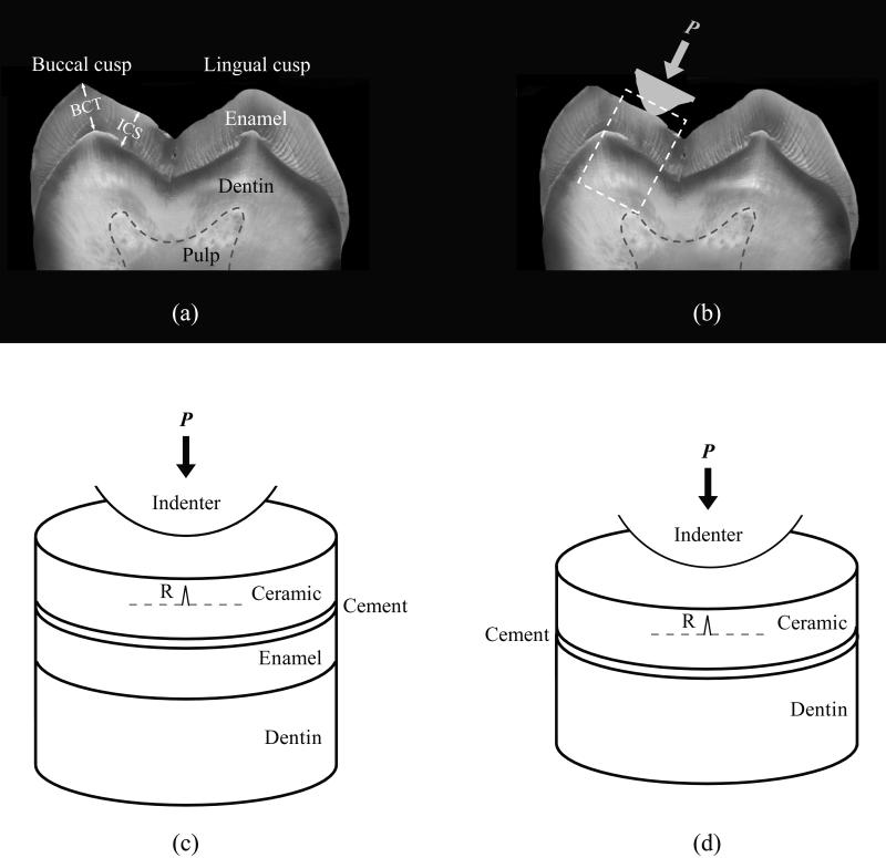
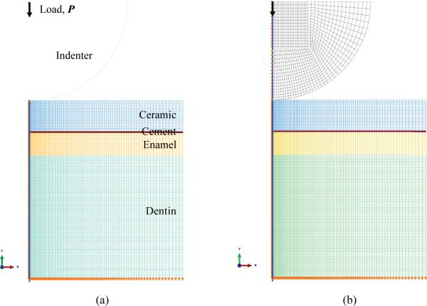
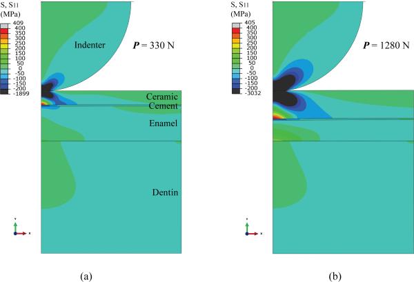
Methods: Ceramic occlusal onlays of various thicknesses cemented to either enamel or dentin were considered. Occlusal load was applied through an enamel-like deformable indenter or a control rigid indenter. Flexural tensile stress at the ceramic intaglio (cementation) surface-a cause for bulk fracture of occlusal onlays-was rigorously analyzed using finite element analysis and classical plate-on-foundation theory.
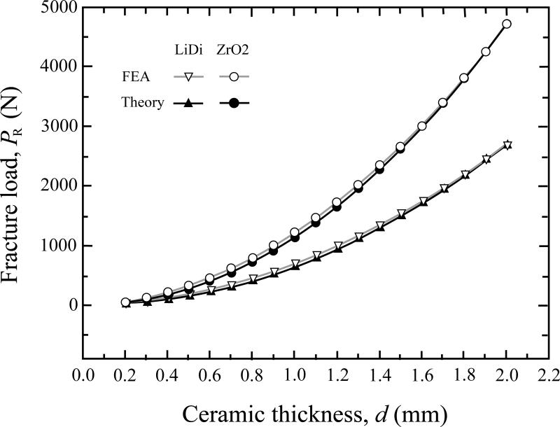
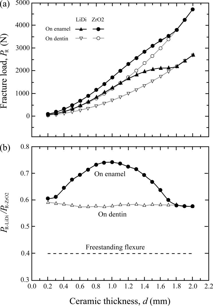
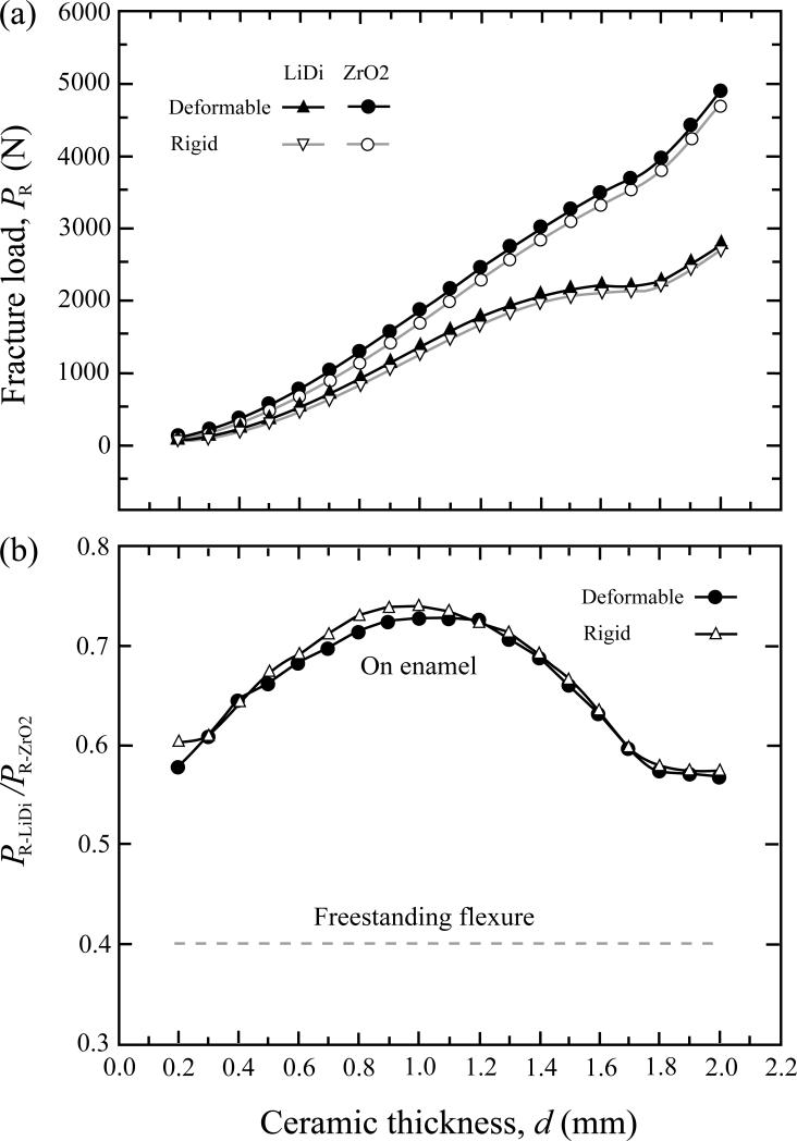
Results: When bonded to enamel (supported by dentin), the load-bearing capacity of lithium disilicate can approach 75% of that of zirconia, despite the flexural strength of lithium disilicate (400MPa) being merely 40% of zirconia (1000MPa). When bonded to dentin (with the enamel completely removed), the load-bearing capacity of lithium disilicate is about 57% of zirconia, still significantly higher than the anticipated value based on its strength. Both ceramics show slightly higher load-bearing capacity when loaded with a deformable indenter (enamel, glass-ceramic, or porcelain) rather than a rigid indenter.
Significance: When supported by enamel, the load-bearing property of minimally invasive lithium disilicate occlusal onlays (0.6-1.4mm thick) can exceed 70% of that of zirconia. Additionally, a relatively weak dependence of fracture load on restoration thickness indicates that a 1.2mm thin lithium disilicate onlay can be as fracture resistant as its 1.6mm counterpart.
Copyright © 2013 Academy of Dental Materials. All rights reserved.
Figures
References
-
- Edelhoff D, Sorensen JA. Tooth structure removal associated with various preparation designs for anterior teeth. The Journal of Prosthetic Dentistry. 2002;87:503–9. - PubMed
-
- Edelhoff D, Sorensen JA. Tooth structure removal associated with various preparation designs for posterior teeth. The International Journal of Periodontics & Restorative Dentistry. 2002;22:241–9. - PubMed
-
- Beier US, Kapferer I, Burtscher D, Dumfahrt H. Clinical performance of porcelain laminate veneers for up to 20 years. The International Journal of Prosthodontics. 2012;25:79–85. - PubMed
-
- Petridis HP, Zekeridou A, Malliari M, Tortopidis D, Koidis P. Survival of ceramic veneers made of different materials after a minimum follow-up period of five years: a systematic review and meta-analysis. The European Journal of Esthetic Dentistry. 2012;7:138–52. - PubMed
-
- Pippin DJ, Mixson JM, Soldan-Els AP. Clinical evaluation of restored maxillary incisors: veneers vs. PFM crowns. Journal of the American Dental Association. 1995;126:1523–9. - PubMed
Publication types
MeSH terms
Substances
Grants and funding
LinkOut - more resources
Full Text Sources
Other Literature Sources
