. 2013 Nov 1;74(9):672-9.
doi: 10.1016/j.biopsych.2013.03.024. Epub 2013 May 14.

Role of prefrontal cortex glucocorticoid receptors in stress and emotion

Affiliations

Role of prefrontal cortex glucocorticoid receptors in stress and emotion

Jessica M McKlveen et al. Biol Psychiatry. .

Abstract

Background: Stress-related disorders (e.g., depression) are associated with hypothalamic-pituitary-adrenocortical axis dysregulation and prefrontal cortex (PFC) dysfunction, suggesting a functional link between aberrant prefrontal corticosteroid signaling and mood regulation.

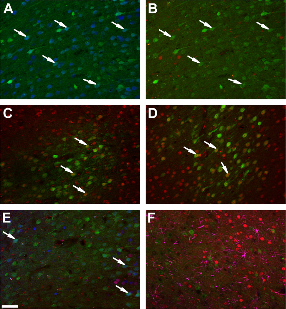
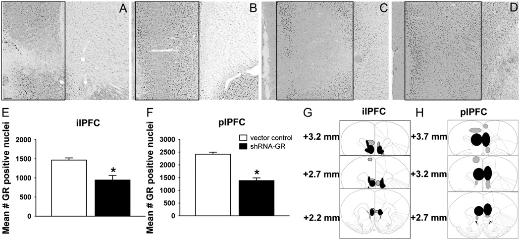
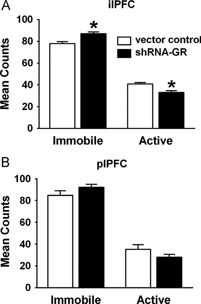
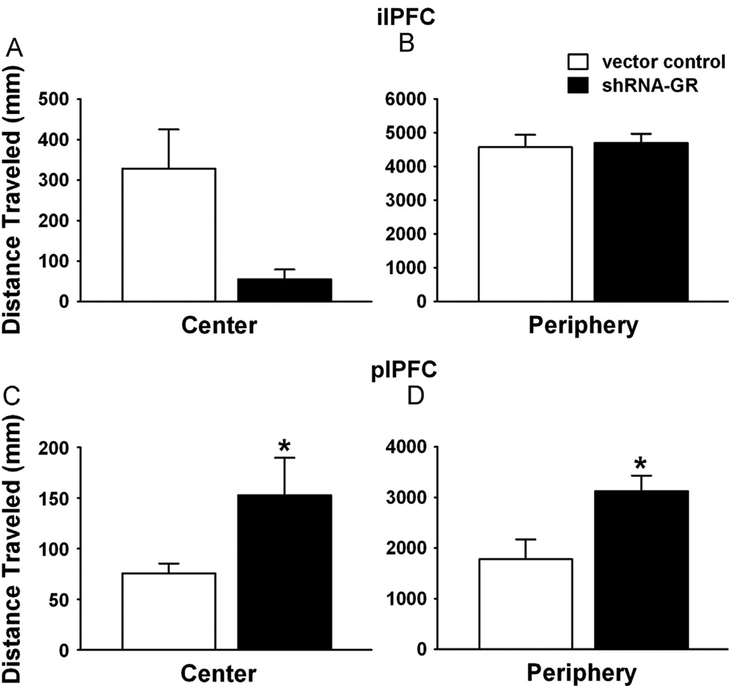
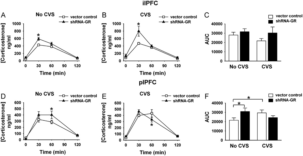
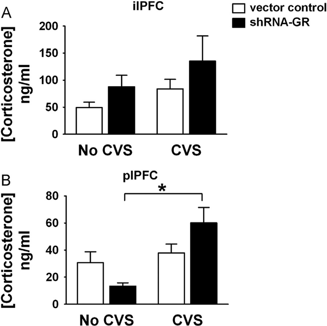
Methods: We used a virally mediated knockdown strategy (short hairpin RNA targeting the glucocorticoid receptor [GR]) to attenuate PFC GR signaling in the rat PFC. Adult male rats received bilateral microinjections of vector control or short hairpin RNA targeting the GR into the prelimbic (n = 44) or infralimbic (n = 52) cortices. Half of the animals from each injection group underwent chronic variable stress, and all were subjected to novel restraint. The first 2 days of chronic variable stress were used to assess depression- and anxiety-like behavior in the forced swim test and open field.

Results: The GR knockdown confined to the infralimbic PFC caused acute stress hyper-responsiveness, sensitization of stress responses after chronic variable stress, and induced depression-like behavior (increased immobility in the forced swim test). Knockdown of GR in the neighboring prelimbic PFC increased hypothalamic-pituitary-adrenocortical axis responses to acute stress and caused hyperlocomotion in the open field, but did not affect stress sensitization or helplessness behavior.

Conclusions: The data indicate a marked functional heterogeneity of glucocorticoid action in the PFC and highlight a prominent role for the infralimbic GR in appropriate stress adaptation, emotional control, and mood regulation.

Keywords: Depression-like behavior; HPA axis; glucocorticoid receptor; prefrontal cortex; rat; stress.


Conflict of interest statement

The authors report no biomedical financial interests or potential conflicts of interest.

Figures

Figure 1
Figure 1
Verification and specificity of a short hairpin RNA targeting the glucocorticoid receptor (shRNA-GR). (A) Representative NeuN (blue) immunolabeled sections after microinjection with shRNA-GR (green). After microinjection of shRNA-GR, NeuN immunoreactivity remained intact in transduced neurons, indicating that neuronal viability was not affected (see arrows). (B) Representative GR (red) immunolabeled sections after microinjection with shRNA-GR (green), (C) shRNA-scrambled control (green), and (D) empty vector control (green). The shRNA-GR reduced GR in green fluorescent protein co-localized cells (B) relative to animals that received microinjections of shRNA-scrambled control (C) or empty vector control (D) (see arrows). (E) Representative dual GR (red) and mineralocorticoid receptor (MR) (blue) immunolabeled sections after intracranial microinjection with shRNA-GR. Mineralocorticoid receptor expression is intact in cells in which GR immunoreactivity is knocked down (as demonstrated in the superficial layers ll/lll on the left of the image and deep layers V/VI of the prelimbic prefrontal cortex on the right of the image, where MR is typically expressed. The agranular layer between layers III and V typically has little MR expression) (see arrows). (F) Representative glial fibrillary acidic protein (purple) and GR (red) immunolabeled sections after intracranial injection with shRNA-GR. The shRNA-GR did not transduce astrocytes (no green fluorescent protein and glial fibrillary acidic protein co-localization) and did not seem to knockdown astrocytic GR. Scale bar = 50 µm.
Figure 2
Figure 2
Selective decreases in glucocorticoid receptor (GR) immunoreactive neurons in the infralimbic prefrontal cortex (ilPFC) and prelimbic prefrontal cortex (plPFC) after short hairpin RNA targeting the GR (shRNA-GR) microinjection. Representative GR-immunolabeled sections from vector control-microinjected animals in (B) the ilPFC and the (D) plPFC and shRNA-GR-microinjected animals in (A) the ilPFC and (C) the plPFC (representative areas of quantification are outlined in panels A–D). (E) Quantified GR expression from vector control-microinjected animals (n = 10) and shRNA-GR-microinjected animals (n = 5) in the ilPFC and (F) vector control-microinjected animals (n = 6) and shRNA-GR-microinjected animals (n = 6) in the plPFC. The GR immunoreactivity was significantly reduced in animals that received shRNA-GR relative to vector control-microinjected animals (p < .05). (G) Extent of GR knockdown in the ilPFC of all shRNA-GR-microinjected animals that were considered “hits” (n = 10) or (H) in the plPFC of all shRNA-GR-microinjected animals that were considered “hits” (n = 16) (reprinted from Paxinos and Watson [16] with permission from Elsevier, copyright 1998). Green fluorescent protein (GFP) expression throughout the plPFC in each animal was traced onto stereotaxic images and compiled into one visual representation. Black circles indicate where GFP expression was most prominent (n ≥ 4 in the ilPFC and n ≥ 8 in the plPFC), whereas gray circles represent areas where GFP was less prominent in animals that received shRNA-GR and were considered “hits” (n ≤ 3 in the ilPFC or n ≤ 7 in the plPFC). Immunoreactive counts are mean ± SEM. Scale bar = 100 µm. *p < .05 vs. vector control-microinjected animals.
Figure 3
Figure 3
Increased helplessness behavior after GR knockdown in the ilPFC. (A) Immobility vs. activity in the modified forced swim test after vector control- or shRNA-GR-microinjection in the ilPFC (n = 10 or 5, respectively) or (B) in the plPFC (n = 11 or 8, respectively). Animals receiving shRNA-GR in the ilPFC but not the plPFC, exhibited increased immobility in the forced swim test relative to vector controls (p < .05). Data are mean ± SEM. *p < .05 vs. vector control-microinjected animals. Abbreviations as in Figure 2.
Figure 4
Figure 4
Increased locomotor activity after GR knockdown in the plPFC relative to vector control-microinjected animals. (A) Locomotor activity in the center and (B) periphery after vector control- or shRNA-GR microinjections in the ilPFC (n = 9–10 or 4, respectively). (C) Locomotor activity in the center and (D) periphery after microinjections of vector control or shRNA-GR in the plPFC (n = 11 or 7–8, respectively). Animals receiving shRNA-GR in the plPFC traveled significantly more throughout the center (C) and periphery (D) than vector controls (p < .05). Data are mean ± SEM. *p < .05 vs. vector control-microinjected animals. Abbreviations as in Figure 2.
Figure 5
Figure 5
Differential impact of GR knockdown in the ilPFC vs. plPFC on hypothalamic-pituitary-adrenal axis reactivity after acute and chronic stress. (A) Corticosterone responses after acute novel restraint in unstressed (no chronic variable stress [CVS]) and (B) CVS animals that received microinjections of vector control (n = 9–11/unstressed or stressed group) or shRNA-GR (n = 5/unstressed or stressed group) in the ilPFC. (C) Integrated area under the curve (AUC) for corticosterone responses (not including baseline values) after vector control-microinjections (n = 9–11/group) or shRNA-GR (n = 5/group) in the ilPFC. (D) Corticosterone responses after acute novel restraint in unstressed (No CVS) and (E) CVS animals that received microinjections of vector control (n = 10–11/unstressed or stress group, respectively) or shRNA-GR (n = 7–9/unstressed or stressed group) in the plPFC. (F) Integrated AUC for corticosterone responses (not including baseline values) after vector control-microinjections (n = 10–11/group) or shRNA-GR (n = 7–9/group) or in the plPFC. After ilPFC microinjection of shRNA-GR, acute stress caused a significant elevation in corticosterone at 30 min compared with vector controls, an effect that is exacerbated in chronically stressed animals relative to acutely stressed shRNA-GR-microinjected animals and controls (p < .05). After microinjection of shRNA-GR in the plPFC, animals acutely stressed in the absence of CVS have significantly elevated corticosterone levels at 60 min compared with vector controls, whereas chronically stressed animals have significantly lower corticosterone responses at 60 min compared with vector control-microinjected animals (p < .05). Data are mean ± SEM. *p < .05 vs. vector control-microinjected animals or between groups indicated by the brackets. Abbreviations as in Figure 2.
Figure 6
Figure 6
The GR knockdown in the plPFC but not NPFC of chronically stressed animals significantly increased baseline corticosterone levels. (A) Baseline corticosterone levels in unstressed and chronically stressed animals receiving vector control (n = 10 or 11/group, respectively) or shRNA-GR (n = 5/group) in the ilPFC and (B) in unstressed and chronically stressed animals receiving vector-control (n = 10 or 11, respectively) or shRNA-GR (n = 7 or 9, respectively) in the plPFC. Baseline corticosterone levels were significantly different in chronically stressed animals receiving shRNA-GR in the plPFC only (relative to acutely stressed animals that received shRNA-GR) (p < .05). Data are mean ± SEM. *p < .05 vs. acutely stressed shRNA-GR-microinjected animals or between groups indicated by the brackets. Abbreviations as in Figures 2 and 5.

Similar articles

Cited by

References

    1. Mayberg HS, Lozano AM, Voon V, McNeely HE, Seminowicz D, Hamani C, et al. Deep brain stimulation for treatment-resistant depression. Neuron. 2005;45:651–660. - PubMed
    1. Vertes RP. Differential projections of the infralimbic and prelimbic cortex in the rat. Synapse. 2004;51:32–58. - PubMed
    1. Reul JM, De Kloet ER. Anatomical resolution of two types of corticosterone receptor sites in rat brain with in vitro autoradiography and computerized image analysis. J Steroid Biochem. 1986;24:269–272. - PubMed
    1. Fuxe K, Wikstrom AC, Okret S, Agnati LF, Harfstrand A, Yu ZY, et al. Mapping of glucocorticoid receptor immunoreactive neurons in the rat tel- and diencephalon using a monoclonal antibody against rat liver glucocorticoid receptor. Endocrinology. 1985;117:1803–1812. - PubMed
    1. Meaney MJ, Sapolsky RM, Aitken DH, McEwen BS. [3H] dexamethasone binding in the limbic brain of the fetal rat. Brain Res. 1985;355:297–300. - PubMed

Publication types

MeSH terms