

Immunohistochemical study of porcine lung lesions associated with Pasteurella multocida
- PMID: 23683857
- PMCID: PMC7128513
- DOI: 10.1016/j.tvjl.2013.03.004
Immunohistochemical study of porcine lung lesions associated with Pasteurella multocida
Abstract
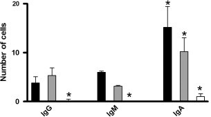
Infectious bronchopneumonia is a widespread disease in modern commercial pig production and Pasteurella multocida is frequently associated with the lesions. To evaluate porcine lung lesions associated with P. multocida, populations of inflammatory cells were examined by immunohistochemistry in necrotic lung lesions from nine pigs and exudative lung lesions from eleven pigs. Lungs from five pigs served as controls. All cases were selected from naturally infected pigs using co-infection based criteria to make them as comparable as possible. The inflammatory cells demonstrated by immunohistochemistry were T-lymphocytes (CD3(+), CD4(+) and CD8(+) subsets), B-lymphocytes, neutrophils, macrophages, and IgA(+), IgM(+) and IgG(+) cells. The results showed that (1) a significant increase in all inflammatory cells was found in lesions associated with P. multocida, (2) necrotic lesions had a larger number of CD3(+) T-lymphocytes and IgA(+) cells, and (3) cases with exudative lesions had a more CD8(+) T-lymphocytes, B-lymphocytes, macrophages and neutrophils. No differences in the numbers of CD4(+) T-lymphocytes, IgG(+) and IgM(+) positive cells were found between necrotic and exudative cases. The results show that P. multocida significantly alters the inflammatory response in the lung and that lesions associated with P. multocida display diverse inflammatory responses according to their distinct morphological pattern.
Keywords: Immunohistochemistry; Inflammatory cells; Pasteurella multocida; Porcine pneumonia.
Copyright © 2013 Elsevier Ltd. All rights reserved.
Figures


Similar articles
-
Occurrence and associated lesions of Pasteurella multocida in porcine bronchopneumonia.Vet Microbiol. 2011 May 12;150(1-2):160-6. doi: 10.1016/j.vetmic.2011.01.005. Epub 2011 Jan 16. Vet Microbiol. 2011. PMID: 21296510
-
Pathology, tissue metalloproteinase transcription and haptoglobin responses in mice after experimental challenge with different isolates of Pasteurella multocida obtained from cases of porcine pneumonia.J Comp Pathol. 2011 Aug-Oct;145(2-3):251-60. doi: 10.1016/j.jcpa.2011.01.002. Epub 2011 Mar 8. J Comp Pathol. 2011. PMID: 21388634
-
Genetic diversity and associated pathology of Pasteurella multocida isolated from porcine pneumonia.Vet Microbiol. 2011 Jun 2;150(3-4):354-61. doi: 10.1016/j.vetmic.2011.02.050. Epub 2011 Mar 3. Vet Microbiol. 2011. PMID: 21439738
-
Localization of Pasteurella multocida antigens in the brains of pigs naturally infected with Pasteurellosis revealing a newer aspect of pathogenesis.Microb Pathog. 2020 Mar;140:103968. doi: 10.1016/j.micpath.2020.103968. Epub 2020 Jan 9. Microb Pathog. 2020. PMID: 31927003
-
Molecular aspects of the virulence of Pasteurella multocida.Can J Vet Res. 1990 Apr;54 Suppl:S45-7. Can J Vet Res. 1990. PMID: 2193704 Review.
Cited by
-
Pasteurella multocida activates Rassf1-Hippo-Yap pathway to induce pulmonary epithelial apoptosis.Vet Res. 2024 Mar 16;55(1):31. doi: 10.1186/s13567-024-01285-y. Vet Res. 2024. PMID: 38493147 Free PMC article.
-
Histopathological and immunohistochemical approaches for the diagnosis of Pasteurellosis in swine population of Punjab.Vet World. 2016 Sep;9(9):989-995. doi: 10.14202/vetworld.2016.989-995. Epub 2016 Sep 18. Vet World. 2016. PMID: 27733801 Free PMC article.
-
Interaction study of Pasteurella multocida with culturable aerobic bacteria isolated from porcine respiratory tracts using coculture in conditioned media.BMC Microbiol. 2021 Jan 9;21(1):19. doi: 10.1186/s12866-020-02071-4. BMC Microbiol. 2021. PMID: 33422011 Free PMC article.
-
Virulence gene profiling of porcine Pasteurella multocida isolates of Assam.Vet World. 2018 Mar;11(3):348-354. doi: 10.14202/vetworld.2018.348-354. Epub 2018 Mar 21. Vet World. 2018. PMID: 29657428 Free PMC article.
-
Case Report: CD8+ T-Lymphocyte Deficit: A Prerequisite for Pasteurella spp. Infection?Front Med (Lausanne). 2021 Apr 27;8:668976. doi: 10.3389/fmed.2021.668976. eCollection 2021. Front Med (Lausanne). 2021. PMID: 33987195 Free PMC article.
References
-
- Ahn K.K., Lee Y.H., Ha Y., Kim D., Chae S., Kim C.H., Lee J.H., Kim S.H., Chae C. Detection by in-situ hybridization of Pasteurella multocida toxin (toxA) gene in the lungs of naturally infected pigs. Journal of Comparative Pathology. 2008;139:51–53. - PubMed
-
- von Altrock A. Occurrence of bacterial infectious agents in pathologically/anatomically altered lungs of pigs and compilation of resistance spectra. Berliner und Münchener Tierärztliche Wochenschrift. 1998;111:164–172. - PubMed
-
- Amass S.F., Clark L.K., van Alstine W.G., Bowersock T.L., Murphy D.A., Knox K.E., Albregts S.R. Interaction of Mycoplasma hyopneumoniae and Pasteurella multocida infections in swine. Journal of the American Veterinary Medical Association. 1994;204:102–107. - PubMed
-
- Bentley O.E., Farrington D.O. Evaluation of an induced Pasteurella multocida swine pneumonia model. American Journal of Veterinary Research. 1980;41:1870–1873. - PubMed
-
- Berndt A., Muller G. Occurrence of T lymphocytes in perivascular regions of the lung after intratracheal infection of swine with Pasteurella multocida. Veterinary Immunology and Immunopathology. 1995;49:143–159. - PubMed
MeSH terms
LinkOut - more resources
Full Text Sources
Other Literature Sources
Medical
Research Materials
Miscellaneous
