. 2013 Aug;132(2):437-45.
doi: 10.1016/j.jaci.2013.03.024. Epub 2013 May 16.

Mechanism of Siglec-8-mediated cell death in IL-5-activated eosinophils: role for reactive oxygen species-enhanced MEK/ERK activation

Affiliations

Mechanism of Siglec-8-mediated cell death in IL-5-activated eosinophils: role for reactive oxygen species-enhanced MEK/ERK activation

Gen Kano et al. J Allergy Clin Immunol. 2013 Aug.

Abstract

Background: Sialic acid-binding immunoglobulin-like lectin (Siglec)-8 is expressed on human eosinophils, where its ligation induces cell death. Paradoxically, Siglec-8-mediated cell death is markedly enhanced by the presence of the activation and survival factor IL-5 and becomes independent of caspase activity.

Objective: In this report we investigate the mechanism of Siglec-8-mediated cell death in activated eosinophils.

Methods: Human peripheral blood eosinophils were treated with agonistic anti-Siglec-8 antibody and IL-5, and cell death was determined by using flow cytometry and morphology. Phosphorylation of mitogen-activated protein kinase (MAPK) was determined by using phosphoLuminex, flow cytometry, and Western blotting. Reactive oxygen species (ROS) accumulation was determined by using dihydrorhodamine fluorescence.

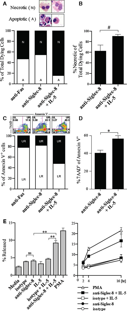
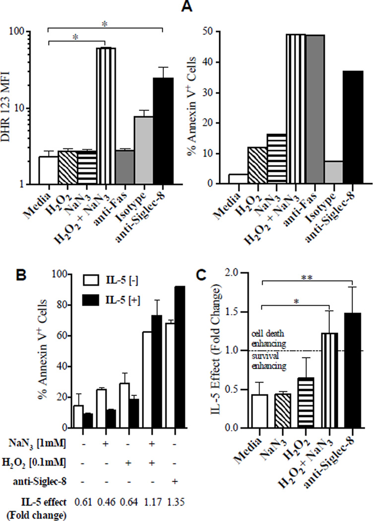
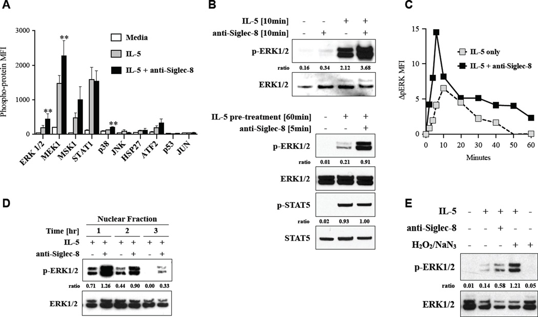
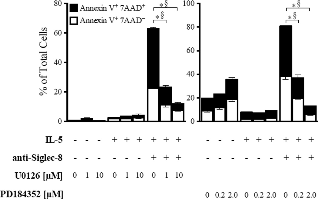
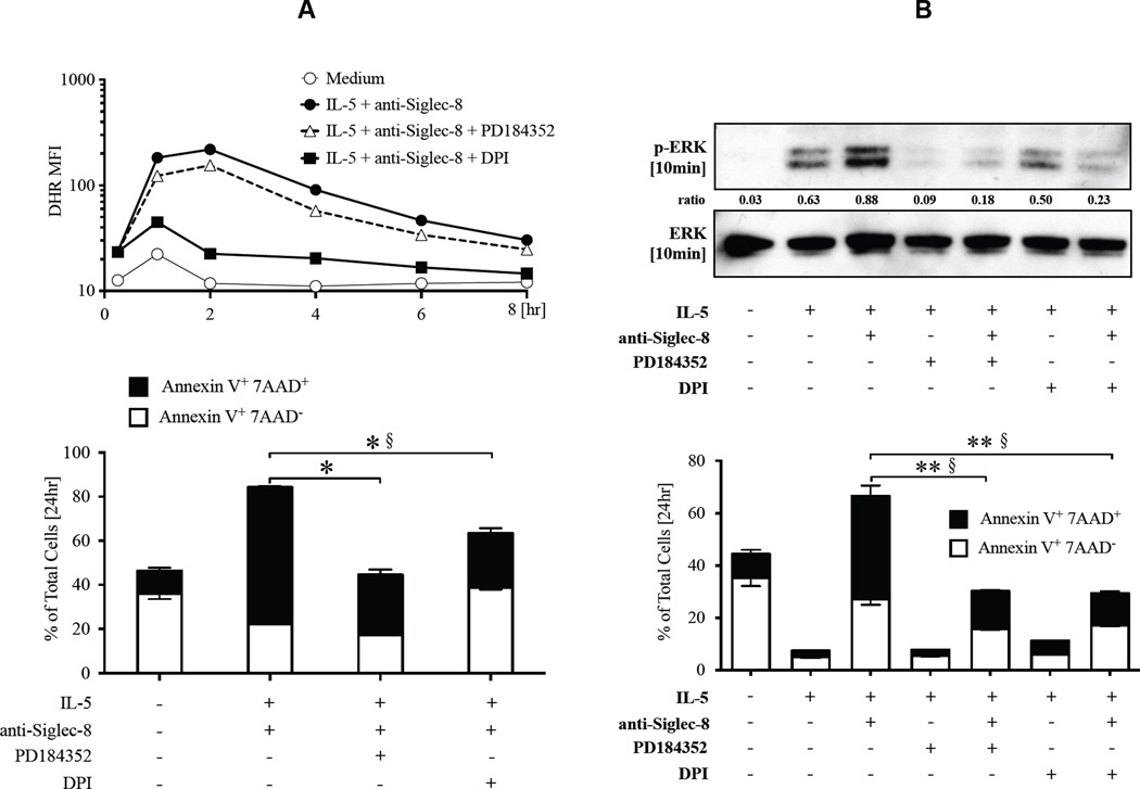
Results: Costimulation with anti-Siglec-8 and IL-5 significantly increased the rate and proportion of cell death by means of necrosis accompanied by granule release compared with that seen after stimulation with anti-Siglec-8 alone, in which apoptosis predominated. Together with the caspase-independent mode of cell death in costimulated cells, these findings suggest the activation of a specific and distinct biochemical pathway of cell death during anti-Siglec-8/IL-5 costimulation. Phosphorylation of extracellular signal-regulated kinase (ERK) 1/2 and MAPK-ERK kinase (MEK) 1 was significantly enhanced and sustained in costimulated cells compared with that seen in cells stimulated with IL-5 alone; anti-Siglec-8 alone did not cause ERK1/2 phosphorylation. MEK1 inhibitors blocked anti-Siglec-8/IL-5-induced cell death. ROS accumulation was induced by Siglec-8 ligation in a MEK-independent manner. In contrast, an ROS inhibitor prevented the anti-Siglec-8/IL-5-induced enhancement of ERK phosphorylation and cell death. Exogenous ROS mimicked stimulation by anti-Siglec-8 and was sufficient to induce enhanced cell death in IL-5-treated cells. Collectively, these data suggest that the enhancement of ERK phosphorylation is downstream of ROS generation.

Conclusions: In activated eosinophils ligation of Siglec-8 leads to ROS-dependent enhancement of IL-5-induced ERK phosphorylation, which results in a novel mode of biochemically regulated eosinophil cell death.

Keywords: 7-Aminoactinomycin D; 7AAD; DPI; DUSP; Diphenyleneiodonium; Dual-specificity phosphatase; EPX; ERK; Eosinophil peroxidase; Eosinophils; Extracellular signal-regulated kinase; H(2)O(2); HRP; Horseradish peroxidase; Hydrogen peroxide; JNK; MAPK; MAPK-ERK kinase; MEK; Mitogen-activated protein kinase; NaN(3); PFA; Paraformaldehyde; ROS; Reactive oxygen species; STAT; Sialic acid–binding immunoglobulin-like lectin; Siglec; Signal transducer and activator of transcription; Sodium azide; c-Jun N-terminal kinase; cell death; signaling.


Conflict of interest statement

Disclosures: Dr. Bochner is a co-inventor on existing and pending Siglec-8-related patents. Dr. Bochner may be entitled to a share of royalties received by the University on the potential sales of such products. Dr. Bochner is also a co-founder of, owns stock in, and is on the scientific advisory board of Allakos, Inc., which makes him subject to certain restrictions under University policy. The terms of this arrangement are being managed by the Johns Hopkins University in accordance with its conflict of interest policies.

Figures

Figure 1
Figure 1. Siglec-8 crosslinking induces a different mode of cell death in activated versus resting eosinophils
A, Representative morphology and the percentage of dead cells that have apoptotic or necrotic morphology in eosinophils cultured for 24 hours with indicated stimuli (n= 6 experiments and donors). B, Mean percentage of dead cells with necrotic morphology in multiple experiments (n = 6). #p = 0.055. C, Representative Annexin V/7AAD staining and the percent of 7AAD+ and 7AADcells among all Annexin V+ cells. D, Mean ratio of 7AAD+ cells among all Annexin V+ cells from multiple experiments (n = 25 experiments with 11 donors; **p <0.01). E, activity of released EPO following incubation with indicated stimuli for 4-hours (left) or over 16 hours (right).
Figure 2
Figure 2. ROS generation is sufficient for eosinophil cell death and IL-5 enhancement of cell death
A, ROS accumulation induced by exogenous H2O2 ± NaN3, crosslinked anti-Siglec-8 or isotype-matched control antibody was measured following 2 hours of incubation and 30 minutes of loading with DHR. Results are shown as mean fluorescence intensity (MFI) values of DHR (left panel). Aliquots of the same cells were also incubated for 24 hours without DHR in order to determine Annexin V-positive staining (right panel). Representative of 3 experiments. B, Representative experiment of eosinophils cultured with (closed bar) or without (open bar) IL-5 in conditions with H2O2 ± NaN3 or anti-Siglec-8. The IL-5 effect was determined by dividing the percentage of Annexin V-positive cells in IL-5[+] with IL-5[-] for each condition. Thus, IL-5 effect <1.0 represents survival enhancement, whereas >1.0 implies cell death enhancement. C, Averaged results from 3 experiments and donors. * p < 0.05, ** p < 0.01.
Figure 3
Figure 3. Up-regulation of MEK/ERK pathway following anti-Siglec-8/IL-5 co-stimulation
A, A phospho-MAPK multiplex bead assay was performed on lysates of eosinophils (n = 4 donors) that were incubated for 10 minutes with indicated stimuli. ** p < 0.01 compared to IL-5. B, Eosinophils incubated in the indicated conditions were analyzed by Western blotting with specific antibodies for phospho-ERK or total ERK (upper panel) and phospho- and total STAT5. Representative data from 3 independent experiments. C, Time course of ERK phosphorylation was determined by flow cytometry. Representative of 3 experiments. D, Western blotting of nuclear fraction from cell lysates of cells treated for 1, 2, or 3 hours. Representative of 3 experiments. Ratio of densitometric value for the band of phospho-kinase to that of total kinase in each lane is indicated. E, Western blotting of nuclear fraction from cell lysates of cells stimulated for 2 hours with indicated stimuli (representative of 3 experiments).
Figure 4
Figure 4. Upregulation of MEK/ERK pathway is required for enhanced cell death induced by anti-Siglec-8/IL-5 co-stimulation
Eosinophils were pre-incubated with A) U0126 or B) PD184352 at the indicated doses and then cultured in media containing IL-5 ± anti-Siglec-8. Annexin V+ and/or 7AAD+ cells were determined after 24 hours. Shown are representative data from 3 experiments with each inhibitor. *p <0.01 and §p <0.01, compared to % Annexin V+ 7AAD+ and % Annexin V+ 7AAD, respectively, in IL-5 + anti-Siglec-8 treated cells without inhibitors.
Figure 5
Figure 5. MEK/ERK up-regulation is downstream of the ROS production induced by anti-Siglec-8/IL-5 co-stimulation
Eosinophils were pre-incubated with or without inhibitors and then cultured in media containing IL-5 and anti-Siglec-8. The lower panel shows Annexin V staining of cells from the same experiment at the 24-hour time point. In A, cells were stained with DHR and ROS level analyzed with flow cytometry (representative of 3 experiments). In B, Western blot of lysates from cells harvested after 10 minutes of stimulation is shown (representative of 3 experiments). Ratio of densitometric value for band of phospho-ERK to that of total ERK in each lane is displayed. *p <0.01 and §p <0.01, compared to % Annexin V+ 7AAD+ and % Annexin V+ 7AAD, respectively, in IL-5 + anti-Siglec-8 treated cells without inhibitors.

Comment in

  • Lysis of primed eosinophils in severe asthma.
    Persson C. Persson C. J Allergy Clin Immunol. 2013 Dec;132(6):1459-60. doi: 10.1016/j.jaci.2013.09.036. Epub 2013 Nov 1. J Allergy Clin Immunol. 2013. PMID: 24184146 No abstract available.

References

    1. Von Gunten S, Bochner BS. Basic and clinical immunology of Siglecs. Ann N Y Acad Sci. 2008;1143:61–82. - PMC - PubMed
    1. O’Reilly MK, Paulson JC. Siglecs as targets for therapy in immune-cell-mediated disease. Trends Pharmacol Sci. 2009;30:240–248. - PMC - PubMed
    1. Floyd H, Ni J, Cornish AL, Zeng Z, Liu D, Carter KC, et al. Siglec-8. A novel eosinophil-specific member of the immunoglobulin superfamily. J Biol Chem. 2000;275:861–866. - PubMed
    1. Nutku E, Aizawa H, Hudson SA, Bochner BS. Ligation of Siglec-8: a selective mechanism for induction of human eosinophil apoptosis. Blood. 2003;101:5014–5020. - PubMed
    1. Kikly KK, Bochner BS, Freeman SD, Tan KB, Gallagher KT, D'Alessio KJ, et al. Identification of SAF-2, a novel siglec expressed on eosinophils, mast cells, and basophils. J Allergy Clin Immunol. 2000;105:1093–1100. - PubMed

Publication types

MeSH terms