. 2014 Jan:29:602-17.
doi: 10.1016/j.jmbbm.2013.03.027. Epub 2013 Apr 20.

Material properties of the posterior human sclera

Affiliations

Material properties of the posterior human sclera

Rafael Grytz et al. J Mech Behav Biomed Mater. 2014 Jan.

Abstract

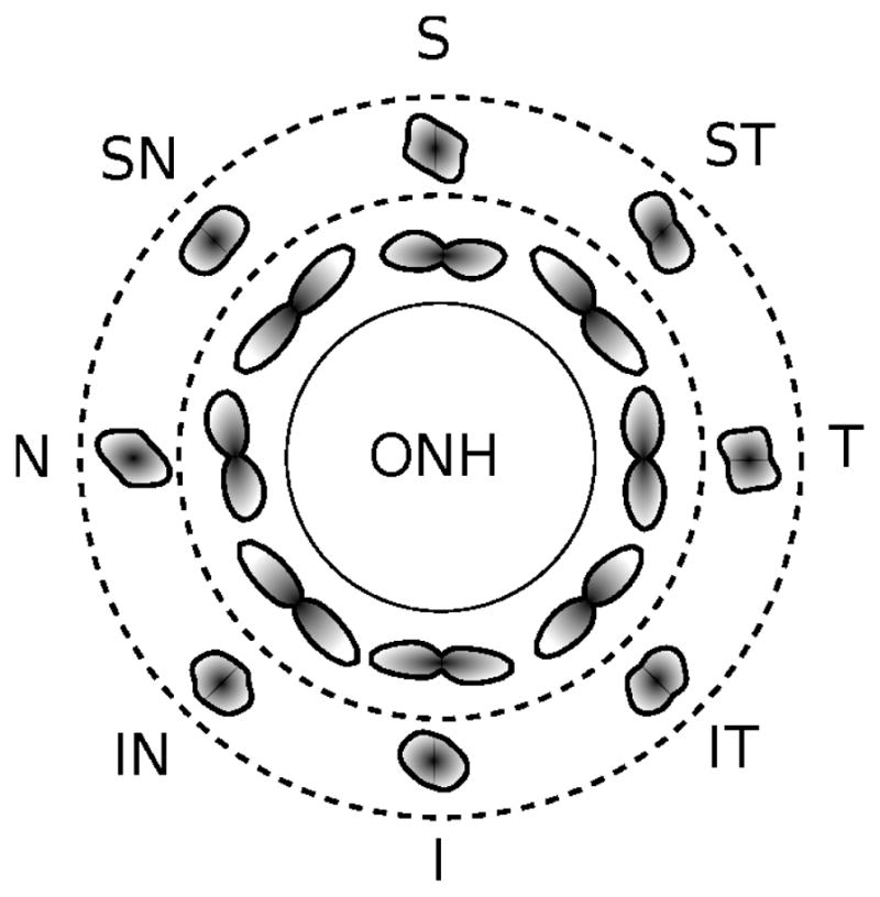
To characterize the material properties of posterior and peripapillary sclera from human donors, and to investigate the macro- and micro-scale strains as potential control mechanisms governing mechanical homeostasis. Posterior scleral shells from 9 human donors aged 57-90 years were subjected to IOP elevations from 5 to 45mmHg and the resulting full-field displacements were recorded using laser speckle interferometry. Eye-specific finite element models were generated based on experimentally measured scleral shell surface geometry and thickness. Inverse numerical analyses were performed to identify material parameters for each eye by matching experimental deformation measurements to model predictions using a microstructure-based constitutive formulation that incorporates the crimp response and anisotropic architecture of scleral collagen fibrils. The material property fitting produced models that fit both the overall and local deformation responses of posterior scleral shells very well. The nonlinear stiffening of the sclera with increasing IOP was well reproduced by the uncrimping of scleral collagen fibrils, and a circumferentially aligned ring of collagen fibrils around the scleral canal was predicted in all eyes. Macroscopic in-plane strains were significantly higher in peripapillary region then in the mid-periphery. In contrast, the meso- and micro-scale strains at the collagen network and collagen fibril level were not significantly different between regions. The elastic response of the posterior human sclera can be characterized by the anisotropic architecture and crimp response of scleral collagen fibrils. The similar collagen fibril strains in the peripapillary and mid-peripheral regions support the notion that the scleral collagen architecture including the circumpapillary ring of collagen fibrils evolved to establish optimal load bearing conditions at the collagen fibril level.

Keywords: Collagen fibril strain; Homeostasis; Inverse analysis; Sclera.


Figures

Figure 1
Figure 1
Multi-scale model of the posterior scleral shell. At the macro-scale, a spherical coordinate system θi is defined that fits the scleral surface best with the polar axis going through the center of the ONH. A1 and A2 are base vectors projected onto the scleral surface following the meridional θ1 and circumferential direction θ2. At the meso-scale, the collagen architecture is represented by one family of collagen fibrils assuming a von Mises distribution of the fibril orientations. φp – the angle that defines the preferred orientation M with respect to the circumferential direction A2; b – the concentration parameter that defines the degree of alignment of the collagen fibrils with respect to the preferred orientation M (b = 0: randomly distributed fibrils in the scleral plane; b = ∞: perfectly aligned fibrils along M). At the micro-scale, collagen fibrils are assumed to crimp into a helix when the sclera is unloaded. λaxial illustrates the stretch in the axis of a collagen fibril, which is used to calculate the average collagen network strain, and εfib illustrates the collagen fibril strain at the micro-scale. θ0 – the crimp angle; R0 – the radius of the helix; r0 – the radius of the collagen fibril crossection.
Figure 2
Figure 2
(a) The eye-specific FE mesh of one eye showing the posterior scleral shell and the lamina cribrosa. The mesh captures the original shape and thickness of each eye. (b) Section through the FE mesh showing the boundary conditions at the clamp, including the simple support at the outer surface and the spring support through the scleral thickness. (c) The extended mesh (red lines) used to model local variations in the collagen fibril architecture by interpolating the meso-structural parameters (φp, b) between the control points (red and green circles). A standard bi-linear interpolation of the control point values is performed in the spherical coordinate space for each element of the extended mesh. The extended mesh extends beyond the posterior scleral boundaries, where control points 13–16 are located at the virtual equator of the eye. d1, d2 represent variable meridional distances. The extended mesh can be rotated by an offset angle offset. The meso-structural parameters (φp, b) are pre-defined at the red control points and fitted at the green control points.
Figure 3
Figure 3
Top: columns show the comparison of experimentally-measured and FE model-predicted displacements for both eyes (both shown in right eye configuration) of one donor (81 years old) for three IOP elevations from 5 to 15, 30, and 45 mmHg. The rows show the comparison between the experimentally measured and computationally predicted meridional, circumferential, and radial surface displacements. Column and row-wise comparisons show that the predicted displacements are in good agreement with both: (i) the overall nonlinear displacement response and (ii) the localized deformation patterns of the experimental measurements. Bottom: the predicted collagen fibril architecture for both eyes showing the concentration of collagen fibrils (contour plot) along their preferred orientations (white lines). A ring of circumferentially aligned collagen fibrils is seen in the peripapillary scleral region around the scleral canal. Exp, Experimental.
Figure 4
Figure 4
Average collagen fibril density distributions in 8 sectors of the peripapillary and mid-peripheral region of the sclera averaged over all eyes, as represented by individual polar plots of the average collagen fibril distribution at each sector location. A high concentration of collagen fibrils in the circumferential direction can be seen across all sectors of the peripapillary region. More randomly organized collagen fibril orientations are observed in the mid-peripheral region.
Figure 5
Figure 5
From top to bottom, IOP versus the average in-plane strain, average collagen network strain, the average collagen fibril strain, and the percentile of locked collagen fibrils plots showing the model responses of all scleral shells (grey lines) and the mean response (black lines) for the peripapillary scleral (left plots) and mid-peripheral region (right plots). The in-plane strain and collagen network strain curves show the nonlinear and IOP-dependent stiffening of the sclera, while the collagen fibril strain increases nearly linear with increasing IOP. The average in-plane strain is higher in the peripapillary region than in the mid-periphery, while the average collagen network and fibril strains are almost identical for both regions. Collagen fibrils start to lock at about 7 mmHg and more fibrils get recruited to bear the load for increasing IOP. All plots show a significant variability in the data set.
Figure 6
Figure 6
Parameter study using the posterior scleral shell from the right eye of Donor 7 and the range of model parameters obtained from the fitting analysis using the full range of fitted parameters found in all the eyes. The influence of the two stiffness parameters (shear modulus of the tissue, elastic modulus of collagen fibrils) and two micro-structural parameters (collagen fibril crimp angle and ratio R0/r0) on the IOP-dependent responses is shown for the peripapillary scleral (red) and mid-peripheral region (blue): from left to right, the average in-plane strain, average collagen network strain, the average collagen fibril strain, and the percentile of locked collagen fibrils. For each parameter, the response for the maximum (dashed line with triangles up), minimum (dashed line with triangles down) and mean values (solid lines) are compared, while keeping the other model parameters at their mean values. The fitted meso-structural parameters (b, φp) were not varied.

References

    1. Başar Y, Weichert D. Nonlinear Continuum Mechanics of Solids. Springer Verlag; Berlin: 2000.
    1. Bhole AP, Flynn BP, Liles M, Saeidi N, Dimarzio CA, Ruberti JW. Mechanical strain enhances survivability of collagen micronet-works in the presence of collagenase: implications for load-bearing matrix growth and stability. Phil Trans R Soc A. 2009 Sep;367 (1902):3339–3362. - PMC - PubMed
    1. Boote C, Sorensen T, Coudrillier B, Myers K, Meek K, Quigley H, Nguyen T. Posterior scleral collagen architecture in normal and glaucoma human eyes, as determined using wide-angle x-ray scattering. ARVO Abstract. 2010;51:4900.
    1. Brest J, Maučec M. Self-adaptive differential evolution algorithm using population size reduction and three strategies. Soft Computing-A Fusion of Foundations, Methodologies and Applications. 2010;15 (11):2157–2174.
    1. Burgoyne CF, Downs JC, Bellezza AJ, Suh JKF, Hart RT. The optic nerve head as a biomechanical structure: a new paradigm for understanding the role of IOP-related stress and strain in the pathophysiology of glaucomatous optic nerve head damage. Prog Retina Eye Res. 2005 Jan;24 (1):39–73. - PubMed

Publication types