. 2013 May 30;3(5):1346-54.
doi: 10.1016/j.celrep.2013.04.014. Epub 2013 May 16.

A genetic screen identifies TCF3/E2A and TRIAP1 as pathway-specific regulators of the cellular response to p53 activation

Affiliations

A genetic screen identifies TCF3/E2A and TRIAP1 as pathway-specific regulators of the cellular response to p53 activation

Zdenek Andrysik et al. Cell Rep. .

Abstract

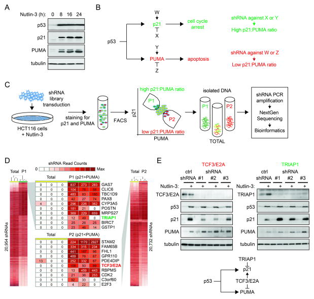
The p53 transcription factor participates in diverse cellular responses to stress, including cell-cycle arrest, apoptosis, senescence, and autophagy. The molecular mechanisms defining the ultimate outcome of p53 activation remain poorly characterized. We performed a genome-wide genetic screen in human cells to identify pathway-specific coregulators of the p53 target gene CDKN1A (p21), an inhibitor of cell-cycle progression, versus BBC3 (PUMA), a key mediator of apoptosis. Our screen identified numerous factors whose depletion creates an imbalance in the p21:PUMA ratio upon p53 activation. The transcription factor TCF3, also known as E2A, drives p21 expression while repressing PUMA across cancer cell types of multiple origins. Accordingly, TCF3/E2A depletion impairs the cell-cycle-arrest response and promotes apoptosis upon p53 activation by chemotherapeutic agents. In contrast, TRIAP1 is a specific repressor of p21 whose depletion slows down cell-cycle progression. Our results reveal strategies for driving cells toward specific p53-dependent responses.


Figures

Figure 1
Figure 1. A genome-wide shRNA screen identifies pathway-specific regulators within the p53 network
A. Western blot assays showing that activation of p53 with Nutlin-3 leads to concurrent induction of p21 and PUMA in HCT116 cells. B. Hypothetical factors W, X, Y and Z function as gene-specific coregulators of p21 and PUMA. C. Experimental design for shRNA screen leading to identification of gene-specific coregulators of p21 and PUMA. D. Heatmaps showing differential counts of shRNAs in the sorted populations dubbed P1 (high p21:PUMA ratio) and P2 (low p21:PUMA ratio) versus the Total population. Numbers represent the shRNA count in the three sets of triplicates. E. Different shRNAs against TCF3/E2A and TRIAP1 change the p21:PUMA expression ratio as predicted by the genetic screen. TCF3/E2A depletion reduces p21 expression and increases PUMA expression without affecting p53 accumulation. TRIAP1 knockdown leads to increased p21 expression with no effects on PUMA or p53 accumulation. See also Figure S1.
Figure 2
Figure 2. TCF3/E2A and TRIAP1 control the p21:PUMA expression ratio in diverse cancer cell types
A. Western blot and Q-RT-PCR assays show that TCF3/E2A functions as a coactivator of p21 expression and a repressor of PUMA expression in HCT116 colorectal carcinoma cells and U2OS osteosarcoma cells. See Figure S2 for data on A549 lung carcinoma cells B. TRIAP1 functions as a gene-specific repressor of p21. C,D. Q-RT-PCR analysis showing the effects of TCF3/E2A and TRIAP1 knockdown on diverse p53 target genes. All Q-RT-PCR experiments are expressed as averages of three biological replicates −/+ s.d. See also Figure S2.
Figure 3
Figure 3. TCF3/E2A affects p21 mRNA stability
A–E. TCF3/E2A binds to the p21 proximal promoters and its depletion does not affect p53 binding or RNAPII transactivation. ChIP analysis of the p21 locus with antibodies against TCF3/E2A (A), p53 (B), total RNAPII (C), Serine 5-phosphorylated RNAPII (S5P, D) and Serine 2-phoshoryalted RNAPII (E), in HCT116 cells expressing control shRNA of shRNA against TCF3/E2A, treated with DMSO or 10 μM Nutlin-3 (24 hours). The p21 gene map is shown at the bottom, with black boxes representing exons and the transcribed region highlighted by a gray box. Numbers at the bottom indicate the position of PCR amplicons used to analyze ChIP-enriched DNA relative to the transcription start site. n indicates the number of ChIPs performed with each antibody. F. TCF3/E2A depletion decreases p21 mRNA half life. mRNA half life assays were performed for the p21 and 14-3-3s (SFN) mRNAs by collecting RNA samples at the indicated times after Actinomycin D treatment in HCT116 cells expressing control or TCF3/E2A shRNAs, treated with DMSO or 10 μM Nutlin-3 (24 hours). Asterisks indicate significant different (p<0.05) between shControl and shTCR3/E2A samples treated with DMSO (blue) or Nutlin-3 (red) within a given time point. Data are expressed as averages of three experiments −/+ s.d.
Figure 4
Figure 4. TCF3/E2A controls the cellular response to p53 activation upon DNA damage in a p21/PUMA-dependent fashion
A. BrdU incorporation assays demonstrate that p21 is required for the cell cycle arrest response observed upon doxorubicin treatment (0.5 μM, 12 h) in HCT116 cells. B. Apoptotic index assays show that p21 attenuates p53/PUMA-dependent apoptosis in response to doxorubicin treatment. C. TCF3/E2A knockdown increases the apoptotic index of HCT116 cells upon doxorubicin treatment. D. Western blot assays show that TCF3/E2A depletion leads to a lower p21:PUMA ratio concurrently with increased levels of activated executioner caspase 3 upon doxorubicin treatment. E. TCF3/E2A knockdown enhances the apoptotic index only in cells expressing p21 and/or PUMA. Data in A, B, C and E are expressed as averages of three experiments −/+ s.d. See also Figure S3.

References

    1. Altura RA, Inukai T, Ashmun RA, Zambetti GP, Roussel MF, Look AT. The chimeric E2A-HLF transcription factor abrogates p53-induced apoptosis in myeloid leukemia cells. Blood. 1998;92:1397–1405. - PubMed
    1. Beckerman R, Donner AJ, Mattia M, Peart MJ, Manley JL, Espinosa JM, Prives C. A role for Chk1 in blocking transcriptional elongation of p21 RNA during the S-phase checkpoint. Genes Dev. 2009;23:1364–1377. - PMC - PubMed
    1. Brown CJ, Lain S, Verma CS, Fersht AR, Lane DP. Awakening guardian angels: drugging the p53 pathway. Nat Rev Cancer. 2009;9:862–873. - PubMed
    1. Bunz F, Dutriaux A, Lengauer C, Waldman T, Zhou S, Brown JP, Sedivy JM, Kinzler KW, Vogelstein B. Requirement for p53 and p21 to sustain G2 arrest after DNA damage. Science. 1998;282:1497–1501. - PubMed
    1. de Pooter RF, Kee BL. E proteins and the regulation of early lymphocyte development. Immunological reviews. 2010;238:93–109. - PMC - PubMed

Publication types

MeSH terms

Substances