. 2013 Jun 3;23(11):1024-30.
doi: 10.1016/j.cub.2013.04.049. Epub 2013 May 16.

Fluid shear stress on endothelial cells modulates mechanical tension across VE-cadherin and PECAM-1

Affiliations

Fluid shear stress on endothelial cells modulates mechanical tension across VE-cadherin and PECAM-1

Daniel E Conway et al. Curr Biol. .

Abstract

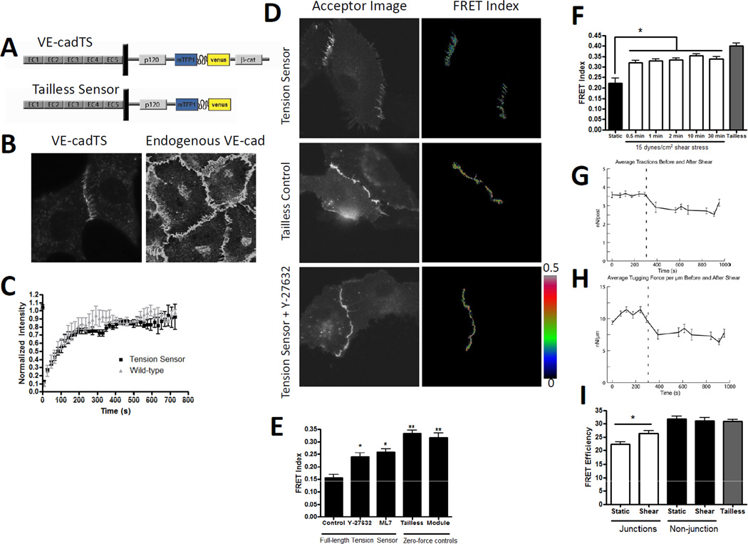
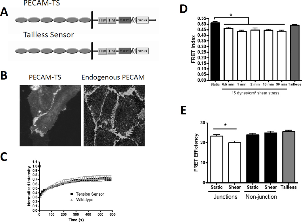
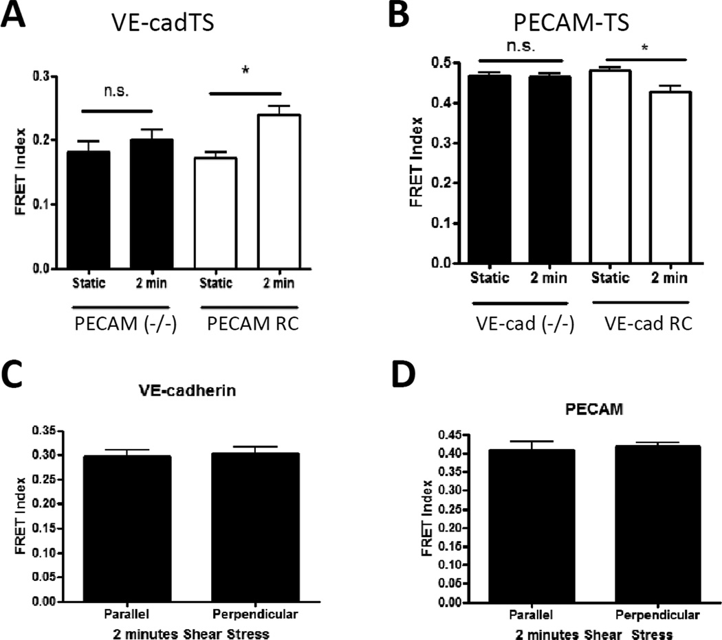
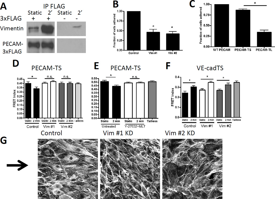
Fluid shear stress (FSS) from blood flow acting on the endothelium critically regulates vascular morphogenesis, blood pressure, and atherosclerosis. FSS applied to endothelial cells (ECs) triggers signaling events including opening of ion channels, activation of signaling pathways, and changes in gene expression. Elucidating how ECs sense flow is important for understanding both normal vascular function and disease. EC responses to FSS are mediated in part by a junctional mechanosensory complex consisting of VE-cadherin, PECAM-1, and VEGFR2. Previous work suggested that flow increases force on PECAM-1, which initiates signaling. Deletion of PECAM-1 blocks responses to flow in vitro and flow-dependent vascular remodeling in vivo. To understand this process, we developed and validated FRET-based tension sensors for VE-cadherin and PECAM-1 using our previously developed FRET tension biosensor. FRET measurements showed that in static culture, VE-cadherin in cell-cell junctions bears significant myosin-dependent tension, whereas there was no detectable tension on VE-cadherin outside of junctions. Onset of shear stress triggered a rapid (<30 s) decrease in tension across VE-cadherin, which paralleled a decrease in total cell-cell junctional tension. Flow triggered a simultaneous increase in tension across junctional PECAM-1, while nonjunctional PECAM-1 was unaffected. Tension on PECAM-1 was mediated by flow-stimulated association with vimentin. These data confirm the prediction that shear increases force on PECAM-1. However, they also argue against the current model of passive transfer of force through the cytoskeleton to the junctions, showing instead that flow triggers cytoskeletal remodeling, which alters forces across the junctional receptors.


Figures

Figure 1
Figure 1. Design and validation of a VE-cadherin tension sensor; effects of flow on junctional forces
(A) Schematic of the VE-cadherin tension sensor (VEcadTS) and the tailless control. The sensor module was inserted into the VE-cadherin cytoplasmic tail between the p120-catenin and beta-catenin binding domains. (B) Localization of VEcadTS in VE-cadherin−/− endothelial cells (left) and endogenous VE-cadherin in HUVECs (right). (C) BAECs were transfected with either VE-cadherin tension sensor or VE-cadherin C-terminal tagged venus. A region in the junction was photobleached and the fluorescence recovery measured. The graph is averaged from four separate photobleaching experiments per condition. (D) Corrected FRET images in pseudocolor (right) or background subtracted acceptor images (left) of BAEC expressing VEcadTS with or without 10 µM ROCK inhibitor Y27632, or the tailless control. (E) Quantification of the FRET index from cell junctions of BAEC expressing VEcadTS untreated or treated with 10 µM Y27632 or 10 µM of MLCK inhibitor ML7 for 30 minutes. The FRET index was also measured from junctions of BAEC expressing the VE-cadherin tailless control and cells expressing soluble TS module. > 20 junctions were measured per condition, values are means ± standard error from one experiment. Similar results were obtained in 3 independent experiments. (F) BAECs expressing VEcadTS were subjected to 15 dynes/cm2 shear stress for the indicated times. FRET index was measured at > 20 cell-cell junctions per condition. VE-cadherin tailless sensor was were used as a zero-force control. Similar results were obtained in 3 independent experiments. (G-H) BAEC on mPAD islands overnight were subjected to shear stress. Each island was imaged under static conditions for 5 minutes, then for 5 min after onset of shear stress at 15 dynes/cm2 (indicated by the dashed vertical line). Traction forces (G) and tugging (junctional) forces (H) were determined as described in the Methods. Values are means ± standard error from 4 independent experiments. (I) FLIM analysis of junctions and non-junctional regions of BAEC expressing VEcadTS with or without shear stress for 2 min. Tailless results are from junctional regions. Values are means ± standard error, n=6–10 junctions per condition.
Figure 2
Figure 2. Design and validation of a PECAM-1 tension sensor; effects of flow on PECAM tension
(A) Schematic of PECAM-TS and tailless control. The sensor module was inserted below the second ITIM domain, before exon 15. (B) Localization of the PECAM-1 tension sensor at cell-cell junctions expressed in BAEC (left) and endogenous PECAM-1 in HUVEC (right). (C) BAECs were transfected with either PECAM-TS or PECAM-1 C-terminal tagged venus. Junctional regions were photobleached and fluorescence recovery measured. Values are means ± standard error, n=4. (D) BAECs expressing the PECAM-TS were subjected to 15 dynes/cm2 shear stress for the indicated times. FRET index was measured at >20 cell-cell junctions per condition. The PECAM-1 tailless sensor were used as a zero-force control. Similar results were obtained in 3 independent experiments. (E) FLIM analysis of junctions and non-junctional regions of BAEC expressing the PECAM-1 sensor from static cultured cells and cells exposed to 2 minutes shear stress. Tailless values are from junctional regions. Values are means ± standard error, n=12 junctions per condition.
Figure 3
Figure 3. Interdependence of force changes on PECAM and VE-cadherin expression and junctional orientation
(A) PECAM−/− cells were co-transfected with VEcadTS plus either empty vector (−/−) or wild-type PECAM (RC). (B) VE-cadherin−/− cells were co-transfected with PECAMTS plus either empty vector (−/−) or wild-type VE-cadherin (RC). Cells were left unstimulated or subjected to shear stress as before. Values are means ± standard error from one experiment. Similar results were obtained in 3 independent experiments. (C-D) FRET index at junctions of BAEC exposed to 2 minutes shear stress were classified based on their orientation relative to the direction of flow. Values are means ± standard error, n=12=17 junctions per condition. No significant difference was observed for either VE-cadherin (C) or PECAM (D) tension between parallel and perpendicular oriented junctions. Similar results were obtained in 3 independent experiments.
Figure 4
Figure 4. Role of vimentin association with PECAM
(A) BAEC expressing PECAM-3xFLAG were immunoprecipitated with anti-FLAG beads and blotted for vimentin and FLAG. Similar results were obtained with 3 different experiments. (B) BAEC expressing human PECAM were seeded on PECAM antibody coated coverslips. Cells with each vimentin knockdown sequence had significantly reduced adhesion as compared to control cells. Graph is an average of four independent experiments. (C) COS-7 cells expressing wild-type (WT) PECAM, PECAM-TS, or PECAM-TL were seeded on PECAM antibody coated coverslips. Cells expressing PECAM-TL had significantly reduced adhesion as compared to PECAM-TS. Graph is an average of four independent experiments. (D) BAEC with vimentin knockdown had no PECAM force change with 2 minutes of shear stress. Similar results were obtained in 3 independent experiments. (E) BAEC pretreated with 10 uM Y-27632 and 10 uM ML7 prior to shear stress had no PECAM force change with 2 minutes shear stress. Similar results were obtained in 3 independent experiments. (F) BAEC with vimentin knockdown had similar decreases in VE-cadherin tension with shear stress as compared to control cells. Similar results were obtained in 3 independent experiments. (G) BAEC with vimentin knockdown and control cells were exposed to 15 dynes/cm2 shear stress for 24 hours. Cells were fixed and stained with phalloidin. The arrow indicates the direction of flow. Similar results were obtained in 3 independent experiments.

References

    1. Hahn C, Schwartz MA. Mechanotransduction in vascular physiology and atherogenesis. Nature reviews. Molecular cell biology. 2009;10:53–62. - PMC - PubMed
    1. Tzima E, Irani-Tehrani M, Kiosses WB, Dejana E, Schultz DA, Engelhardt B, Cao G, DeLisser H, Schwartz MA. A mechanosensory complex that mediates the endothelial cell response to fluid shear stress. Nature. 2005;437:426–431. - PubMed
    1. Osawa M, Masuda M, Kusano K, Fujiwara K. Evidence for a role of platelet endothelial cell adhesion molecule-1 in endothelial cell mechanosignal transduction: is it a mechanoresponsive molecule? The Journal of cell biology. 2002;158:773–785. - PMC - PubMed
    1. Chiu Y-J, McBeath E, Fujiwara K. Mechanotransduction in an extracted cell model: Fyn drives stretch- and flow-elicited PECAM-1 phosphorylation. The Journal of cell biology. 2008;182:753–763. - PMC - PubMed
    1. Chen Z, Rubin J, Tzima E. Role of PECAM-1 in arteriogenesis and specification of preexisting collaterals. Circulation research. 2010;107:1355–1363. - PMC - PubMed

Publication types

MeSH terms