. 2013 Jun 3;23(11):937-46.
doi: 10.1016/j.cub.2013.04.028. Epub 2013 May 16.

Condensin and the spindle midzone prevent cytokinesis failure induced by chromatin bridges in C. elegans embryos

Affiliations

Condensin and the spindle midzone prevent cytokinesis failure induced by chromatin bridges in C. elegans embryos

Joshua N Bembenek et al. Curr Biol. .

Abstract

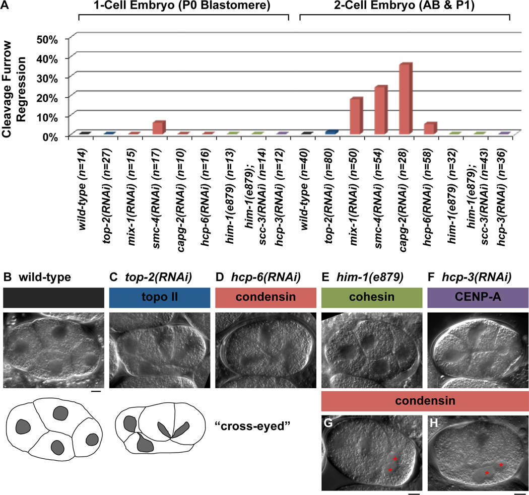
Background: During cell division, chromosomes must clear the path of the cleavage furrow before the onset of cytokinesis. The abscission checkpoint in mammalian cells stabilizes the cleavage furrow in the presence of a chromatin obstruction. This provides time to resolve the obstruction before the cleavage furrow regresses or breaks the chromosomes, preventing aneuploidy or DNA damage. Two unanswered questions in the proposed mechanistic pathway of the abscission checkpoint concern factors involved in (1) resolving the obstructions and (2) coordinating obstruction resolution with the delay in cytokinesis.

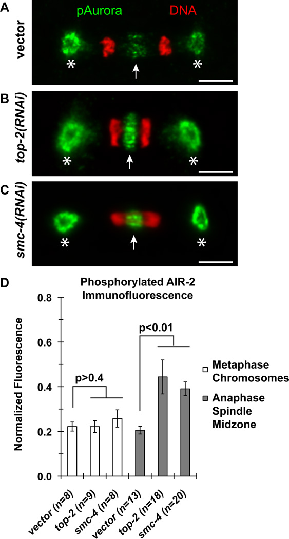
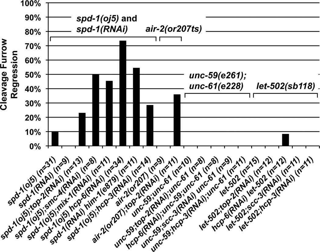
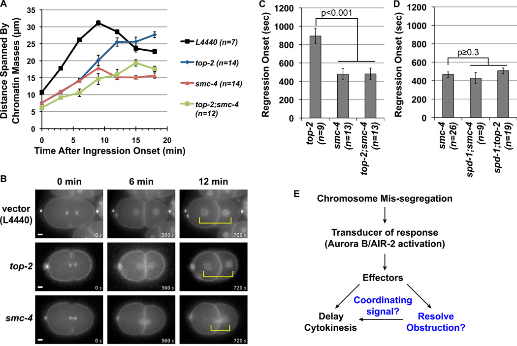
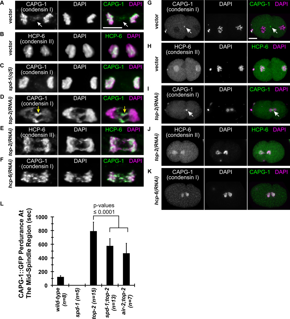
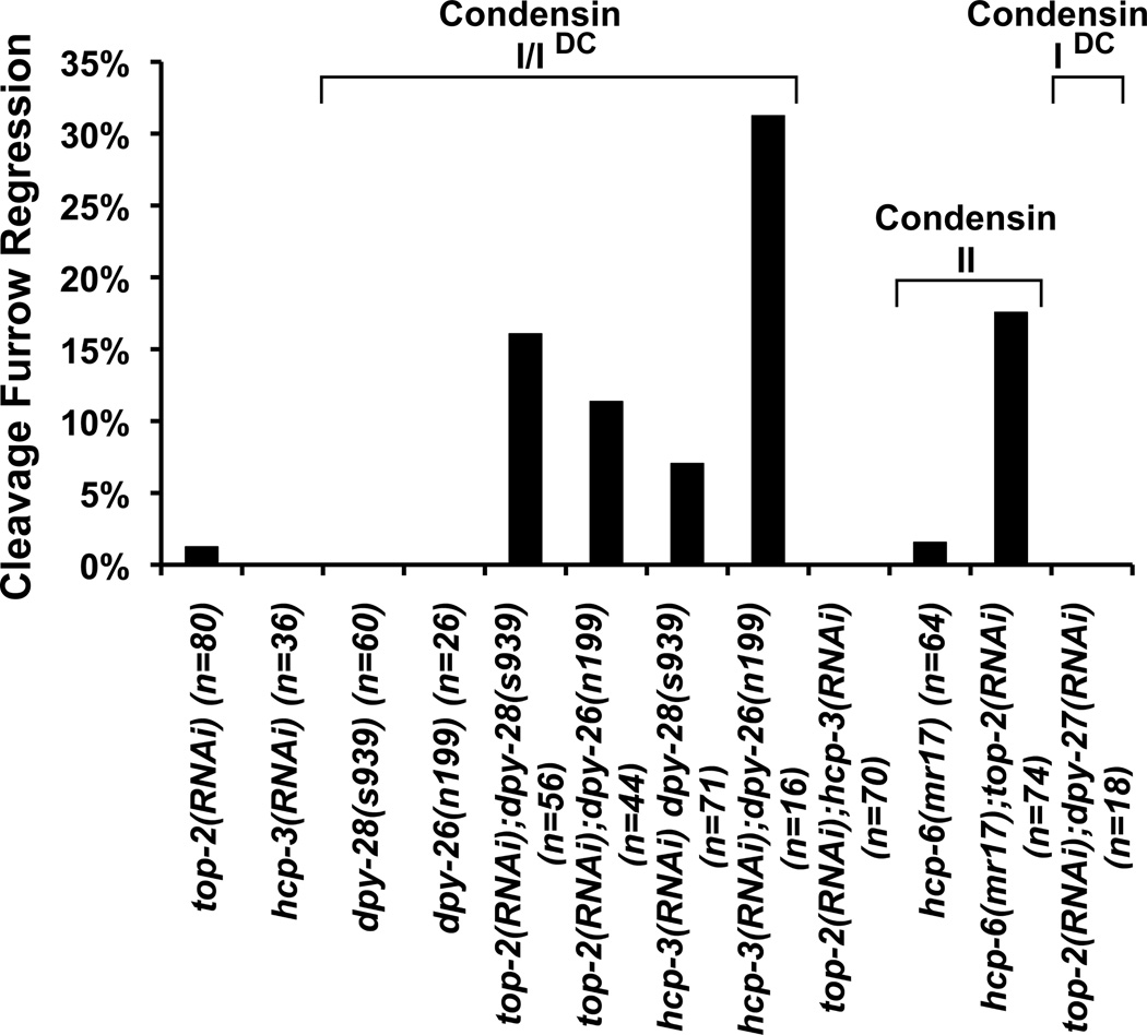
Results: We found that the one-cell and two-cell C. elegans embryos suppress furrow regression following depletion of essential chromosome-segregation factors: topoisomerase II(TOP-2), CENP-A(HCP-3), cohesin, and to a lesser degree, condensin. Chromatin obstructions activated Aurora B(AIR-2) at the spindle midzone, which is needed for the abscission checkpoint in other systems. Condensin I, but not condensin II, localizes to the spindle midzone in anaphase and to the midbody during normal cytokinesis. Interestingly, condensin I is enriched on chromatin bridges and near the midzone/midbody in an AIR-2-dependent manner. Disruption of AIR-2, the spindle midzone, or condensin leads to cytokinesis failure in a chromatin-obstruction-dependent manner. Examination of the condensin-deficient embryos uncovered defects in both the resolution of the chromatin obstructions and the maintenance of the stable cleavage furrow.

Conclusions: We postulate that condensin I is recruited by Aurora B(AIR-2) to aid in the resolution of chromatin obstructions and also helps generate a signal to maintain the delay in cytokinesis.


Figures

Figure 1
Figure 1
Nomarski DIC time-lapse analysis of cell division in the 1-cell and 2-cell embryos. (A) Graph represents the percentages of P0 and AB/P1 cell divisions in wild-type and RNAi-treated embryos that exhibited a cytokinesis cleavage furrow regression defect in the presence of chromatin obstruction as monitored indirectly by the cross-eyed nuclei phenotype. The number of P0 or AB/P1 cell divisions (n) examined per condition is provided. (B–F) Representative still images of 4-cell embryos taken from Nomarski DIC time-lapse movies. Cartoon tracings show a wild-type embryo with each nucleus (grey) centered within the cell, compared with the top-2(RNAi) embryo which exhibited the defective cross-eyed nuclei morphology. (G–H) Nomarski DIC images of a condensin (G) mix-1 RNAi embryo and (H) smc-4 RNAi embryo after the P1 cell division had failed resulting in a binucleated cell (red asterisks). Scale bar, 5 µm.
Figure 2
Figure 2
Quantitative immunofluorescence microcopy of phosphorylated AIR-2 in 1-cell embryos. (A–C) Representative micrographs of phosphorylated Aurora kinase immunostaining recognizing phosphorylated AIR-1 at the centrosomes (asterisks) and phosphorylated AIR-2 at the spindle midzone (white arrows) of the 1-cell P0 blastomere division at anaphase. Phosphorylated AIR-1 and AIR-2 staining are shown in green and DAPI-DNA fluorescence shown in red. Scale bars, 5 µm. (D) Graph represents the average level of phosphorylated AIR-2 staining on metaphase chromosomes and at the anaphase spindle midzone normalized to the level of phosphorylated AIR-1 staining detected at the anterior spindle pole. The number of embryos (n) quantified per condition is provided. Error bars, SEM. The p-values are determined by two-tailed t-Tests.
Figure 3
Figure 3
Nomarski DIC time-lapse analysis of cell division. Graph represents the percentages of cell divisions exhibiting the cleavage furrow regression defect after chromosome mis-segregation were induced in the spd-1(RNAi) spd-1(oj5) mutant, the air-2(or207) mutant (acute inactivation), the unc-59(e261);unc-61(e228) mutant, and the let-502(sb118) mutant backgrounds in the 1-cell embryo. The number of cell divisions (n) examined per condition is indicated.
Figure 4
Figure 4
Quantification of the extent of chromosome separation and the onset of the cleavage furrow regression in 1-cell P0 blastomeres. (A) Graph represents the average distance spanned by the separating chromosomes (y-axis) as a function of time after the onset of cleavage furrow ingression (x-axis). (B) Representative still images from the mCherry fluorescence time-lapse movies used for the quantification in (A). The movies are synchronized to the onset of ingression as t = 0 min. (C and D) Graphs represent the average time after the onset of ingression when the cleavage furrow began to regress. The p-values are determined by two-tailed t-Tests in comparisons to top-2(RNAi) and smc-4(RNAi) as indicated. For all graphs (A, C and D) the error bars represent SEM and n indicates the number of embryos examined for each test condition. (E) A schematic model for the response to chromosome mis-segregation mediated by AIR-2 activation to halt cytokinesis and facilitate obstruction resolution. The putative functions for condensin in aiding obstruction resolution and providing a signal to maintain the stable cleavage furrow are shown in blue.
Figure 5
Figure 5
Localization of condensin I but not condensin II specific subunits at the midzone and midbody. Immunofluorescence micrographs using antibodies to condensin I CAPG-1 and condensin II HCP-6 subunits in RNAi-treated embryos at anaphase in the P0 division (A-F, 5 µm scale bar) and at the midbody between the AB and P1 blastomeres (G-K, 10 µm scale bar). (L) The graph represents the average time duration that CAPG-1::GFP was detected at the mid-spindle region from the onset of anaphase. The number of embryos (n) examined per condition is indicated. Error bars, SD. The p-values are determined by two-tailed t-Tests.
Figure 6
Figure 6
Condensin I and II are both required for robust cytokinesis in presence of chromatin obstruction. The graph represents the percentages of mutant and RNAi-treated embryos that exhibited a cytokinesis cleavage furrow regression defect in the AB and P1 blastomere divisions as monitored by Nomarski DIC time-lapse microscopy. The number of AB and P1 cell divisions (n) examined is provided for each test condition.

References

    1. Steigemann P, Wurzenberger C, Schmitz MH, Held M, Guizetti J, Maar S, Gerlich DW. Aurora B-mediated abscission checkpoint protects against tetraploidization. Cell. 2009;136:473–484. - PubMed
    1. Carlton JG, Caballe A, Agromayor M, Kloc M, Martin-Serrano J. ESCRT-III governs the Aurora B-mediated abscission checkpoint through CHMP4C. Science. 2012;336:220–225. - PMC - PubMed
    1. Norden C, Mendoza M, Dobbelaere J, Kotwaliwale CV, Biggins S, Barral Y. The NoCut pathway links completion of cytokinesis to spindle midzone function to prevent chromosome breakage. Cell. 2006;125:85–98. - PubMed
    1. Carmena M, Ruchaud S, Earnshaw WC. Making the Auroras glow: regulation of Aurora A and B kinase function by interacting proteins. Curr. Opin. Cell Biol. 2009;21:796–805. - PMC - PubMed
    1. Ruchaud S, Carmena M, Earnshaw WC. Chromosomal passengers: conducting cell division. Nat. Rev. Mol. Cell Biol. 2007;8:798–812. - PubMed

Publication types

MeSH terms

LinkOut - more resources