

Nerve injury-induced changes in Homer/glutamate receptor signaling contribute to the development and maintenance of neuropathic pain
- PMID: 23685007
- PMCID: PMC6371789
- DOI: 10.1016/j.pain.2013.03.035
Nerve injury-induced changes in Homer/glutamate receptor signaling contribute to the development and maintenance of neuropathic pain
Abstract
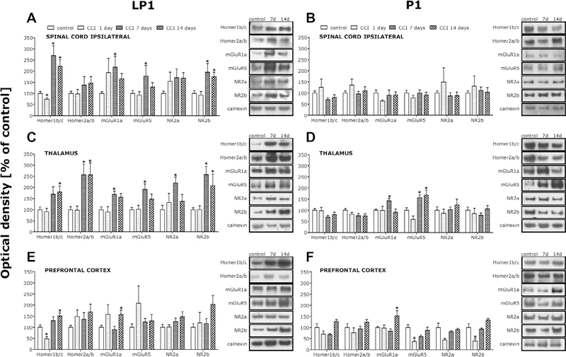
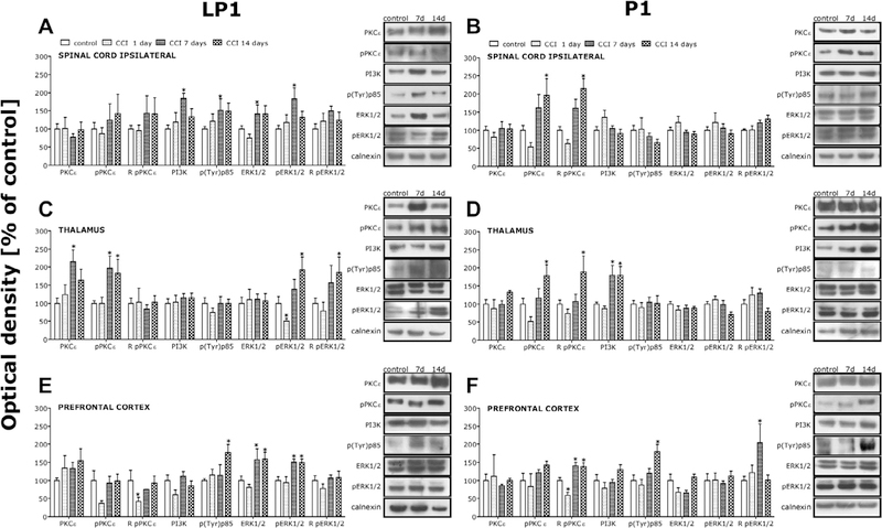
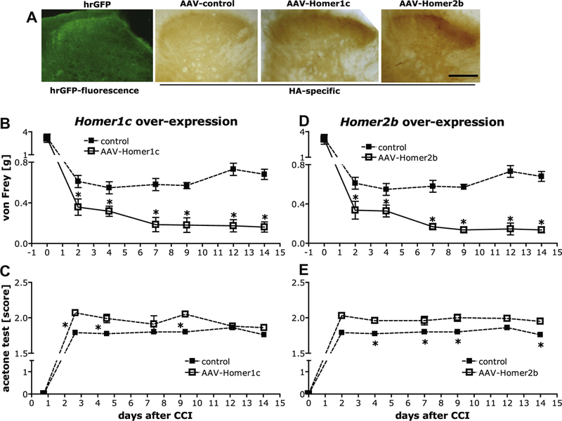
While group 1 metabotropic glutamate receptors (mGluRs) and ionotropic N-methyl-d-aspartate (NMDA) receptors regulate nociception, the precise molecular mechanism(s) contributing to glutamate signaling in chronic pain remain unclear. Here we not only confirmed the key involvement of Homer proteins in neuropathic pain, but also distinguished between the functional roles for different Homer family members and isoforms. Chronic constriction injury (CCI) of the sciatic nerve induced long-lasting, time-dependent increases in the postsynaptic density expression of the constitutively expressed (CC) isoforms Homer1b/c and/or Homer2a/b in the spinal dorsal horn and supraspinal structures involved in nociception (prefrontal cortex, thalamus), that co-occurred with increases in their associated mGluRs, NR2 subunits of the NMDA receptor, and the activation of downstream kinases. Virus-mediated overexpression of Homer1c and Homer2b after spinal (intrathecal) virus injection exacerbated CCI-induced mechanical and cold hypersensitivity, however, Homer1 and Homer2 gene knockout (KO) mice displayed no changes in their neuropathic phenotype. In contrast, overexpression of the immediate early gene (IEG) Homer1a isoform reduced, while KO of Homer1a gene potentiated neuropathic pain hypersensitivity. Thus, nerve injury-induced increases in CC-Homers expression promote pain in pathological states, but IEG-Homer induction protects against both the development and maintenance of neuropathy. Additionally, exacerbated pain hypersensitivity in transgenic mice with reduced Homer binding to mGluR5 supports also an inhibitory role for Homer interactions with mGluR5 in mediating neuropathy. Such data indicate that nerve injury-induced changes in glutamate receptor/Homer signaling contribute in dynamic but distinct ways to neuropathic pain processing, which has relevance for the etiology of chronic pain symptoms and its treatment.
Keywords: Group 1 metabotropic glutamate receptors; Homer proteins; NMDA receptors; Neuropathic pain; Spinal cord.
Copyright © 2013 International Association for the Study of Pain. Published by Elsevier B.V. All rights reserved.
Conflict of interest statement
Conflict of interest statement
The authors disclose no conflict of interest in respect of this work.
Figures


Similar articles
-
Homers at the Interface between Reward and Pain.Front Psychiatry. 2013 Jun 7;4:39. doi: 10.3389/fpsyt.2013.00039. eCollection 2013. Front Psychiatry. 2013. PMID: 23761764 Free PMC article.
-
Early changes in Homer1 proteins in the spinal dorsal horn are associated with loose ligation of the rat sciatic nerve.Anesth Analg. 2009 Dec;109(6):2000-7. doi: 10.1213/ANE.0b013e3181beea9b. Anesth Analg. 2009. PMID: 19923532 Free PMC article.
-
Imbalances in prefrontal cortex CC-Homer1 versus CC-Homer2 expression promote cocaine preference.J Neurosci. 2013 May 8;33(19):8101-13. doi: 10.1523/JNEUROSCI.1727-12.2013. J Neurosci. 2013. PMID: 23658151 Free PMC article.
-
The Role of Stress-Induced Changes of Homer1 Expression in Stress Susceptibility.Biochemistry (Mosc). 2021 Jun;86(6):613-626. doi: 10.1134/S0006297921060018. Biochemistry (Mosc). 2021. PMID: 34225586 Review.
-
The Homer family and the signal transduction system at glutamatergic postsynaptic density: potential role in behavior and pharmacotherapy.Psychopharmacol Bull. 2003 Summer;37(3):51-83. Psychopharmacol Bull. 2003. PMID: 14608240 Review.
Cited by
-
Neuroplasticity of ascending and descending pathways after somatosensory system injury: reviewing knowledge to identify neuropathic pain therapeutic targets.Spinal Cord. 2016 May;54(5):330-40. doi: 10.1038/sc.2015.225. Epub 2016 Jan 12. Spinal Cord. 2016. PMID: 26754470 Review.
-
AMPAkines and morphine provide complementary analgesia.Behav Brain Res. 2017 Sep 15;334:1-5. doi: 10.1016/j.bbr.2017.07.020. Epub 2017 Jul 19. Behav Brain Res. 2017. PMID: 28734765 Free PMC article.
-
Targeting the Microglial Signaling Pathways: New Insights in the Modulation of Neuropathic Pain.Curr Med Chem. 2016;23(26):2908-2928. doi: 10.2174/0929867323666160607120124. Curr Med Chem. 2016. PMID: 27281131 Free PMC article. Review.
-
Pain hypersensitivity in a pharmacological mouse model of attention-deficit/hyperactivity disorder.Proc Natl Acad Sci U S A. 2022 Jul 26;119(30):e2114094119. doi: 10.1073/pnas.2114094119. Epub 2022 Jul 19. Proc Natl Acad Sci U S A. 2022. PMID: 35858441 Free PMC article.
-
Overexpression of Homer1a in the basal and lateral amygdala impairs fear conditioning and induces an autism-like social impairment.Mol Autism. 2016 Feb 29;7:16. doi: 10.1186/s13229-016-0077-9. eCollection 2016. Mol Autism. 2016. PMID: 26929812 Free PMC article.
References
-
- Aira Z, Buesa I, Gallego M, García del Caño G, Mendiable N, Mingo J, Rada D, Bilbao J, Zimmermann M, Azkue JJ. Time-dependent cross talk between spinal serotonin 5-HT2A receptor and mGluR1 subserves spinal hyperexcitability and neuropathic pain after nerve injury. J Neurosci 2012;32:13568–81. - PMC - PubMed
-
- Ango F, Prézeau L, Muller T, Tu JC, Xiao B, Worley PF, Pin JP, Bockaert J, Fagni L. Agonist-independent activation of metabotropic glutamate receptors by the intracellular protein Homer. Nature 2001;411:962–5. - PubMed
-
- Bennett GJ, Xie YK. A peripheral mononeuropathy in rat that produces disorders of pain sensation like those seen in man. PAIN® 1988;33:87–107. - PubMed
Publication types
MeSH terms
Substances
Grants and funding
LinkOut - more resources
Full Text Sources
Other Literature Sources
Research Materials
