. 2013 Jun 4;21(6):1042-50.
doi: 10.1016/j.str.2013.04.010. Epub 2013 May 16.

Crystal structure of the DNA cytosine deaminase APOBEC3F: the catalytically active and HIV-1 Vif-binding domain

Affiliations

Crystal structure of the DNA cytosine deaminase APOBEC3F: the catalytically active and HIV-1 Vif-binding domain

Markus-Frederik Bohn et al. Structure. .

Abstract

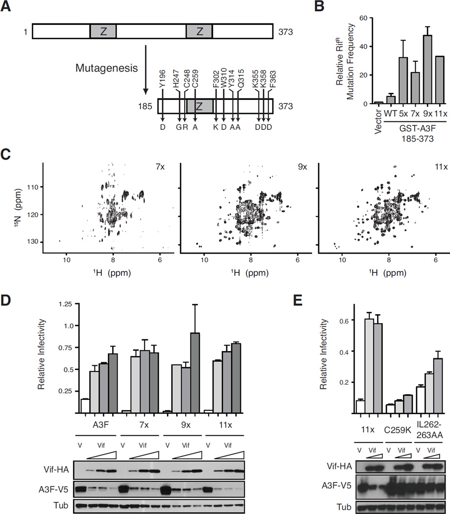
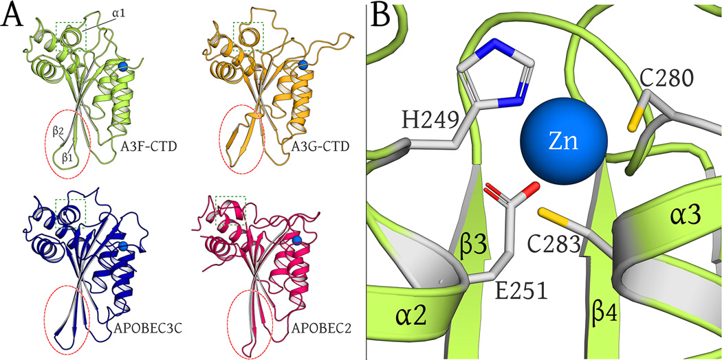
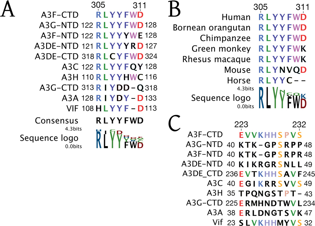
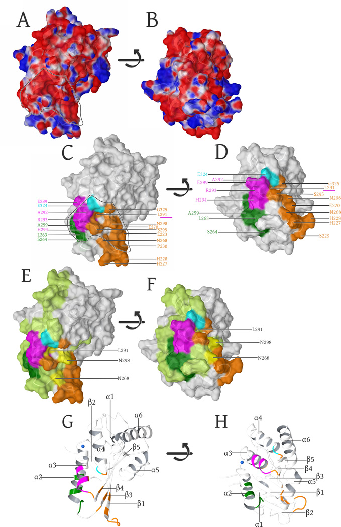
Human APOBEC3F is an antiretroviral single-strand DNA cytosine deaminase, susceptible to degradation by the HIV-1 protein Vif. In this study the crystal structure of the HIV Vif binding, catalytically active, C-terminal domain of APOBEC3F (A3F-CTD) was determined. The A3F-CTD shares structural motifs with portions of APOBEC3G-CTD, APOBEC3C, and APOBEC2. Residues identified to be critical for Vif-dependent degradation of APOBEC3F all fit within a predominantly negatively charged contiguous region on the surface of A3F-CTD. Specific sequence motifs, previously shown to play a role in Vif susceptibility and virion encapsidation, are conserved across APOBEC3s and between APOBEC3s and HIV-1 Vif. In this structure these motifs pack against each other at intermolecular interfaces, providing potential insights both into APOBEC3 oligomerization and Vif interactions.


Figures

Fig. 1
Fig. 1. Derivation of an active A3F variant for structural studies
(A) Schematic of the N-terminal deletion and amino acid substitutions rendered on wildtype A3F to yield the soluble variant A3F185-373-11x. (B) Relative capacity of GST-A3F185-373 (WT) and indicated derivatives to trigger RifR mutation in E. coli. A3F185-373-5x has C259A, F302K, W310K, Y314A, and Q315A, 7x is 5x plus Y196D and F363D, 9x is 7x plus K355D and K358D and W310D instead of W310K, and 11x is 9x plus H247G and C248R. Data are reported as the mean ± SEM of the median mutation frequencies acquired from multiple independent experiments (relative to each experiment’s vector-only control set to 1). (C) NMR spectra of 1H, 15N-labeled A3F-CTD 7x (~30 µM), 9x (~30 µM), and 11x (~100 µM). HSQC spectra were recorded at 298 K on a Bruker Avance 700 MHz instrument. (D & E) Single-cycle HIV-1 infectivity data. Histogram graphs showing the infectivity of Vif-deficient HIVIIIB produced in the presence of a fixed amount of each indicated full-length A3F-V5 construct and vector control (V) or increasing amounts of Vif-HA. Each bar represents the mean ± SD of duplicate infections normalized to the corresponding infectivity of virus produced in the absence of A3F. The 11x used in panel (D) has F363, whereas the 11x in panel (E) has the same set of mutations (D363) as those in the final crystallized construct depicted in panel (A) however this change does not affect restriction or Vif sensitivity.
Fig. 2
Fig. 2. Crystallographic Asymmetric unit, crystallographic binding and potential oligomerization interfaces
(A): The A3F-CTD crystal structure asymmetric unit comprises of four chains, illustrated in identical colors schematically and in the ribbon representation. Chains A/B and C/D form the heterologous interface and chains B/D form the isologous interface. The active-site catalytic zinc atoms are illustrated as blue spheres. (B): The A3F-CTD has a canonical cytosine deaminase fold with five β-strands and six α-helices. The conformational plasticity in β1-β2 loop region is highlighted (inset). (C): The isologous interface, formed between chains B (cyan) and D (yellow), is the largest interface in terms of surface area, burying a total of 657 Å2. (D): The isologous interface is splayed open to show interfacial residues (red/orange), with chains B and D turned by +90°/−90° respectively. Residues from the two chains B and D that are major contributors to this interface are labeled. (C): The heterologous interface formed between chains A (green) and B (cyan), and identical to the interface between chains C and D, is the second largest interface in terms of surface area, burying a total of 569 Å2. (D): The heterologous interface splayed open to show interfacial residues (orange/red), with chain A turned by +180°. Residues from the two chains A and B that are major contributors to this interface are labeled.
Fig. 3
Fig. 3. Structural Alignment of APOBEC family members
(A): Comparison of ribbon representations of A3F-CTD, A3G-CTD (3V4K), A3C (3VOW) and APOBEC2 (2NYT) crystal structures (chain A of all). The β2/ β2-β2’ region is circled in red to highlight similarities across all proteins except A3G-CTD. The α1 helix region is boxed in green to highlight similarities across all proteins except APOBEC2. (B): The catalytic zinc (blue sphere) is coordinated at the active site by H249, C280, C283 and indirectly via a water molecule (view occluded by zinc atom) with E251.
Fig. 4
Fig. 4. Sequence Conservation of A3F loops with regions of HIV-1 Vif
(A): The 305-RLYYFWD-311 motif of the A3F-CTD β4-α4 loop is conserved across human APOBEC3 protein domains and reappears in HIV-1 Vif. (B): Sequence alignment of the 305-RLYYFWD-311 motif of the A3-CTD β4-α4 loop across a wide array of mammalian APOBEC3 proteins. (C): The 223-EVVKHHSPVS-232 motif of the A3F-CTD β1-β2 loop is conserved in HIV-1 Vif.
Fig. 5
Fig. 5. Surface representation of the A3F-CTD structure
(A): Surface representation, colored by electrostatic potential (blue is positive, red is negative: − 0.05 -to- +0.05 kcal/mol (Maestro 9/Schrodinger, LLC)). The negatively charged groove region, electrostatically favored to bind the positively charged Vif is delineated by the dashed line and highlights the overlap with known Vif-binding regions in (C). (B). Same representation as in (A), with the surface rotated +90° (bottom facing out) along the horizontal axis in the image plane. (C): α3-helix (289–294, magenta) (Smith and Pathak, 2010), the α4 (324, cyan (Albin et al., 2010b), β3-strand (Y269) including part of the α2-helix and the adjacent loop region (L255, F258, C259, I262-S264, dark green) (Kitamura et al., 2012) are known Vif-binding regions that adjoin the β1-β2 (E223, H227-P230), β3(N268, E270), α3(L291, S295), β4(N298) and the α4-β5 (G325) regions (orange) involved in forming the interface. The α3 (L291) residue involved in our intermolecular interface as well as the previously characterized Vif-binding region (Smith and Pathak, 2010) is underlined in magenta. (D). Same coloring and labels as in (C), with the surface rotated +90° (bottom facing out) along the horizontal axis in the image plane. (E): Highlights of the complete mutational scan (Kitamura et al., 2012) of A3C (light green) projected onto our A3F-CTD structure. The regions that have were shown to be important for HIV-1 Vif binding are highlighted as above in (C). Yellow highlights and labels show the three residues that overlap between our interface and the A3C mutational scan not tested in A3F by (Kitamura et al., 2012), However L291 was independently probed (Smith and Pathak, 2010)) and found to be important in A3F binding to Vif. (F). Same coloring and labels as in (E), with the surface rotated +90° (bottom facing out) along the horizontal axis in the image plane. (G): Secondary structure of these views labeled for reference orientation, with the highlights colored as per (C). (H). Same coloring and labels as in (G), with the structure rotated +90° (bottom facing out) along the horizontal axis in the image plane.

References

    1. Albin JS, Hache G, Hultquist JF, Brown WL, Harris RS. Long-term restriction by APOBEC3F selects human immunodeficiency virus type 1 variants with restored Vif function. J Virol. 2010a;84:10209–10219. - PMC - PubMed
    1. Albin JS, Harris RS. Interactions of host APOBEC3 restriction factors with HIV-1 in vivo: implications for therapeutics. Expert reviews in molecular medicine. 2010;12:e4. - PMC - PubMed
    1. Albin JS, LaRue RS, Weaver Ja, Brown WL, Shindo K, Harjes E, Matsuo H, Harris RS. A single amino acid in human APOBEC3F alters susceptibility to HIV-1 Vif. The Journal of biological chemistry. 2010b;285:40785–40792. - PMC - PubMed
    1. Altschul SF, Madden TL, Schaffer AA, Zhang J, Zhang Z, Miller W, Lipman DJ. Gapped BLAST and PSI-BLAST: a new generation of protein database search programs. Nucleic Acids Res. 1997;25:3389–3402. - PMC - PubMed
    1. Auclair JR, Green KM, Shandilya S, Evans JE, Somasundaran M, Schiffer CA. Mass spectrometry analysis of HIV-1 Vif reveals an increase in ordered structure upon oligomerization in regions necessary for viral infectivity. Proteins. 2007;69:270–284. - PMC - PubMed

Publication types

MeSH terms

Substances