. 2013 May;189(1-2):43-53.
doi: 10.1016/j.molbiopara.2013.05.004. Epub 2013 May 17.

Identification of functional modules of AKMT, a novel lysine methyltransferase regulating the motility of Toxoplasma gondii

Affiliations

Identification of functional modules of AKMT, a novel lysine methyltransferase regulating the motility of Toxoplasma gondii

Senthilkumar Sivagurunathan et al. Mol Biochem Parasitol. 2013 May.

Abstract

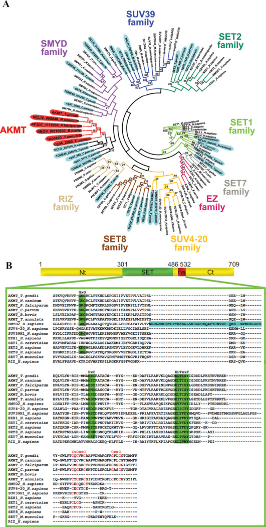
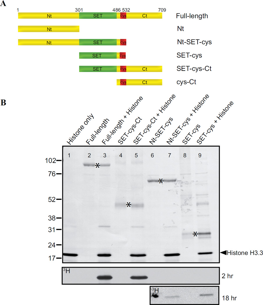
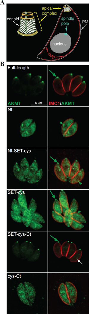
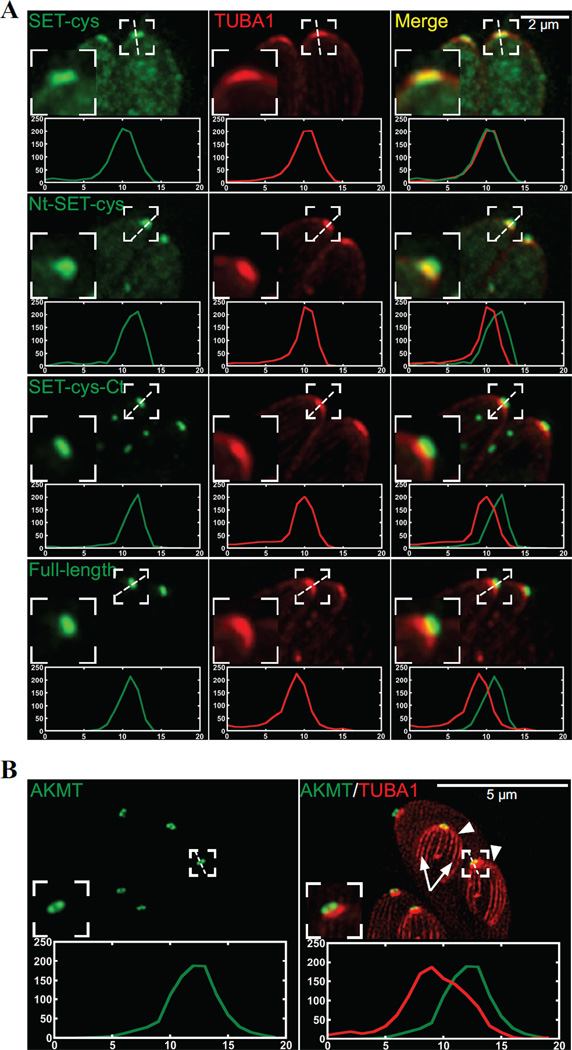
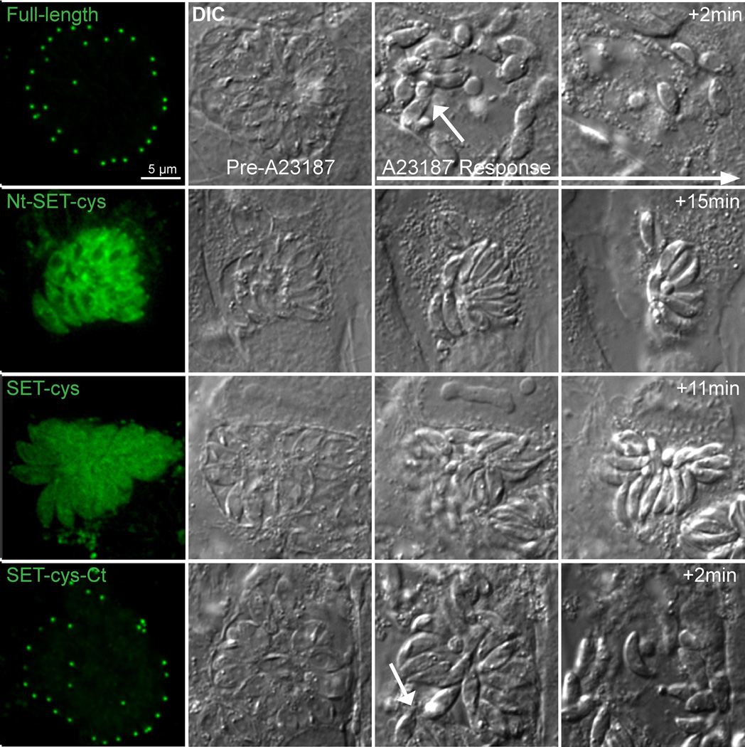
The intracellular parasite Toxoplasma gondii is a leading cause of congenital neurological defects. To cause disease, it must reiterate its lytic cycle through host cell invasion, replication, and parasite egress. This requires the parasite to sense changes in its environment and switch between the non-motile (for replication) and motile (for invasion and egress) states appropriately. Recently, we discovered a previously unknown mechanism of motility regulation in T. gondii, mediated by a lysine methyltransferase, AKMT (for Apical complex lysine (K) methyltransferase). When AKMT is absent, activation of motility is inhibited, which compromises parasite invasion and egress, and thus severely impairs the lytic cycle. Although the methyltransferase activity of AKMT has been established, the phylogenetic relationship of AKMT with other better studied lysine methyltransferases (KMTs) was not known. Also unknown was the functional relationships between different domains of AKMT. In this work we carried out phylogenetic analyses, which show that AKMT orthologs form a new subfamily of KMTs. We systematically generated truncation mutants of AKMT, and discovered that the predicted enzymatic domain alone is a very poor enzyme and cannot complement the function of AKMT in vivo. Interestingly, the N- and C-terminal domains of the AKMT have drastically different impacts on its enzyme activity, localization as well as in vivo function. Our results thus reveal that AKMT is an unusual, parasite-specific enzyme and identified regions and interactions within this novel lysine methyltransferase that can be used as drug targets.


Figures

Figure 1
Figure 1. Phylogenetic analyses of KMTs
A) Phylogenetic analysis of SET and post-SET domains from AKMT orthologs and KMTs representing several other subfamilies of SET-domain containing methyltransferases. The tree shows that AKMT orthologs form a distinct clade close to the SMYD subfamily. Gene identifiers used are either accession numbers or, when available, annotated gene names in publicly accessible databases (e.g. GenBank, EuPathDB or ToxoDB). The names for apicomplexan KMTs are shaded (AKMTs in red and others in light blue). The name and branches of each subfamily are color-coded. Bootstrap percentages higher than 20 are displayed. B) Protein sequence alignment of AKMT SET and post-SET domains with representative members of the KMT superfamily. Green highlighting indicates highly conserved motifs found in SET domain-containing proteins. Letters highlighted in red indicate cysteine cluster. Note that although AKMTs and SMYDs clusters are closely placed in the phylogenetic analysis, they each have some distinct features. SMYDs have a MYND zinc finger domain (highlighted in blue) within the SET domain, a unique and fundamental feature of this subfamily. The post-SET cysteine cluster in AKMTs (CXCX2CX11CX2C) contains 2 more cysteine residues and is significantly longer than that (CXCX2C) in SMYDs. Accession numbers of the proteins used in the alignment are: AKMT_T.gondii: TGME49_016080, AKMT_N.caninum: NCLIV_060040, AKMT_P.falciparum: PF3D7_1115200, AKMT_C.parvum: cgd4_2090, AKMT_B.bovis: BBOV_IV010830, AKMT_T.annulata: TA06820, SMYD2_H.sapiens: NP_064582, SUV4-20H1_H.sapiens: NP_060105, SUV39H1_H.sapiens: NP_003164, EZH1_H.sapiens: NP_001982, SET1_S.cerevisiae: EDN62358, SETD2_H.sapiens: NP_054878, SETD8_H.sapiens: Q9NQR1, SETD7_Mus musculus: NP_542983, RIZ_H.sapiens: Q13029.
Figure 2
Figure 2. The effect of free calcium concentration on AKMT lysine methyltransferase activity
In vitro methyltransferase assays were performed with 2 µg Xenopus histone H3.3, 2.5 µM 3H-SAM and 0.2 µg of recombinant FLAG-AKMT-full-length in the presence of < 0.001 µM , 10 µM, 30 µM, 90 µM, and 180 µM free Ca2+. Top: Blot stained with amido black showing total histone H3.3. Bottom: Autoradiograph showing 3H signals.
Figure 3
Figure 3. In vitro analyses of the lysine methyltransferase activity of AKMT truncations
A) Schematic representation of the full-length and truncated AKMT proteins used in the study. Full-length: 2–709 aa; Nt: 2–300 aa; Nt-SET-cys: 2–532 aa; SET-cys: 301–532 aa; SET-cys-Ct: 301–709 aa; cys-Ct: 487–709 aa. B) In vitro methyltransferase assays were performed on histone H3.3 with FLAG tagged recombinant full-length and truncated AKMT constructs that contain the putative enzymatic core, the SET-cys domain. Top: Blot stained with amido black showing total protein in the reactions: histone H3.3 alone (lane 1), full-length alone (lane 2), full-length + histone H3.3 (lane 3), SET-cys-Ct alone (lane 4), SET-cys-Ct + histone H3.3 (lane 5), Nt-SET-cys alone (lane 6), Nt-SET-cys + histone H3.3 (lane 7), SET-cys alone (lane 8), and SET-cys + histone H3.3 (lane 9). The protein truncations are indicated by an asterisk and the position of histone H3.3 is indicated by the arrowhead. Bottom: Autoradiographs showing 3H signals of the same blot after 2 hour (for all lanes) and 18 hour (for lanes 6–9) exposures. The irregularly-shaped dark spot observed (lane 6) in the 18 hr exposure panel is an artifact.
Figure 4
Figure 4. Cellular localization of the AKMT truncations
A) Cartoon drawing showing several membrane and cytoskeletal structures referred to in the text. For clarity, cortical microtubules of the parasite are not shown. PM: Plasma Membrane. IMC: Inner Membrane Complex B) Immunofluorescence of intracellular parasites expressing eGFP-tagged full-length, Nt, Nt-SET-cys, SET-cys, SET-cys-Ct, or cys-Ct in the Δakmt background. Similar to full-length AKMT, SET-cys-Ct is incorporated into the apical complex (green arrow) very efficiently. SET-cys-Ct also shows faint nuclear localization (white arrow). Nt, Nt-SET-cys and SET-cys are predominantly distributed in the cytoplasm and found in a perinuclear foci (green arrowheads), likely to be the spindle pole. Nt-SET-cys is incorporated into the apical complex in all transfected cells examined, but the apical localization of SET-cys was only detectable in some (as shown), but not others (data not shown). cys-Ct is cytoplasmic. Each image is the sum projection of a deconvolved 3D stack. Red: anti-IMC1, outlining the parasite periphery; Green: eGFP fluorescence. The host cells occupy the dark space. They are not visible in these images, because they are not fluorescently labeled.
Figure 5
Figure 5. Colocalization of the AKMT truncations and T. gondii α1- tubulin
A) Intracellular Δakmt parasites co-expressing mTAG-RFP-TgTUBA1 with eGFP tagged SET-cys, Nt-SET-cys, SET-cys-Ct, or full-length. To facilitate the line-scan analyses, maximum intensity projections of deconvolved 3-D stacks are used. Red: mTAG-RFP-TgTUBA1; Green: eGFP fluorescence. Insets: 2X. B) Structured illumination-based super-resolution imaging of dividing intracellular parasites co-expressing mCherryFP-TgTUBA1 and eGFP-AKMT-full length in the Δakmt background. Arrowheads: daughters. Arrows: daughter cortical microtubules. Contrast of the mCherryFP-TgTUBA1 image is adjusted to display the daughter cortical microtubules. Insets: 2X. Line-scans represent fluorescence intensity variation along the region indicated by the white dotted lines. Green:AKMT. Red:TUBA1. X-axis: distance in pixels. Y-axis: relative intensity in arbitrary units.
Figure 6
Figure 6. Functional complementation by AKMT truncations in Δakmt parasites
Images show the representative results from induced egress experiments of Δakmt parasites expressing eGFP tagged full-length, Nt-SET-cys, SET-cys, SET-cys-Ct. Active dispersion occurred in most vacuoles containing parasites expressing eGFP-tagged full-length or SET-cys-Ct upon 5 µM A23187 exposure. In these vacuoles, egress and membrane permeabilization occurred nearly simultaneously (White arrows in the full-length and SET-cys-Ct “A23187 response” panels indicate moving parasites). However, most parasites expressing eGFP tagged SET-cys and Nt-SET-cys failed to actively exit the vacuole despite efficient vacuole permeabilization (as evidenced by the increase in refractivity of the culture).

Similar articles

Cited by

References

    1. Levine ND. Progress in taxonomy of the Apicomplexan protozoa. J Protozool. 1988;35:518–520. - PubMed
    1. Holliman RE. Toxoplasmosis and the acquired immune deficiency syndrome. J Infect. 1988;16:121–128. - PubMed
    1. Fleming AF. Opportunistic infections in AIDS in developed and developing countries. Trans R Soc Trop Med Hyg. 1990;84(Suppl 1):1–6. - PubMed
    1. Belanger F, Derouin F, Grangeot-Keros L, Meyer L. Incidence and risk factors of toxoplasmosis in a cohort of human immunodeficiency virus-infected patients: 1988–1995. HEMOCO and SEROCO Study Groups. Clin Infect Dis. 1999;28:575–581. - PubMed
    1. Vastagh I, Jelencsik I, Barsi P, Balint K, Szlavik J, Szirmai I. Single cerebral Toxoplasma abscess: first manifestation of HIV infection. Eur J Neurol. 1999;6:725–726. - PubMed

Publication types

LinkOut - more resources