. 2013 Jul;19(7):902-15.
doi: 10.1261/rna.039024.113. Epub 2013 May 17.

Molecular dissection of step 2 catalysis of yeast pre-mRNA splicing investigated in a purified system

Affiliations

Molecular dissection of step 2 catalysis of yeast pre-mRNA splicing investigated in a purified system

Thomas Ohrt et al. RNA. 2013 Jul.

Abstract

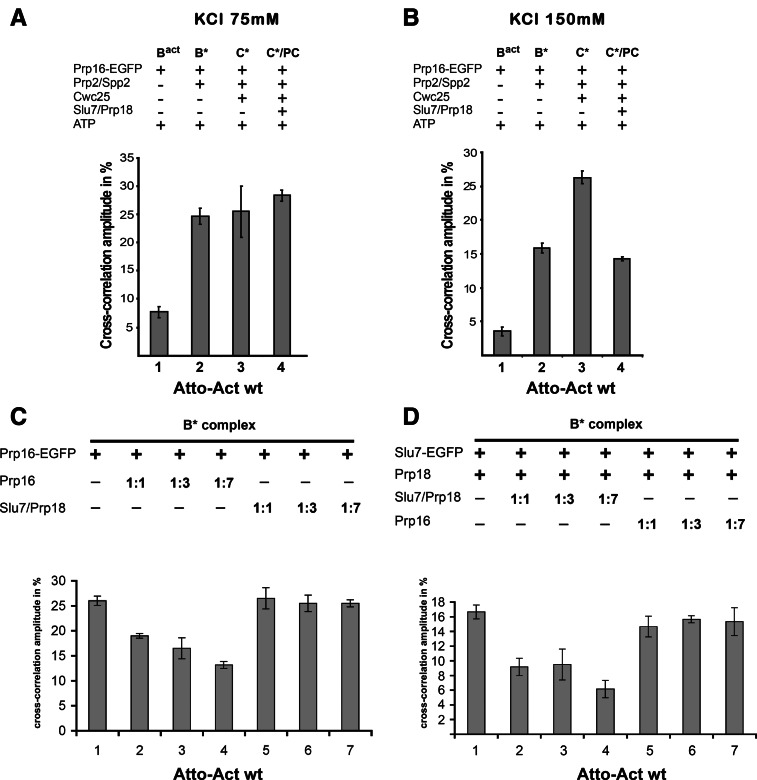
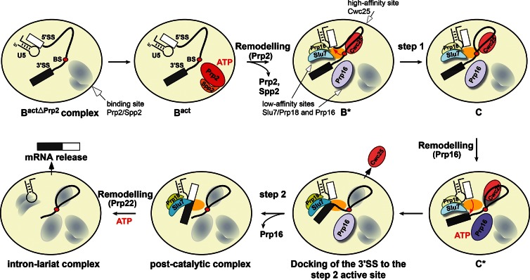
Step 2 catalysis of pre-mRNA splicing entails the excision of the intron and ligation of the 5' and 3' exons. The tasks of the splicing factors Prp16, Slu7, Prp18, and Prp22 in the formation of the step 2 active site of the spliceosome and in exon ligation, and the timing of their recruitment, remain poorly understood. Using a purified yeast in vitro splicing system, we show that only the DEAH-box ATPase Prp16 is required for formation of a functional step 2 active site and for exon ligation. Efficient docking of the 3' splice site (3'SS) to the active site requires only Slu7/Prp18 but not Prp22. Spliceosome remodeling by Prp16 appears to be subtle as only the step 1 factor Cwc25 is dissociated prior to step 2 catalysis, with its release dependent on docking of the 3'SS to the active site and Prp16 action. We show by fluorescence cross-correlation spectroscopy that Slu7/Prp18 and Prp16 bind early to distinct, low-affinity binding sites on the step-1-activated B* spliceosome, which are subsequently converted into high-affinity sites. Our results shed new light on the factor requirements for step 2 catalysis and the dynamics of step 1 and 2 factors during the catalytic steps of splicing.

Keywords: Prp22; Slu7 Prp18; spliceosome; step 2 catalysis; step 2 factors/Prp16.


Figures

FIGURE 1.
FIGURE 1.
Spliceosomal factor requirements for step 2 catalysis of Bact spliceosomes assembled on actin pre-mRNAs. (A) Extent of splicing in vitro catalyzed by affinity-purified spliceosomes assembled on the Actwt pre-mRNA substrate. The Bact complex (lane 1) was affinity-purified by a three-step procedure (see Supplemental Methods). Peak fractions of the second glycerol gradient were used for complementation assays with recombinant proteins at a 30-fold molar excess over the Bact complex (10 fmol) as indicated (lanes 29). Reaction mixtures were incubated (45 min at 23°C) in the presence of ATP. RNAs were extracted, separated by denaturing polyacrylamide gel electrophoresis (PAGE), and visualized by autoradiography. (B) Bact complexes assembled on Act7 pre-mRNA were purified and eluted from the amylose matrix. Subsequently, the complexes were complemented with the recombinant proteins indicated and were further incubated in the presence of ATP. Symbols for pre-mRNA, splicing intermediates, and products are indicated on the right: the 5′ exon (with the three MS2 loops shown) in gray, intron as a thin black line, 3′ exon in black. (*) Uncharacterized pre-mRNA bands.
FIGURE 2.
FIGURE 2.
ATP-dependent Prp16 activity is sufficient to generate stable, catalytically activated C* complexes in the absence of Slu7, Prp18, and Prp22. (A) Bact complexes were purified and complemented with Prp2, Spp2, and Cwc25 on the amylose matrix to generate C complexes. One-half of the reaction mixture was supplemented with buffer, the other half with Prp16 and ATP to generate C* complexes. After incubation, complexes were washed with GK75 buffer and eluted with maltose. Complexes (specific activity 100 cpm/fmol) were sedimented on glycerol gradients containing 75 mM KCl. Fractions were analyzed by Cherenkov counting. (B) Fraction 15 of both complexes was analyzed directly (lanes 2,6) or was additionally complemented with Slu7/Prp18 and Prp16 +/− ATP as indicated. After incubation, RNAs were extracted, separated by denaturing PAGE, and visualized by autoradiography. RNA species (right), see Figure 1.
FIGURE 3.
FIGURE 3.
Displacement of the step 1 factor Cwc25 from the spliceosome by Slu7/Prp18 as analyzed by dcFCCS. (A) Affinity-purified Bact complexes assembled on Atto-Actwt pre-mRNA were complemented on the matrix with Prp2, Spp2, ATP, and Cwc25-Alexa. The reaction mixture was incubated and then depleted of ATP. After washing of the matrix, reconstituted C complexes were eluted with maltose and complemented in solution as indicated above each column, and dcFCCS measurements were performed. (B) Affinity-purified Bact complexes assembled on Atto-ActACAC 3′SS mutant (see text) were complemented on the matrix as described in A. The reaction mixture was incubated, C complexes were eluted and complemented in solution as indicated above each column, and dcFCCS measurements were performed. Affinity-purified Bact complexes assembled on Atto-Act7wt (C) and Atto-Act7ACAC 3′SS mutant (D) were complemented on the matrix to generate C complexes. Eluted C complexes were then complemented in solution as indicated above each column, and dcFCCS measurements were performed. Cross-correlation amplitudes derived from two independent experiments are shown for each complex. Error bars indicate the standard deviation from two independent measurements.
FIGURE 4.
FIGURE 4.
A low-affinity binding site is created for Slu7-EGFP during catalytic activation of the spliceosome by Prp2 as analyzed by dcFCCS. (A) Affinity-purified Bact complexes assembled on Atto-Actwt were complemented on the matrix with recombinant Slu7-EGFP plus Prp18 (column 1, Bact) and additionally with Prp2 and Spp2 (column 2, B*), Cwc25 (column 3, C), Prp16 (column 4, C*/PC), or without Prp18 (column 5, C*). C*/PC indicates that upon Slu7/Prp18 binding to the C* complex, the latter is transformed into the post-catalytic complex (PC). After incubation, complexes were washed with GK75 buffer or GK150 buffer (B) and then eluted from the matrix with maltose. dcFCCS measurements were then performed at complex concentrations of 1.0 nM. Cross-correlation amplitudes derived from two independent experiments are shown for each complex. Error bars indicate the standard deviation from two independent measurements. (C) Reconstituted C complexes were incubated on the matrix with Slu7/Prp18 at 75 or 150 mM KCl, respectively. Complexes were washed with buffers containing 75 or 150 mM KCl, eluted from the matrix in GK75 buffer and mock-treated (lanes 2,5) or incubated in solution with Prp16 plus ATP (lanes 3,6). After incubation, RNAs were extracted, separated by denaturing PAGE, and visualized by autoradiography. Symbols for RNA species (as in Fig. 1) are shown on the right. The intensities of the intermediate and mRNA product signals were quantified using the ImageQuant software (Molecular Dynamics). The signals in lane 3 were used as 100%.
FIGURE 5.
FIGURE 5.
A low-affinity binding site is created for Prp16-EGFP during catalytic activation of the spliceosome by Prp2 as analyzed by dcFCCS. Affinity-purified Bact complexes assembled on Atto-Actwt were complemented on the matrix with recombinant Prp16-EGFP plus ATP (column 1, Bact) and additionally with the proteins indicated on the left. After incubation, complexes were washed with GK75 buffer (A) or GK150 buffer (B) and then eluted from the matrix with maltose. dcFCCS measurements were then performed at complex concentrations of 1.0 nM. C*/PC indicates that upon Slu7/Prp18 binding to the C* complex, the latter is transformed into the post-catalytic complex (PC). (C) The B* complex was complemented on the matrix with Prp16-EGFP and incubated at 23°C, the matrix was washed with GK75 buffer, and supplemented with increasing amount of untagged Prp16 (columns 2–4) or Slu7/Prp18 (columns 5–7). The matrix was washed, complexes were eluted with maltose, and cross-correlation amplitudes were measured by dcFCCS. (D) The B* complex was complemented on the matrix with Slu7-EGFP and Prp18 and incubated at 23°C. The matrix was then washed with GK75 buffer, and the complexes were supplemented with an increasing amount of untagged Slu7 and Prp18 (columns 2–4) or Prp16 (columns 5–7). After washing, complexes were eluted with maltose and cross-correlation amplitudes were measured by dcFCCS. Cross-correlation amplitudes derived from two independent experiments are shown for each complex. Error bars indicate the standard deviation from two independent measurements.
FIGURE 6.
FIGURE 6.
Binding and activity of step 1 and step 2 factors during the catalytic phase of the spliceosome. Schematic representation of the substantial remodeling that the precatalytic Bact spliceosome undergoes during the transition from step 1 to step 2 catalysis. The ATPases Prp2 and Prp16 are required for activating the spliceosome for the first and second catalytic steps, respectively. The ATPase Prp2 plus ATP creates a high-affinity binding site for Cwc25, which stabilizes the step 1 conformation of the catalytic center, thereby facilitating step 1 catalysis. Prp2 also creates low-affinity binding sites for Prp16 and Slu7; the latter binds to the spliceosome only together with Prp18. The ATPase Prp16 plus ATP is required for remodeling the spliceosome for step 2 catalysis and to stabilize the binding of Slu7/Prp18, which is needed to dock efficiently the 3′SS into the step 2 active site, leading to displacement of Cwc25 before exon ligation. The exons are then ligated and the post-catalytic spliceosome is generated. Proteins are oval-shaped, shadowed, and highlighted with light colors (low binding affinity) or with dark colors without a shadow (high binding affinity). The yellow oval in the middle of the spliceosome represents the active site. The conserved U5 stem–loop I is also shown.

References

    1. Ansari A, Schwer B 1995. SLU7 and a novel activity, SSF1, act during the PRP16-dependent step of yeast pre-mRNA splicing. EMBO J 14: 4001–4009 - PMC - PubMed
    1. Aronova A, Bacikova D, Crotti LB, Horowitz DS, Schwer B 2007. Functional interactions between Prp8, Prp18, Slu7, and U5 snRNA during the second step of pre-mRNA splicing. RNA 13: 1437–1444 - PMC - PubMed
    1. Brys A, Schwer B 1996. Requirement for SLU7 in yeast pre-mRNA splicing is dictated by the distance between the branchpoint and the 3′ splice site. RNA 2: 707–717 - PMC - PubMed
    1. Burgess SM, Guthrie C 1993. A mechanism to enhance mRNA splicing fidelity: The RNA-dependent ATPase Prp16 governs usage of a discard pathway for aberrant lariat intermediates. Cell 73: 1377–1391 - PubMed
    1. Burgess S, Couto JR, Guthrie C 1990. A putative ATP binding protein influences the fidelity of branchpoint recognition in yeast splicing. Cell 60: 705–717 - PubMed

Publication types

MeSH terms

LinkOut - more resources