. 2013 Jul 2;128(1):9-18.
doi: 10.1161/CIRCULATIONAHA.112.001238. Epub 2013 May 17.

Perivascular adipose tissue potentiates contraction of coronary vascular smooth muscle: influence of obesity

Affiliations

Perivascular adipose tissue potentiates contraction of coronary vascular smooth muscle: influence of obesity

Meredith Kohr Owen et al. Circulation. .

Abstract

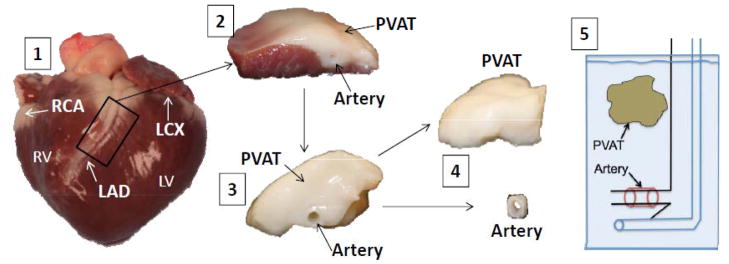
Background: This investigation examined the mechanisms by which coronary perivascular adipose tissue (PVAT)-derived factors influence vasomotor tone and the PVAT proteome in lean versus obese swine.

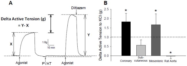
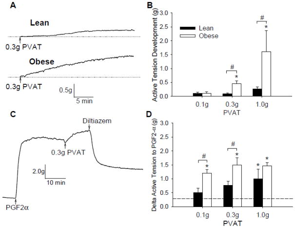
Methods and results: Coronary arteries from Ossabaw swine were isolated for isometric tension studies. We found that coronary (P=0.03) and mesenteric (P=0.04) but not subcutaneous adipose tissue augmented coronary contractions to KCl (20 mmol/L). Inhibition of CaV1.2 channels with nifedipine (0.1 µmol/L) or diltiazem (10 µmol/L) abolished this effect. Coronary PVAT increased baseline tension and potentiated constriction of isolated arteries to prostaglandin F2α in proportion to the amount of PVAT present (0.1-1.0 g). These effects were elevated in tissues obtained from obese swine and were observed in intact and endothelium denuded arteries. Coronary PVAT also diminished H2O2-mediated vasodilation in lean and, to a lesser extent, in obese arteries. These effects were associated with alterations in the obese coronary PVAT proteome (detected 186 alterations) and elevated voltage-dependent increases in intracellular [Ca(2+)] in obese smooth muscle cells. Further studies revealed that the Rho-kinase inhibitor fasudil (1 µmol/L) significantly blunted artery contractions to KCl and PVAT in lean but not obese swine. Calpastatin (10 μmol/L) also augmented contractions to levels similar to that observed in the presence of PVAT.

Conclusions: Vascular effects of PVAT vary according to anatomic location and are influenced by an obese phenotype. Augmented contractile effects of obese coronary PVAT are related to alterations in the PVAT proteome (eg, calpastatin), Rho-dependent signaling, and the functional contribution of K(+) and CaV1.2 channels to smooth muscle tone.

Keywords: adipose tissue; coronary disease; muscle, smooth; obesity; vasoconstriction.


Conflict of interest statement

Conflict of Interest Disclosures: The authors have no conflicts to disclose.

Figures

Figure 1
Figure 1
Representative picture illustrating isolation of coronary artery PVAT and isometric tension methodology. RV (right ventricle), LV (left ventricle), RCA (right coronary artery), LCX (left circumflex artery), LAD (left anterior descending artery), PVAT (perivascular adipose tissue). 1) Lean and obese hearts were excised upon sacrifice and perfused with Ca2+-free Krebs to remove excess blood; 2) Arteries and PVAT were grossly isolated from the heart; 3) the myocardium was removed; 4) arteries were further isolated and surrounding PVAT dissected away; 5) 3 mm lean and obese arteries were mounted in organ baths at 37°C.
Figure 2
Figure 2
Representative tracing of paired experiments to assess the vascular effects of PVAT from different anatomical depots. A, Representative wire myograph tracing of tension generated by arteries before (x) and after (y) the addition of PVAT to the organ bath. Upward deflections indicate an increase in tension (constriction). The difference in tension generated by each artery before (x) and after (y) PVAT is expressed as Delta Active Tension (g) and is independent of changes in baseline with PVAT. B, Delta active tension (g) of coronary arteries before and after exposure to coronary PVAT, subcutaneous adipose or mesenteric PVAT (0.3 g each). *P < 0.05 vs. average of paired time controls (represented by dashed line; 1.01 ± 0.21 g).
Figure 3
Figure 3
Effect of PVAT on baseline tension and response to PGF2α. A, Representative tracings of a lean and obese artery after addition of 0.3 g PVAT for 30 min. B, Addition of coronary PVAT (0.1–1.0g) to the organ bath increased tension in both lean and obese arteries and was dependent on the amount of coronary PVAT added to the bath. C, Representative tracing of a lean artery contracted with PGF2α to plateau, incubation with PVAT and treatment with diltiazem (10 μM). D, Delta active tension of arteries stimulated with PGF2α before and after the addition of coronary PVAT (0.1–1.0 g). *P < 0.05 vs. average of paired time controls (represented by dashed line; 0.29 ± 0.08 g). #P < 0.05 lean vs. obese, same amount of PVAT.
Figure 4
Figure 4
KCl dose-response curves in intact and denuded coronary arteries in the presence and absence of PVAT. Cumulative dose-response data of lean (A) and obese (B) arteries to KCl (10–60 mM) before and after coronary PVAT incubation (30 min). Arteries were incubated with coronary PVAT from the same animal on the same day. Cumulative dose-response data from denuded lean (C) and obese (D) vessels before and after PVAT incubation. *P < 0.05 vs. no PVAT-control at same KCl concentration.
Figure 5
Figure 5
Effect of PVAT on coronary vasodilation to H2O2. A, Representative tracings of H2O2-induced relaxations of lean control arteries pre-constricted with 1 μM U46619 in the absence and presence of PVAT. Average percent relaxation of lean (B) and obese (C) control and PVAT-treated arteries to H2O2 after pre-constriction with either U46619 (1 μM) or KCl (60 mM). *P < 0.05 vs. control at same H2O2 concentration.
Figure 6
Figure 6
Vascular effects of lean vs. obese coronary PVAT. A, Representative tracings of lean arteries treated with 20 mM KCl, exposed to either lean or obese PVAT. B, Delta active tension (g) to 20 mM KCl of lean arteries exposed to time control, lean or obese PVAT. *P < 0.05 vs. control. C, Delta active tension (g) to 20 mM KCl after exposure to SERCA inhibition with CPA (10 μM) P < 0.05 vs. control. D, F360/F380 ratio of fura-2 experiments after stimulation of isolated lean (n = 4) and obese (n = 5) coronary vascular smooth muscle with 80 mM KCl. *P < 0.05 obese vs. lean.
Figure 7
Figure 7. Effects of Rho kinase signaling and calpastatin on coronary artery contractions to KCl
Lean (A) and obese (B) arteries were incubated with 1 μM fasudil for 10 min prior to dose-responses to KCl (10–60 mM) in the absence and presence of coronary PVAT. *P < 0.05 vs. no PVAT-control at same KCl concentration. C, Delta active tension (g) in response to 20 mM KCl in lean and obese PVAT control and PVAT + fasudil-treated arteries *P < 0.05 vs. respective PVAT control. D, Delta active tension (g) to 20 mM KCl after incubation with increasing concentrations of calpastatin (1–10 μM) or scrambled calpastatin peptide (10 μM Neg Cnt) for 30 min. *P < 0.05 relative to time control.

References

    1. Mahabadi AA, Reinsch N, Lehmann N, Altenbernd J, Kalsch H, Seibel RM, Erbel R, Mohlenkamp S. Association of pericoronary fat volume with atherosclerotic plaque burden in the underlying coronary artery: a segment analysis. Atherosclerosis. 2010;211:195–9. - PubMed
    1. Iacobellis G, Willens HJ. Echocardiographic epicardial fat: a review of research and clinical applications. J Am Soc Echocardiogr. 2009;22:1311–9. - PubMed
    1. Greif M, Becker A, von ZF, Lebherz C, Lehrke M, Broedl UC, Tittus J, Parhofer K, Becker C, Reiser M, Knez A, Leber AW. Pericardial adipose tissue determined by dual source CT is a risk factor for coronary atherosclerosis. Arterioscler Thromb Vasc Biol. 2009;29:781–6. - PubMed
    1. Boydens C, Maenhaut N, Pauwels B, Decaluwe K, Van d V. Adipose tissue as regulator of vascular tone. Curr Hypertens Rep. 2012;14:270–8. - PubMed
    1. Tesauro M, Cardillo C. Obesity, blood vessels and metabolic syndrome. Acta Physiol (Oxf) 2011;203:279–86. - PubMed

Publication types

MeSH terms