. 2013 Aug;62(8):1335-45.
doi: 10.1007/s00262-013-1436-4. Epub 2013 May 18.

Gamma delta T cells are activated by polysaccharide K (PSK) and contribute to the anti-tumor effect of PSK

Affiliations

Gamma delta T cells are activated by polysaccharide K (PSK) and contribute to the anti-tumor effect of PSK

Carol Inatsuka et al. Cancer Immunol Immunother. 2013 Aug.

Abstract

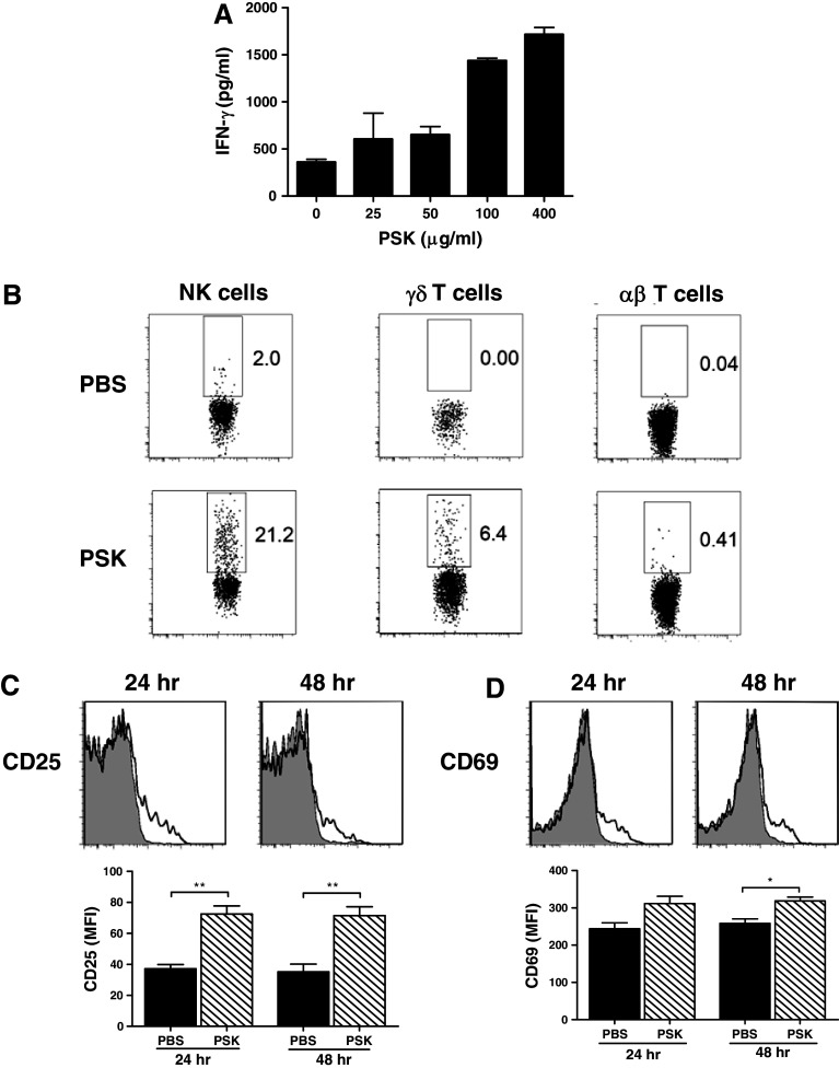
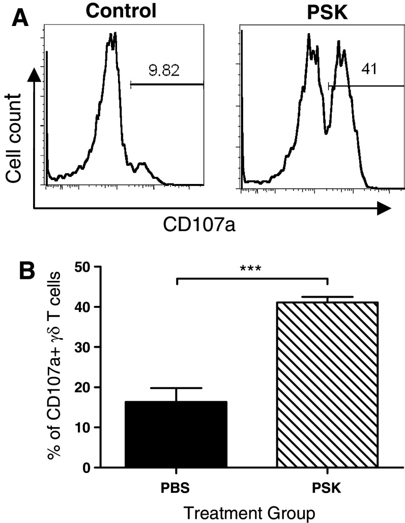
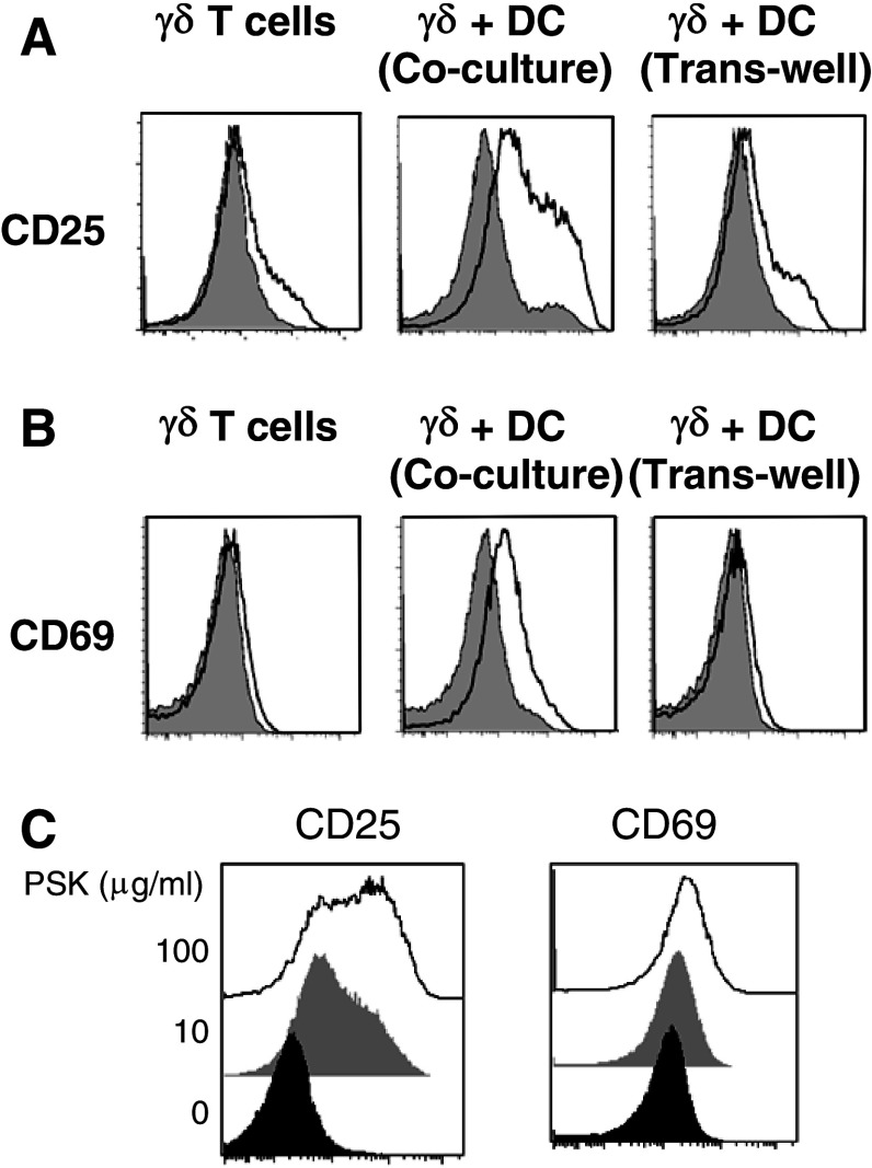
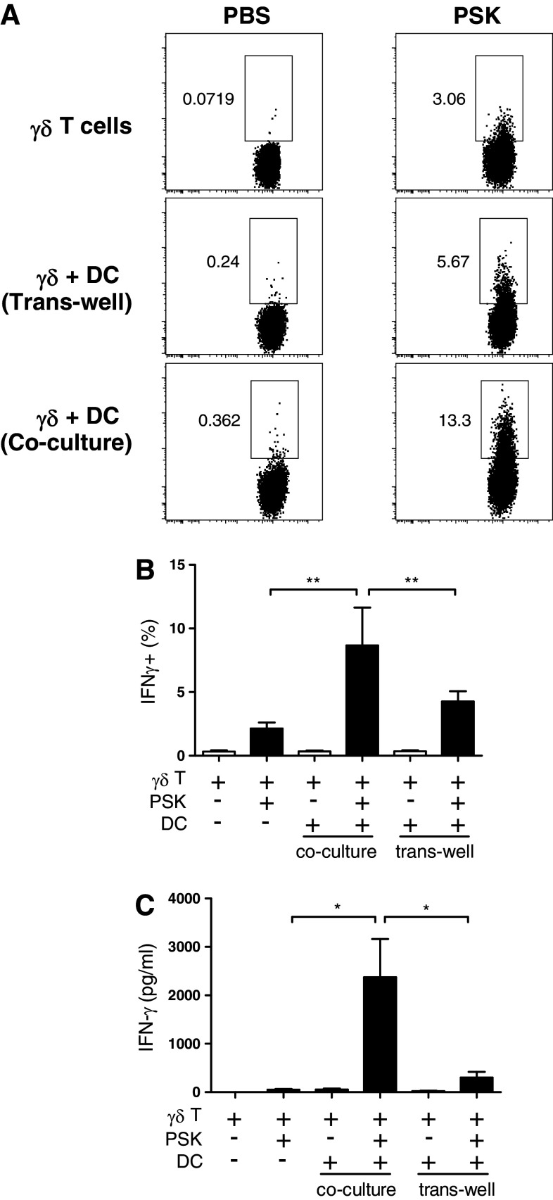
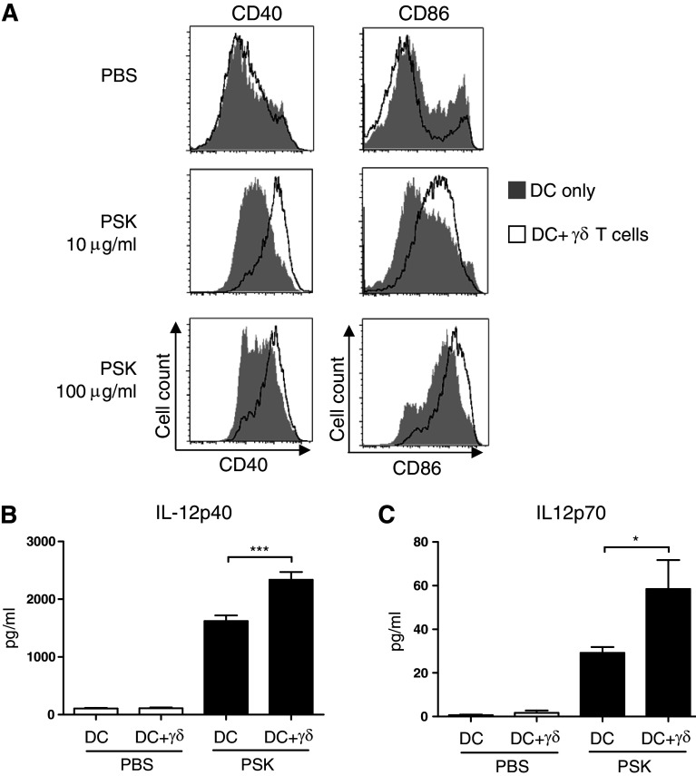
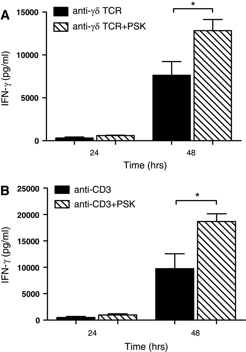
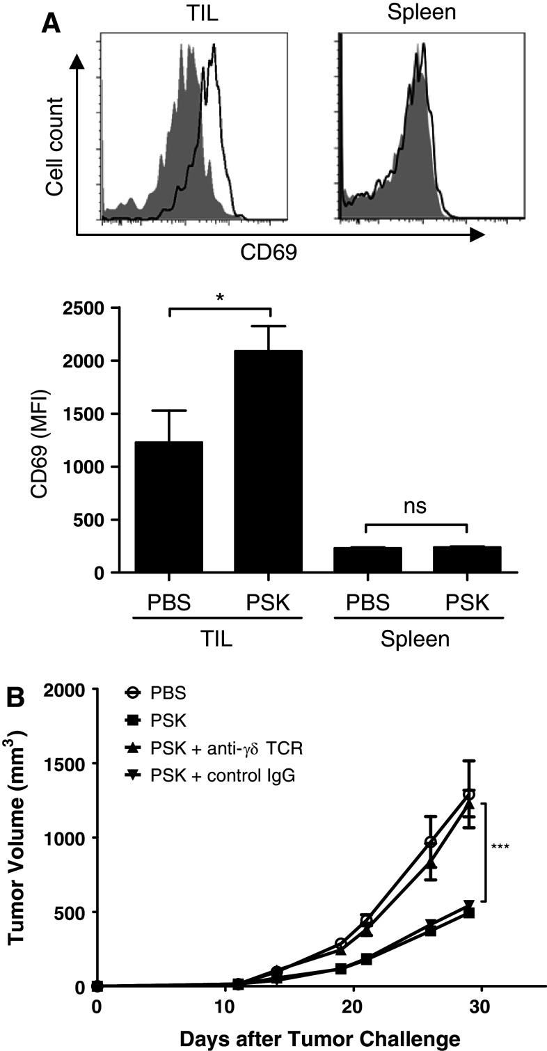
Polysaccharide K (PSK) is a widely used mushroom extract that has shown anti-tumor and immunomodulatory effects in both preclinical and clinical studies. Therefore, it is important to understand the mechanism of actions of PSK. We recently reported that PSK can activate toll-like receptor 2 and enhances the function of NK cells. The current study was undertaken to study the effect of PSK on gamma delta (γδ) T cells, another important arm of the innate immunity. In vitro experiments using mouse splenocytes showed that γδ T cells produce IFN-γ after treatment with PSK and have up-regulated expression of CD25, CD69, and CD107a. To investigate whether the effect of PSK on γδ T cells is direct or indirect, purified γδ T cells were cultured either alone or together with bone marrow-derived DC in a co-culture or trans-well system and then stimulated with PSK. Results showed that direct cell-to-cell contact between γδ T cells and DC is required for optimal activation of γδ T cells. There was also reciprocal activation of DC by PSK-activated γδ T cells, as demonstrated by higher expression of costimulatory molecules and enhanced production of IL-12 by DC in the presence of γδ T cells. PSK can also co-stimulate γδ T cells with anti-TCR and anti-CD3 stimulation, in the absence of DC. Finally, in vivo treatment with PSK activates γδ T cells among the tumor infiltrating lymphocytes, and depleting γδ T cells during PSK treatment attenuated the anti-tumor effect of PSK. All together, these results demonstrated that γδ T cells are activated by PSK and contribute to the anti-tumor effect of PSK.


Conflict of interest statement

The authors declare they have no conflict of interest.

Figures

Fig. 1
Fig. 1
Gamma delta T cells are activated by PSK to produce IFN-γ. a Dose–response of PSK-stimulated IFN-γ production by splenocytes. Shown are mean ± SD of IFN-γ levels in triplicate culture wells treated with serial dilutions of PSK for 48 h, as determined by ELISA. Similar results were obtained from three independent experiments. b Representative FACS dot plots showing intracellular staining of IFN-γ in NK cells, γδ T cells, or αβ T cells in splenocytes from neu-transgenic mice. Splenocytes were treated with PSK (100 μg/ml) for 48 h. Similar results were obtained from three independent experiments. c, d Expression of CD25 and CD69 on γδ T cells in PSK-treated splenocytes. Shown are representative overlay histograms. Gray histogram: control; empty histogram: PSK-treated γδ T cells. The bar graphs summarize the results from three independent experiments. *p < 0.05, **p < 0.01 by two-tailed Student’s t test
Fig. 2
Fig. 2
PSK induces CD107a degranulation in γδ T cells. a Representative histograms showing the percentages of CD107a+ γδ T cells in control and PSK-treated splenocytes. b Summary graph showing the percentages of CD107a+ cells among total γδ T cells. The bars represent mean ± sem of results from three independent experiments. ***p < 0.001 by Student’s t test
Fig. 3
Fig. 3
Direct contact between DC and γδ T cells is required for optimal activation of γδ T cells by PSK. a Representative overlay histograms and summary graphs showing CD25 expression in PSK-treated purified γδ T cells, γδ + DC co-culture, and γδ + DC trans-well culture. b Representative overlay histograms and summary graphs showing CD69 expression in PSK-treated purified γδ T cells, γδ + DC co-culture, and γδ + DC trans-well culture. Gray histogram: control; empty histogram: PSK-treated γδ T cells. c Dose-dependent induction of CD25 and CD69 on γδ T cells in γδ + DC co-culture. Black histogram: control; gray histogram: low-dose PSK (10 μg/ml); white histogram: high-dose PSK (100 μg/ml)
Fig. 4
Fig. 4
Direct contact between DC and γδ T cells is required for PSK-induced IFN-γ production. a Representative dot plots showing the expression of IFN-γ in control PBS- and PSK-treated γδ T cells in different culture systems: γδ T cell alone, γδ + DC trans-well culture, or γδ + DC co-culture. b Bar graph summarizing the percentage of IFN-γ+ γδ T cells in PBS- and PSK-treated γδ T cells in different culture systems. The graph summarizes the results from three independent experiments. c Bar graph summarizing the level of IFN-γ in PBS- and PSK-treated γδ T cells in different culture systems. The graph summarizes the results from three independent experiments. *p < 0.05, **p < 0.01, by two-tailed Student’s t test
Fig. 5
Fig. 5
Reciprocal activation of DC in the BMDC-γδ T cell co-culture leads to increased expression of co-stimulatory molecules and enhanced production of IL-12. BMDC and BMDC-γδ T cell co-culture were treated with PSK for 24 h, and the cells were harvested for FACS analysis of DC activation markers. The culture supernatant was analyzed for IL-12 using ELISA. a Expression of CD40 and CD86 in BMDC treated with control RPMI medium or PSK (10 or 100 μg/ml). Shaded histograms: DC alone culture; empty histograms: DC + γδ T cell co-culture. b, c The levels of IL-12p40 and IL-12p70 in control untreated DC or DC + γδ T cell co-culture (white columns) and PSK-treated DC or DC + γδ T cell co-culture (black columns). *p < 0.05, by two-tailed Student’s t test
Fig. 6
Fig. 6
PSK co-stimulates purified γδ T cells with anti-γδ TCR mAb or anti-CD3 mAb to produce IFN-γ. a Purified γδ T cells were cultured on anti-γδ TCR mAb-coated plates in the presence or absence of PSK (100 μg/ml). The presence of PSK resulted in higher production of IFN-γ, as measured by ELISA. The bar graph summarizes results from three independent experiments. b Purified γδ T cells were cultured on anti-CD3 mAb-coated plates in the presence or absence of PSK (100 μg/ml). The presence of PSK resulted in higher production of IFN-γ, as measured by ELISA. The bar graph summarizes results from three independent experiments. *p < 0.05, by two-tailed Student’s t test
Fig. 7
Fig. 7
In vivo PSK treatment activates γδ T cells in TIL, and γδ T cell contributes to the anti-tumor effect of PSK. a Intratumoral injection of PSK results in CD69 upregulation on γδ T cells in TIL, but not in spleen. Shown are representative overlay histograms of CD69 expression on γδ T cells in TIL or spleen from control PBS- and PSK-treated mice. Shaded histogram PBS-treated mice (control); empty histogram: PSK-treated mice. The summary bar graph shows the mean fluorescence intensity (MFI) of CD69 expression in γδ T cells (mean ± sem) in TIL and spleen in PBS or PSK group (n = 8 mice per group). *p < 0.05, by two-tailed Student’s t test. b Depletion of γδ T cells during PSK treatment decreased the anti-tumor effect of PSK. Mice received anti-γδ TCR mAb or a control hamster IgG during PSK treatment. PSK by itself significantly inhibited tumor growth, and the effect is significantly attenuated when mice received anti-γδ TCR mAb. **p < 0.01 between PSK + γδ T cell depletion group (filled triangle) and PSK group (open square); ***p < 0.0001 between control PBS (open circle) and PSK group (open square). There was no difference between PSK + control IgG (filled inverted triangle) and PSK group (open square). N = 5 mice per group. Similar results were obtained from two independent experiments

References

    1. Ernst E, Cassileth BR. The prevalence of complementary/alternative medicine in cancer: a systematic review. Cancer. 1998;83:777–782. doi: 10.1002/(SICI)1097-0142(19980815)83:4<777::AID-CNCR22>3.0.CO;2-O. - DOI - PubMed
    1. Cassileth BR, Vickers AJ. High prevalence of complementary and alternative medicine use among cancer patients: implications for research and clinical care. J Clin Oncol. 2005;23:2590–2592. doi: 10.1200/JCO.2005.11.922. - DOI - PubMed
    1. Hyodo I, Amano N, Eguchi K, Narabayashi M, Imanishi J, Hirai M, Nakano T, Takashima S. Nationwide survey on complementary and alternative medicine in cancer patients in Japan. J Clin Oncol. 2005;23:2645–2654. doi: 10.1200/JCO.2005.04.126. - DOI - PubMed
    1. Kidd PM. The use of mushroom glucans and proteoglycans in cancer treatment. Altern Med Rev. 2000;5:4–27. - PubMed
    1. Fisher M, Yang LX. Anticancer effects and mechanisms of polysaccharide-K (PSK): implications of cancer immunotherapy. Anticancer Res. 2002;22:1737–1754. - PubMed

Publication types

MeSH terms

LinkOut - more resources