. 2013 Aug;126(2):189-205.
doi: 10.1007/s00401-013-1129-2. Epub 2013 May 18.

N-truncated amyloid β (Aβ) 4-42 forms stable aggregates and induces acute and long-lasting behavioral deficits

Affiliations

N-truncated amyloid β (Aβ) 4-42 forms stable aggregates and induces acute and long-lasting behavioral deficits

Yvonne Bouter et al. Acta Neuropathol. 2013 Aug.

Abstract

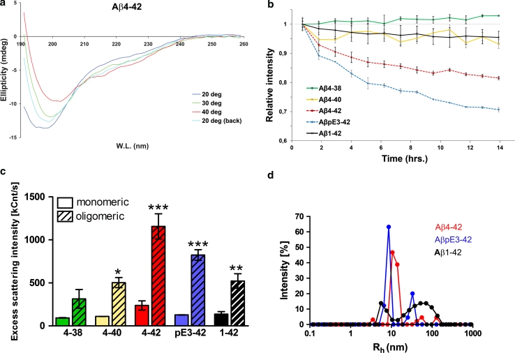
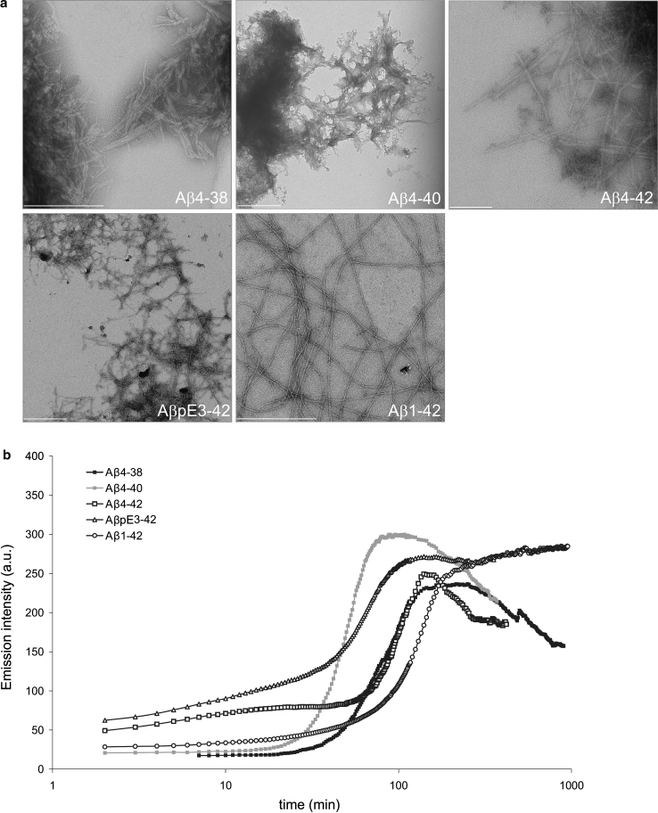
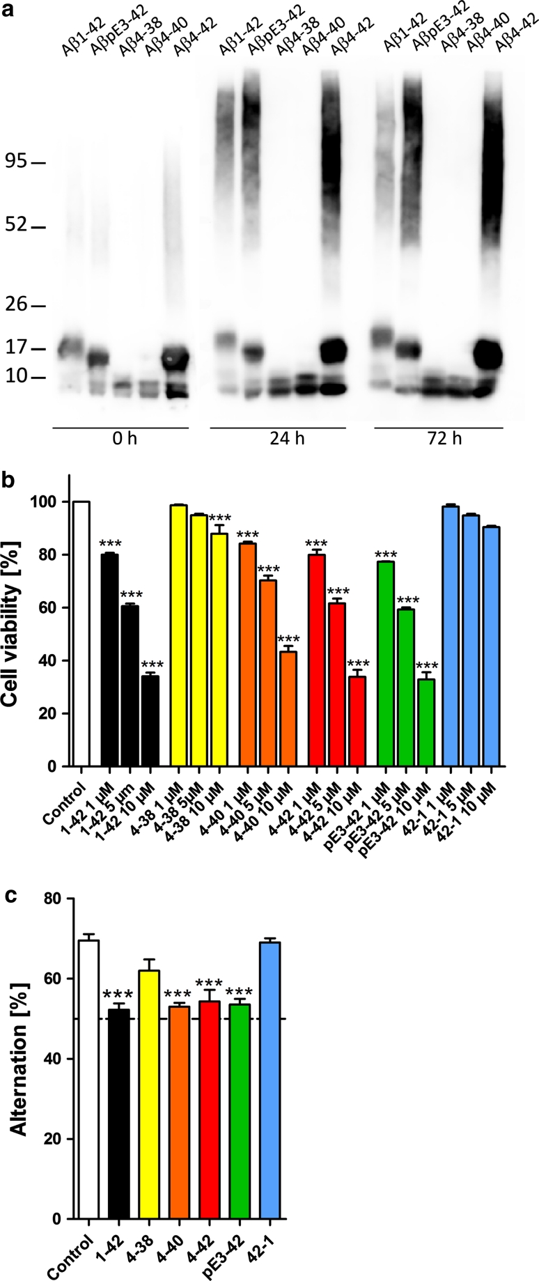
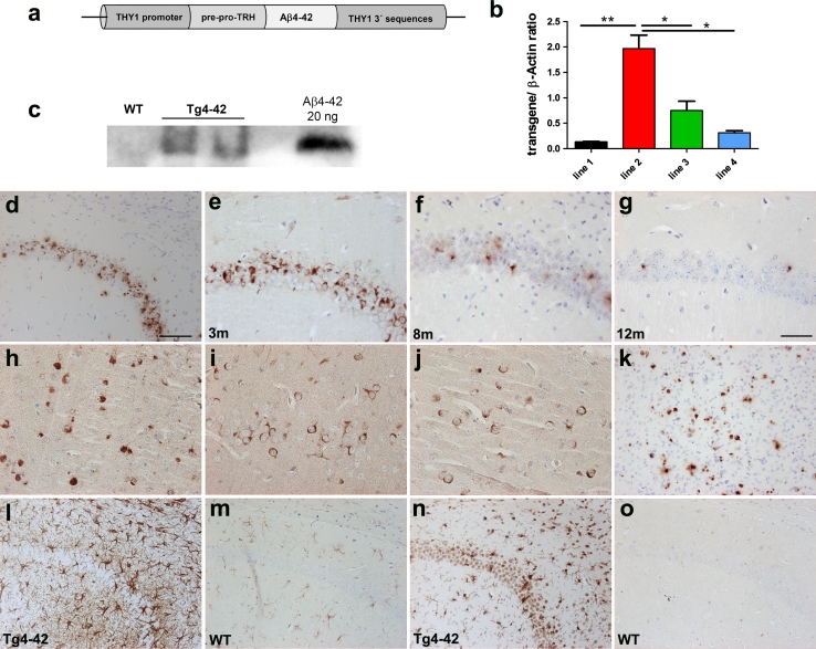
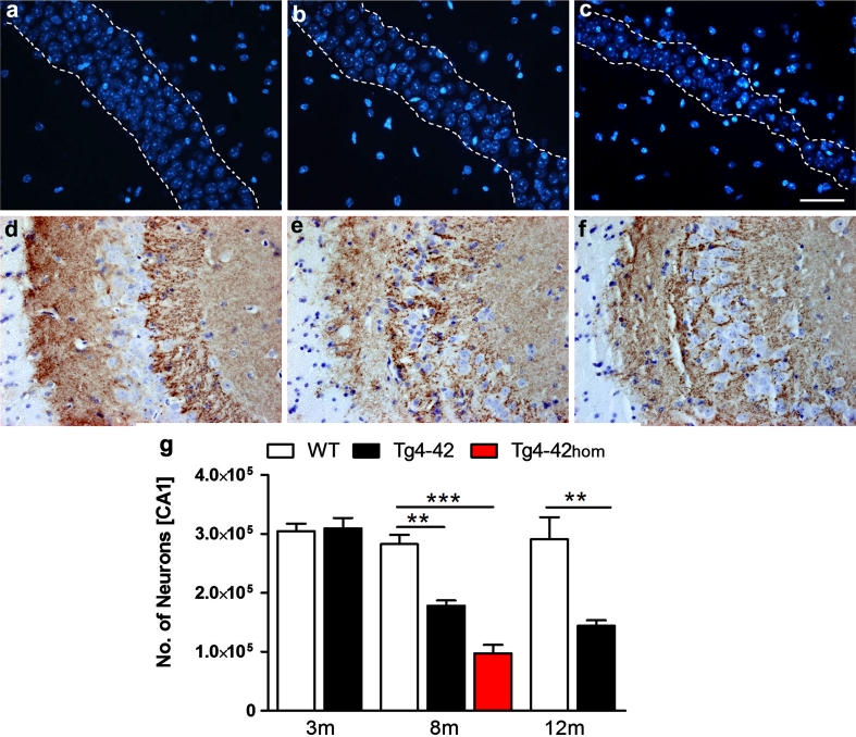
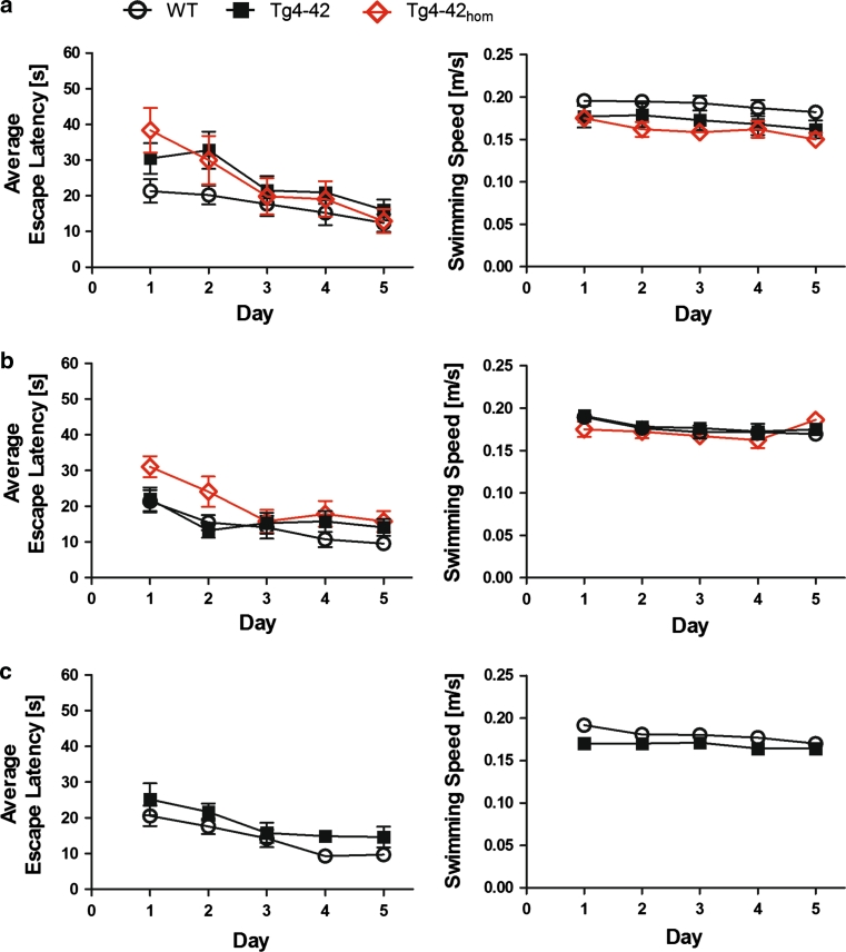
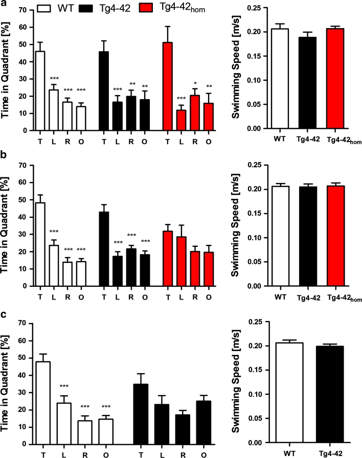
N-truncated Aβ4-42 is highly abundant in Alzheimer disease (AD) brain and was the first Aβ peptide discovered in AD plaques. However, a possible role in AD aetiology has largely been neglected. In the present report, we demonstrate that Aβ4-42 rapidly forms aggregates possessing a high aggregation propensity in terms of monomer consumption and oligomer formation. Short-term treatment of primary cortical neurons indicated that Aβ4-42 is as toxic as pyroglutamate Aβ3-42 and Aβ1-42. In line with these findings, treatment of wildtype mice using intraventricular Aβ injection induced significant working memory deficits with Aβ4-42, pyroglutamate Aβ3-42 and Aβ1-42. Transgenic mice expressing Aβ4-42 (Tg4-42 transgenic line) developed a massive CA1 pyramidal neuron loss in the hippocampus. The hippocampus-specific expression of Aβ4-42 correlates well with age-dependent spatial reference memory deficits assessed by the Morris water maze test. Our findings indicate that N-truncated Aβ4-42 triggers acute and long-lasting behavioral deficits comparable to AD typical memory dysfunction.


Figures

Fig. 1
Fig. 1
Structural properties of N-terminally truncated Aβ. a Temperature dependence of far-UV CD spectra of Aβ4-42. Measurements were performed at 20, 30 and 40 °C. 20 ° back indicates the measurement after cooling down from 40 to 20 °C. b Rates of monomer consumption for different Aβ peptides, probed through a decay in relative intensity of methyl (0.50–1.05 ppm) and aromatic (6.5–8.0 ppm) signals in their 1D 1H NMR spectra. c Excess light scattering intensity of Aβ variants measured before (solid bars) and after (striped bars) 24 h of aggregation through dynamic light scattering (DLS). d Distribution of hydrodynamic radius (R h) of Aβ aggregates, derived from their scattering intensity autocorrelation curve in DLS experiments. For Aβ4-42 and AβpE3-42, the main aggregated species had an R h of approximately 10 nm
Fig. 2
Fig. 2
a Transmission electron micrograph of Aβ peptides after 3 days of incubation in an aggregation-promoting condition. All Aβ variants formed fibrillar aggregates which tended to clump together in all preparations but Aβ1-42. b Temporal evolution of Thioflavin T (ThT) fluorescence emission intensity during aggregation of various Aβ peptides. Despite some variation in the kinetics, all five Aβ variants were capable of forming ThT-reactive aggregates. Scale bars 200 nm (Aβ4-42), 500 nm (all other Aβ variants)
Fig. 3
Fig. 3
Cellular toxicity of N-truncated Aβ peptides. a Freshly prepared Aβ4-38, Aβ4-40,Aβ4-42, AβpE3-42 and Aβ1-42 rapidly formed stable aggregates. All peptides displayed dimeric oligomers and monomers under reducing conditions, while Aβ1-42, AβpE3-42 and Aβ4-42 also developed SDS-stable tri- or tetrameric oligomers. Aged Aβ1-42, AβpE3-42 and Aβ4-42 peptides retained this pattern and exhibited in addition higher molecular weight aggregates. SDS-PAGE Western blot of Aβ peptides using the polyclonal antiserum 24311. b In vitro toxicity with short-term exposure. Primary neurons were treated with Aβ peptides at different concentrations and analyzed by a cell toxicity assay. Aβ1-42pE3-42 and Aβ4-42 demonstrated comparable toxicity profiles followed by Aβ4-40. Aβ4-38 was only toxic at 10 μM. No toxicity was observed with vehicle control and reverse Aβ42-1. c In vivo toxicity of short-term exposure in wildtype mouse brain. Working memory was assessed after intraventricular injection of Aβ4-38, Aβ4-40, Aβ4-42, AβpE3-42 and Aβ1-42 as well as vehicle and reverse peptide control. Mice injected with Aβ4-40, Aβ4-42pE3-42 and Aβ1-42 performed at chance level and showed a significant and robust deficit in working memory. Mice treated with Aβ4-38 demonstrated normal working memory. The same is true for the reverse peptide Aβ42-1 and vehicle control. One-way analysis of variance (ANOVA) followed by Bonferroni multiple comparisons. ***p < 0.001
Fig. 4
Fig. 4
Analysis of transgenic mice expressing Aβ4-42. a Scheme of transgene expression vector with THY1 promoter, signal peptide of pre-pro-thyrotropin releasing hormone (TRH), Aβ4-42 and THY1 3′ sequences. b Quantitative RT-PCR analysis of four different transgenic mouse lines producing Aβ4-42 showing variable transgene expression levels. Line 2 exhibited significantly the highest transgene expression (p < 0.01 unpaired t test compared to the other lines). This line was chosen for further breeding and renamed Tg4-42. c Western blot analysis of whole brain TBS lysates (40 μg total protein loaded) using pan-Aβ antibody 4G8 of 5-month-old hemizygous Tg4-42 mice and a wildtype control. d, on Immunohistochemical staining profile of Tg4-42 mice expressing Aβ4-42. d Abundant intraneuronal Aβ immunoreactivity was found in the CA1 pyramidal cell layer of the hippocampus in 3-month-old hemizygous Tg4-42 mice (polyclonal antiserum 24311). Aβ42 immunostaining in CA1 at 3 e, 8 f and 12 g months of age in hemizygous Tg4-42 mice with an age-dependent reduction in positive cells. Other brain regions with Aβ42 staining were occipital cortex (h), piriform cortex i, striatum j and superior colliculus k. In addition, increased astrogliosis with GFAP staining l and microgliosis with IBA1 staining n was observed as early as 2 months of age in hemizygous Tg4-42 mice. No significant astro and microgliosis were seen in wildtype (WT) controls (GFAP m, IBA1 o). Scale bar in d 100 μm (d, ko); scale bar in g: 50 μm (ej)
Fig. 5
Fig. 5
Age- and dose-dependent neuron loss in hippocampus in Tg4-42 mice. DAPI staining (ac) revealed a loss in neuron number in the CA1 layer of the hippocampus at the age of 8 months between hemizygous b Tg4-42 and a age-matched WT mice. c A more pronounced neuron loss was apparent in homozygous Tg4-42 mice. df Synaptophysin staining showed an altered synaptic patterning in the CA3 region of the hippocampus in hemizygous and more pronounced in homozygous Tg4-42 mice at the age of 8 months. g Quantification using unbiased stereology. Scale bars ac 100 μm; df 50 μm; one-way analysis of variance (ANOVA) followed by Bonferroni multiple comparisons. *p < 0.05, **p < 0.01
Fig. 6
Fig. 6
Spatial learning was assessed using acquisition training of the Morris water maze. Heterozygous Tg4-42 mice and wildtype (C57BL/6 J) littermate controls were tested at a 3, b 8 and c 12 months of age. In addition, homozygous Tg4-42 mice (Tg4-42hom) were assessed at 3 and 8 months. Each group was sex- and age-matched and contained 10–15 animals. Animals tested underwent acquisition training to learn to use distal and proximal cues to navigate a direct path to a hidden platform. ac Escape latencies decreased progressively over 5 days of training for wildtype, Tg4-42 and Tg4-42hom. Swimming speed was not affected in all mice tested. m age in months
Fig. 7
Fig. 7
Memory deficits in aged Tg4-42 mice shown in the probe trial of the Morris water maze. Hemizygous Tg4-42 mice and WT (C57BL/6 J) littermate controls were tested at a 3, b 8 and c 12 months of age. In addition, homozygous Tg4-42 (Tg4-42hom) mice were assessed at 3 and 8 months. Each group was sex- and age-matched and contained 10–15 mice. The probe trial was given at the end of the learning phase (acquisition training) to assess spatial reference memory. Quadrant preference and swimming speed for the first 30 s of the probe trial were analyzed. a Tg4-42, Tg4-42hom and WT mice showed no impairment in spatial reference memory at 3 months of age. Both groups spent a significant greater percentage of the time in the target quadrant. b The probe trial revealed significantly reduced learning behavior for Tg4-42hom mice at 8 months of age as they showed no preference for the target quadrant. In contrast, hemizygous Tg4-42 and WT mice had no learning deficits at this age. c At 12 months of age, hemizygous Tg4-42 mice showed no quadrant preference revealing an impaired spatial reference memory. However, WT mice still learned, as they had a significant preference for the target quadrant. ac No differences in swimming speed between WT, Tg4-42 and Tg4-42hom were detected at any tested age. T target quadrant, L left quadrant, R right quadrant, O opposite quadrant, m age in months. Quadrant preference: Two-way analysis of variance (ANOVA) followed by Bonferroni multiple comparisons. Swimming speed: unpaired t-test. ***p < 0.001; **p < 0.01

References

    1. Alexandru A, Jagla W, Graubner S, et al. Selective Hippocampal neurodegeneration in transgenic mice expressing small amounts of truncated Aβ is induced by pyroglutamate–Aβ formation. J Neurosci. 2011;31:12790–12801. doi: 10.1523/JNEUROSCI.1794-11.2011. - DOI - PMC - PubMed
    1. Barrow CJ, Zagorski MG. Solution structures of beta peptide and its constituent fragments: relation to amyloid deposition. Science. 1991;253:179–182. doi: 10.1126/science.1853202. - DOI - PubMed
    1. Benilova I, Karran E, De Strooper B. The toxic A[beta] oligomer and Alzheimer’s disease: an emperor in need of clothes. Nat Neurosci. 2012;29:349–357. doi: 10.1038/nn.3028. - DOI - PubMed
    1. Breyhan H, Wirths O, Duan K, Marcello A, Rettig J, Bayer TA. APP/PS1KI bigenic mice develop early synaptic deficits and hippocampus atrophy. Acta Neuropathol. 2009;117:677–685. doi: 10.1007/s00401-009-0539-7. - DOI - PubMed
    1. Broadbent NJ, Squire LR, Clark RE. Spatial memory, recognition memory, and the hippocampus. Proc Natl Acad Sci USA. 2004;101:14515–14520. doi: 10.1073/pnas.0406344101. - DOI - PMC - PubMed

MeSH terms