Review
. 2013 Jul 1;5(7):a012997.
doi: 10.1101/cshperspect.a012997.

Human papillomavirus infections: warts or cancer?

Affiliations
Review

Human papillomavirus infections: warts or cancer?

Louise T Chow et al. Cold Spring Harb Perspect Biol. .

Abstract

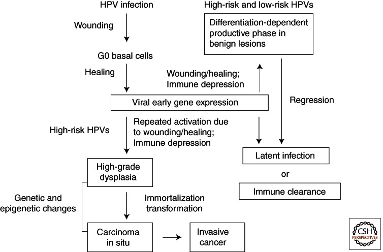
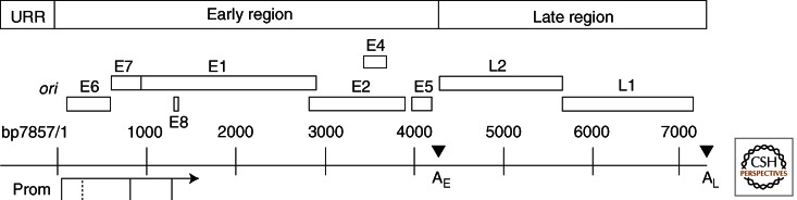
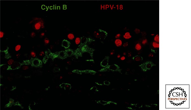
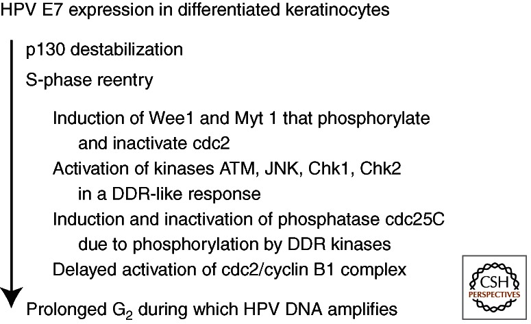
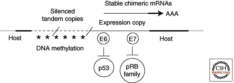
Human papillomaviruses (HPVs) are prevalent pathogens of mucosal and cutaneous epithelia. Productive infections of squamous epithelia lead to benign hyperproliferative warts, condylomata, or papillomas. Persistent infections of the anogenital mucosa by high-risk HPV genotypes 16 and 18 and closely related types can infrequently progress to high-grade intraepithelial neoplasias, carcinomas-in-situ, and invasive cancers in women and men. HPV-16 is also associated with a fraction of head and neck cancers. We discuss the interactions of the mucosotropic HPVs with the host regulatory proteins and pathways that lead to benign coexistence and enable HPV DNA amplification or, alternatively, to cancers that no longer support viral production.


Figures

Figure 1.
Figure 1.
Natural history of HPV infections. Most infections are subclinical and are quickly cleared by host immune surveillance. Some infections become active but regress to a latent state or are cleared within a year. Immune suppression or cycles of wounding and healing, conditions that induce viral early gene expression in proliferating undifferentiated cells, may reactivate the infections. In tissues persistently infected by high-risk HPVs, repeated wounding and healing lead to recurring and extended inactivation of tumor suppressor proteins by the viral oncoproteins and hence excessive basal cell proliferation and progression to high-grade lesions. These cells may accumulate mutations and epigenetic changes, resulting over time in cell immortalization and transformation, integration of the viral DNA into host chromosomes (in most cases), and carcinomas.
Figure 2.
Figure 2.
The genome organization of HPV-18. The upstream regulatory region (URR) contains transcription regulatory elements as well as the origin of DNA replication (ori), which overlaps the major promoter preceding E6. Major promoters (Prom) are indicated (solid vertical lines connected to an arrow, which denotes the direction of transcription). Open reading frames (ORFs) are depicted by open boxes. The early (E) and late (L) gene blocks are each followed by a polyadenylation site (AE and AL). This genome organization is largely conserved among HPVs. Some mucosotropic genotypes have one or two E5 ORFs whereas many cutaneous HPVs have no recognizable E5 ORF. The HR HPVs use the E6 promoter to express both E6 and E7 and possibly E1 and E2. Alternative E6 intragenic mRNA splicing allows efficient expression of the E7 protein. The LR HPV types use a separate promoter located in E6 (dotted vertical line) to express E7. Downstream ORFs are accessed by alternative RNA splicing. Transcripts from the promoter located within E7 encode E1, E2, E1ME2C (a fusion of the amino-terminal portion of E1 to the middle of E2), E1E4 (a fusion of the amino terminus of E1 to E4) and E5, as well as L2 and L1. The minor promoter embedded within E1 generates a short E8 exon, which is spliced to the middle of the E2 ORF, creating E8E2C. The expression of late genes from the promoter in E7 occurs only in the superficial squamous cells after viral DNA amplification and requires elongation of transcripts through AE to AL and stabilization of the late 3′ UTR. The functions of encoded proteins and regulatory elements are detailed in Table 1.
Figure 3.
Figure 3.
The initiation of viral DNA amplification in G2 phase cells. A formalin-fixed, paraffin-embedded section of an HPV-18 containing PHK raft culture held at the air–medium interface for 12 days was simultaneously probed for viral DNA (red) by in situ hybridization and cyclin B1 protein (green) with an antibody. Low viral DNA signal was detected in cells with cytoplasmic G2 cyclin B1. Cells with high viral DNA signal were negative for cyclin B1 (and cyclin A, not shown) as these cells have already exit the cell cycle. (Adapted from Wang et al. 2009a.)
Figure 4.
Figure 4.
The activities of E7 in differentiated keratinocytes. The expression of E7 in the differentiated keratinocytes destabilizes p130, leading to S-phase reentry. Wee1 and Myt 1 kinases are also activated, hyperphosphorylating and inactivating cdc2. Moreover, E7 further induces a DNA damage repair (DDR)-like response in that ATM, Chk1, Chk2, and JNK are each activated, resulting in the inactivation of the cdc25C phosphatase, disabling it from removing the inhibitory phosphates from cdc2. The inactive cdc2 and cyclin B1 accumulate in the cytoplasm in a prolonged G2 phase during which viral DNA amplifies (see Fig. 3). However, an antibody to γH2AX detected only sporadic signals (LT Chow and TR Broker, unpubl.), inconsistent with widespread DNA damage.
Figure 5.
Figure 5.
Epigenetic regulation of HPV transcription in a clonally selected cancer cell with tandemly integrated viral DNA. Thick dotted lines represent host sequences. Thin dashed lines represent HPV genomes integrated in tandem arrays. The predominant viral transcripts are chimeric. They originate from the URR promoter of the downstream integration junction copy, which is disrupted in E1 or E2. The E6/E7 transcripts have 3′ UTR and polyA site sequence derived from the host. All upstream viral DNA copies are silenced by DNA methylation (Wentzensen et al. 2002; Van Tine et al. 2004b) such that other viral genes are no longer expressed.

References

    1. Abbate EA, Berger JM, Botchan MR 2004. The X-ray structure of the papillomavirus helicase in complex with its molecular matchmaker E2. Genes Dev 18: 1981–1986 - PMC - PubMed
    1. Banerjee NS, Genovese N, Noya F, Chien W-M, Broker TR, Chow LT 2006. Inducible E7 proteins of high-risk and low-risk HPVs promote S phase re-entry in post-mitotic differentiated keratinocytes in organotypic cultures. J Virol 80: 6517–6524 - PMC - PubMed
    1. Banerjee NS, Wang H-K, Broker TR, Chow LT 2011. HPV E7 protein induces prolonged G2 following S-phase reentry in differentiated human keratinocytes. J Biol Chem 286: 11473–11482 - PMC - PubMed
    1. Belleudi F, Leone L, Purpura V, Cannella F, Scrofani C, Torrisi MR 2011. HPV16 E5 affects the KGFR/FGFR2b-mediated epithelial growth through alteration of the receptor expression, signaling and endocytic traffic. Oncogene 30: 4963–4876 - PubMed
    1. Bernard H-U 2002. Gene expression of genital human papillomaviruses and considerations on potential antiviral approaches. Antiviral Ther 7: 219–237 - PubMed

Publication types