Review
. 2013 May 17;5(5):1622-47.
doi: 10.3390/nu5051622.

Iron absorption in Drosophila melanogaster

Affiliations
Review

Iron absorption in Drosophila melanogaster

Konstantinos Mandilaras et al. Nutrients. .

Abstract

The way in which Drosophila melanogaster acquires iron from the diet remains poorly understood despite iron absorption being of vital significance for larval growth. To describe the process of organismal iron absorption, consideration needs to be given to cellular iron import, storage, export and how intestinal epithelial cells sense and respond to iron availability. Here we review studies on the Divalent Metal Transporter-1 homolog Malvolio (iron import), the recent discovery that Multicopper Oxidase-1 has ferroxidase activity (iron export) and the role of ferritin in the process of iron acquisition (iron storage). We also describe what is known about iron regulation in insect cells. We then draw upon knowledge from mammalian iron homeostasis to identify candidate genes in flies. Questions arise from the lack of conservation in Drosophila for key mammalian players, such as ferroportin, hepcidin and all the components of the hemochromatosis-related pathway. Drosophila and other insects also lack erythropoiesis. Thus, systemic iron regulation is likely to be conveyed by different signaling pathways and tissue requirements. The significance of regulating intestinal iron uptake is inferred from reports linking Drosophila developmental, immune, heat-shock and behavioral responses to iron sequestration.


Figures
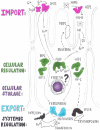
Figure 1
Figure 1
Simplified scheme of iron absorption in mammals. A typical enterocyte of the duodenum of the mammalian intestine has uptake transporters for iron (DMT1) and heme (HCP1) localized in the apical membrane. An iron export transporter (ferroportin) is localized in the basolateral membrane. Ferric iron is reduced by Dcytb prior to import and oxidized by Hephaestin upon export. Iron is stored locally in the enterocyte in ferritin. Whether the iron chaperone PCBP has a role in iron absorption remains to be determined (indicated by a question mark). Heme oxygenases release iron from heme. The large byproduct of this reaction (biliverdin) is modified and secreted into the gut lumen though the Multidrug Resistant Protein-2 (MRP2) transporter. Iron absorption is regulated at the systemic level by hepcidin, which is secreted by the liver hepatocytes in response to various physiologic stimuli. Local cellular regulation also occurs via the Hypoxia Inducible Factors (HIFs) and Iron Regulatory Proteins (IRPs) and may be influenced by circulating levels of erythropoietin (EPO).
Figure 2
Figure 2
Iron absorption likely takes place in the anterior midgut where all transport proteins studied to date are found. Copper cells are acid-secreting cells, which have also been shown to secrete iron-loaded ferritin in the intestinal lumen. The iron cells express ferritin constitutively and likely serve an iron storage function. Iron cells regulate iron homeostasis independently of IRP-1A and ferritin independently of iron. The posterior midgut appears to be involved with iron homeostasis only in conditions of iron overload. How the different intestinal domains interact with each other remains unknown. For higher resolution images of the different cell types in this diagram the readers are referred to a beautiful representation based on ultrastructure studies performed recently for this tissue [57]. Our diagram is also based on the now 61-year-old study of iron and copper localization in insects [17]. The most posterior part of the midgut (not shown) has no involvement in iron homeostasis, but is a compartment specialized in zinc storage [58,59].

Similar articles

Cited by

References

    1. Law J.H. Insects, oxygen, and iron. Biochem. Biophys. Res. Commun. 2002;292:1191–1195. doi: 10.1006/bbrc.2001.2015. - DOI - PubMed
    1. Missirlis F., Kosmidis S., Brody T., Mavrakis M., Holmberg S., Odenwald W.F., Skoulakis E.M., Rouault T.A. Homeostatic mechanisms for iron storage revealed by genetic manipulations and live imaging of Drosophila ferritin. Genetics. 2007;177:89–100. doi: 10.1534/genetics.107.075150. - DOI - PMC - PubMed
    1. Warburg O. Iron, the oxygen-carrier of respiration-ferment. Science. 1925;61:575–582. - PubMed
    1. Brown N.C., Eliasson R., Reichard P., Thelander L. Nonheme iron as a cofactor in ribonucleotide reductase from E. coli. Biochem. Biophys. Res. Commun. 1968;30:522–527. doi: 10.1016/0006-291X(68)90083-1. - DOI - PubMed
    1. Clark D.V. Molecular and genetic analyses of Drosophila Prat, which encodes the first enzyme of de novo purine biosynthesis. Genetics. 1994;136:547–557. - PMC - PubMed

Publication types

LinkOut - more resources