. 2013 Aug;20(8):1116-23.
doi: 10.1038/cdd.2013.49. Epub 2013 May 17.

Evidence for a common progenitor of epithelial and mesenchymal components of the liver

Affiliations

Evidence for a common progenitor of epithelial and mesenchymal components of the liver

A Conigliaro et al. Cell Death Differ. 2013 Aug.

Abstract

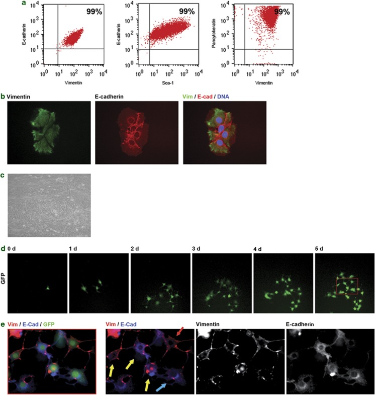
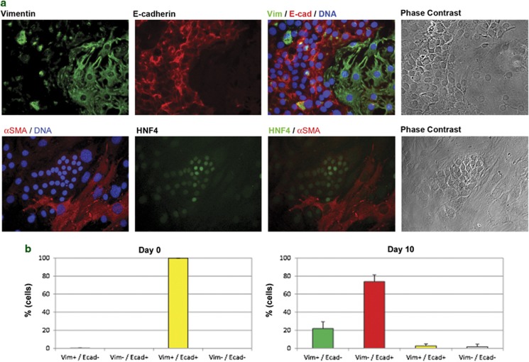
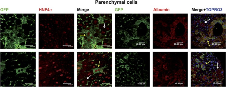
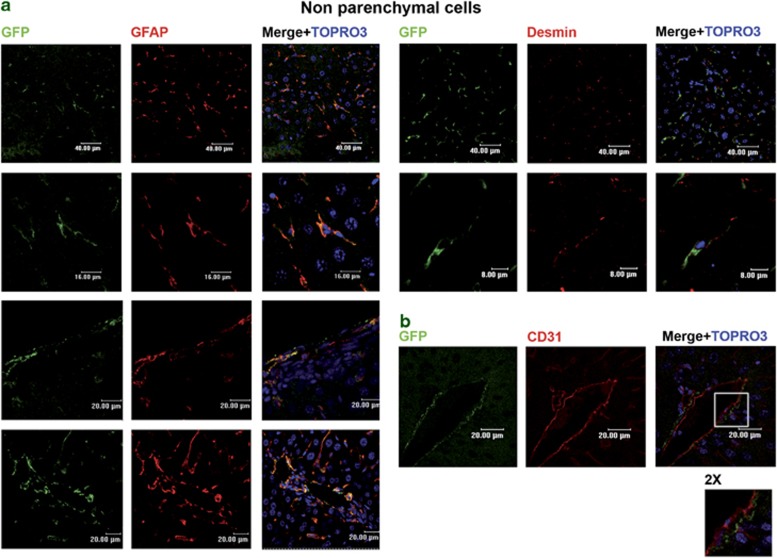
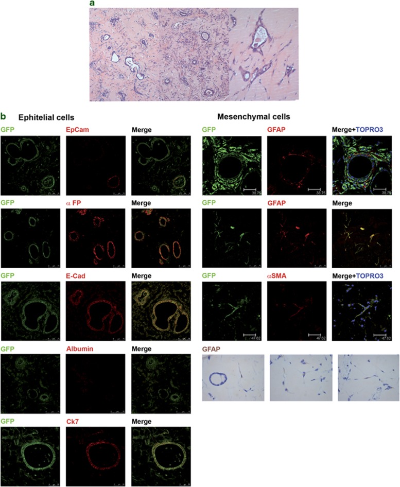
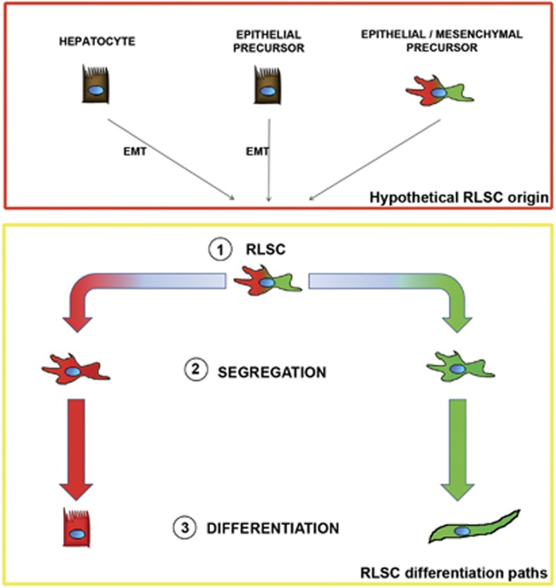
Tissues of the adult organism maintain the homeostasis and respond to injury by means of progenitor/stem cell compartments capable to give rise to appropriate progeny. In organs composed by histotypes of different embryological origins (e.g. the liver), the tissue turnover may in theory involve different stem/precursor cells able to respond coordinately to physiological or pathological stimuli. In the liver, a progenitor cell compartment, giving rise to hepatocytes and cholangiocytes, can be activated by chronic injury inhibiting hepatocyte proliferation. The precursor compartment guaranteeing turnover of hepatic stellate cells (HSCs) (perisinusoidal cells implicated with the origin of the liver fibrosis) in adult organ is yet unveiled. We show here that epithelial and mesenchymal liver cells (hepatocytes and HSCs) may arise from a common progenitor. Sca+ murine progenitor cells were found to coexpress markers of epithelial and mesenchymal lineages and to give rise, within few generations, to cells that segregate the lineage-specific markers into two distinct subpopulations. Notably, these progenitor cells, clonally derived, when transplanted in healthy livers, were found to generate epithelial and mesenchymal liver-specific derivatives (i.e. hepatocytes and HSCs) properly integrated in the liver architecture. These evidences suggest the existence of a 'bona fide' organ-specific meso-endodermal precursor cell, thus profoundly modifying current models of adult progenitor commitment believed, so far, to be lineage-restricted. Heterotopic transplantations, which confirm the dual differentiation potentiality of those cells, indicates as tissue local cues are necessary to drive a full hepatic differentiation. These data provide first evidences for an adult stem/precursor cell capable to differentiate in both parenchymal and non-parenchymal organ-specific components and candidate the liver as the instructive site for the reservoir compartment of HSC precursors as yet non-localized in the adult.


Figures

Figure 1
Figure 1
RLSC characterization and in vitro differentiation. (a) FACS analysis for the indicated antigens highlighted as the RLSC starting population homogeneously expresses the stemness marker SCA1, the epithelial markers Pan-CK and E-cad and the mesenchymal marker Vimentin. (b) Immunocytochemical analysis for the indicated markers highlighted as the RLSC starting population coexpresses epithelial and mesenchymal markers. (c) Within 10 days, RLSCs in low-serum culture condition gave rise to derivatives that acquired distinct epithelial and mesenchymal phenotypes, as observed by phase contrast (original magnification × 20). (d–e) Time-lapse analysis (see also video 1). Within 5 days single EGFP-RLSC cells, in low-serum culture condition, gave rise to clonal progeny composed of cells coexpressing Vimentin and E-cadherin (yellow arrows) and cells expressing E-cadherin (blue arrow) or Vimentin (red arrow)
Figure 2
Figure 2
RLSC derivatives segregation of epithelial and mesenchymal markers into distinct subpopulations. (a) Immunocytochemical analysis for the indicated markers highlighted as the RLSCs, in low-serum culture condition for 10 days, undergo into either epithelial or mesenchymal differentiation. The morphology of the two different cellular subpopulations is coherent with the expression of specific markers. (b) Cells were scored in four categories based on their Vimentin and E-Cadherin expression. Data show percentage of cells for each category. Average of three experiments. N=500 cells were counted for each experiment. Data are shown as mean±S.E.M.
Figure 3
Figure 3
RLSC parenchymal derivatives in orthotopic transplants. Immunohistochemical analysis of RLSC-engrafted livers. The RLSC EGFP progeny was detected in untreated CD1 null mice injected at day 1 after birth and killed after 2 months. EGFP parenchymal cells were found integrated into the epithelial cords expressing hepatocyte markers HNF4 and Albumin. Yellow arrows indicate exogenous hepatocytes; white arrows indicate endogenous cells
Figure 4
Figure 4
RLSC non-parenchymal derivatives in orthotopic transplants. Immunohistochemical analysis of RLSC-engrafted livers. The RLSC EGFP progeny was detected in untreated CD1 null mice injected at day 1 after birth and killed after 2 months. EGFP non-parenchymal cells expressing GFAP and Desmin were found both (a) scattered in the parenchyma and (b) in subendothelial positions. The endothelium was decorated with CD31 antibody
Figure 5
Figure 5
RLSC derivatives in heterotopic transplants. H&E, Immunohistochemical and Immunofluorescence analysis of RLSCs engrafted in SCID adult null mice. RLSCs were injected in Matrigel in the epifascial region and animals were analyzed 1 month after injection. (a) H&E revealed epithelial-like RLSC derivatives found either arranged in compact islands or delimiting empty spaces. Mesenchymal-like RLSC derivatives were found scattered in the matrigel scaffold. (b) The EGFP epithelial cells were found to express the hepatic progenitor/hepatocyte markers, EpCAM, αFP, E-Cad, Albumin and CK7. (c) The EGFP mesenchymal cells were found to express GFAP and αSMA by IF and IHC (bottom panel)
Figure 6
Figure 6
Schematic representation of hypothetical RLSC origin and of observed RLSC differentiation capacity. RLSC have been obtained from single-cell cloning of liver explants. Red box: cells hypothetically giving rise to RLSCs may include epithelial cells that underwent to EMT during the immortalization procedure, thus indicating a broad reprogramming potential of parenchymal cells. Alternatively, an epithelial/mesenchymal RLSC precursor counterpart may exist in vivo. Yellow box: RLSCs were found in cell culture, in heterotopic and in orthotopic transplantations, to undergo into two mutually exclusive differentiations having as ending point mesenchymal or epithelial cells

References

    1. Dellavalle A, Sampaolesi M, Tonlorenzi R, Tagliafico E, Sacchetti B, Perani L, et al. Pericytes of human skeletal muscle are myogenic precursors distinct from satellite cells. Nat Cell Biol. 2007;9:255–267. - PubMed
    1. Tanaka EM, Ferretti P. Considering the evolution of regeneration in the central nervous system. Nat Rev Neurosci. 2009;10:713–723. - PubMed
    1. Li L, Clevers H. Coexistence of quiescent and active adult stem cells in mammals. Science. 2010;327:542–545. - PMC - PubMed
    1. Nagy P, Bisgaard HC, Thorgeirsson SS. Expression of hepatic transcription factors during liver development and oval cell differentiation. J Cell Biol. 1994;126:223–233. - PMC - PubMed
    1. Sell S. Liver stem cells. Mod Pathol. 1994;7:105–112. - PubMed

Publication types

MeSH terms

Substances