Review
. 2013:371:229-57.
doi: 10.1007/978-3-642-37765-5_9.

Interplay between DNA tumor viruses and the host DNA damage response

Affiliations
Review

Interplay between DNA tumor viruses and the host DNA damage response

Karyn McFadden et al. Curr Top Microbiol Immunol. 2013.

Abstract

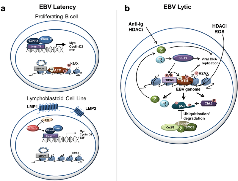
Viruses encounter many challenges within host cells in order to replicate their nucleic acid. In the case of DNA viruses, one challenge that must be overcome is recognition of viral DNA structures by the host DNA damage response (DDR) machinery. This is accomplished in elegant and unique ways by different viruses as each has specific needs and sensitivities dependent on its life cycle. In this review, we focus on three DNA tumor viruses and their interactions with the DDR. The viruses Epstein-Barr virus (EBV), Kaposi's sarcoma-associated herpesvirus (KSHV), and human papillomavirus (HPV) account for nearly all of the virus-associated human cancers worldwide. These viruses have also been excellent models for the study of oncogenic virus-mediated cell transformation. In this review, we will discuss how each of these viruses engage and subvert aspects of the host DDR. The first level of DDR engagement is a result of the genetic linkage between the oncogenic potential of these viruses and their ability to replicate. Namely, the promotion of cells from quiescence into the cell cycle to facilitate virus replication can be sensed through aberrant cellular DNA replication structures which activate the DDR and hinder cell transformation. DNA tumor viruses subvert this growth-suppressive DDR through changes in viral oncoprotein expression which ultimately facilitate virus replication. An additional level of DDR engagement is through direct detection of replicating viral DNA. These interactions parallel those observed in other DNA virus systems in that the need to subvert these intrinsic sensors of aberrant DNA structure in order to replicate must be in place. DNA tumor viruses are no exception. This review will cover the molecular features of DNA tumor virus interactions with the host DDR and the consequences for virus replication.


Figures

Figure 1
Figure 1. Schematic diagram of the ATM and ATR DNA damage response signaling pathways.
a) ATM activation responding to a DNA double-stranded break (DSB). The initial complex of Mre11, Rad50, and Nbs1 targets the DSB and recruits ATM to activate its kinase domain. This leads to phosphorylation and monomerization, which then triggers a cascade of target phosphorylations. H2AX, the histone H2A variant, is first phosphorylated and provides a scaffold for MDC1, which recruits the ring finger Ub ligases RNF8 and RNF168 to ubiquitinate H2AX. Concomitantly, enzymes such as Tip60 are recruited that can facilitate both ATM activation and chromatin de-compaction through acetylation of ATM directly and histone tails, respectively. The ubiquitination of H2AX further promotes recruitment and retention of factors such as 53BP1 and Brca1 that establish stable DDR foci propagating ATM signaling to effector molecules such as the checkpoint kinase Chk2, the transcription factor p53, and others. b) ATR activation responding to ssDNA exposure following DNA replicative stress. Following replication fork collapse, ssDNA exposure is sensed by the protein RPA that coats these regions and serves as a scaffold to recruit the kinase ATR through its co-factor ATRIP. The critical ATR activating factor TopBP1 is recruited through the Rad9-Rad1-Hus1 (9-1-1) complex at the replication fork. Finally, Claspin recruitment to the complex enables ATR phosphorylation of Chk1 and many other downstream effectors. A common downstream target is p53, which can be phosphorylated by both ATM and ATR leading to phenotypic outcomes ranging from DNA repair to apoptosis and senescence depending on the strength and duration of the DNA damaging signal.
Figure 2
Figure 2. Epstein-Barr virus and the DNA damage response.
a) DDR activation during EBV latency. Top, Early after B cell infection with EBV, cells hyper-proliferate dependent on the expression of the viral EBNA2 and EBNA-LP proteins driving c-Myc and other S phase-promoting genes. This results in an activated DDR including ATM and H2AX phosphorylation. Bottom, in later divisions after infection, and also in immortalized lymphoblastoid cell lines (LCLs), the expression of the EBNA3C protein mitigates the DDR, presumably through attenuating the CDK inhibitor p16. Additional viral proteins are expressed such as LMP1 and LMP2 that contribute to the LCL survival phenotype. B) DDR activation during EBV lytic infection. Reactivation from diverse stimuli converge on expression of the critical viral trans-activator Z. In the case of anti-Ig treatment of latently-infected B cells or pan-HDAC inhibition, these effects are direct on the Z promoter (top left). However, other molecules such as DNA damaging agents like reactive oxygen species (ROS) can directly promote ATM activation. The consequence of this activation is also Z expression through the ATM-dependent regulation of viral lytic promoters and Tip60-mediated acetylation of viral chromatin. Expression of the viral kinase BGLF4 further supports this mechanism through direct activation of Tip60, ATM, and H2AX. Ultimately the downstream signaling from ATM can lead to the deleterious effect of p53-mediated apoptosis. However, the viral Z protein can form an E3 ubiquitin ligase complex to promote p53 degradation at late stages during viral lytic replication.
Figure 3
Figure 3. Kaposi’s sarcoma-associated herpesvirus and the DNA damage response.
a) DDR activation during KSHV latency. The induction of cell cycle progression by the viral cyclin (v-cyclin) in complex with CDK6 leads to the activation of an ATM-mediated DDR.The consequence of this activation is p53 induction, oncogene-induced senescence (OIS), and autophagy. However, co-expression of the KSHV v-FLIP protein overrides v-cyclin-mediated (OIS) by suppressing autophagy. b) KSHV and the DDR during lytic reactivation. Little is understood about KSHV lytic viral DNA replication and the DDR. However, the expression of the lytic cycle vIRF1 gene is capable of suppressing p53 activity and, in particular, the ability of ATM to activate p53. This suggests that if ATM were to be activated by KSHV lytic DNA replication (as occurs in EBV, HPV, and MHV68 infection), then a plausible mechanism to overcome checkpoint activation or apoptosis would be in place.
Figure 4
Figure 4. Human papillomavirus and the DNA damage response.
a) HPV infection of basal layer epithelial cells and DDR activation. The infection of basal epithelial cells by HPV leads to a constitutive ATM-dependent DDR. This may be due to the role of HPV E1 in promoting replication of integrated HPV genomes or perhaps promoting DNA damage at other sites through the host genome (left). Normally, in this layer, E2 protein suppresses the expression of E6 and E7. If E2 is abrogated due to integration, then E7 up-regulation could lead to increased S-phase entry promoting the DDR or through direct engagement and activation of ATM (bottom, right). b) During vegetative DNA replication upon differentiation of the basal keratinocyte layer the ATM pathway is activated and DDR factors co-localize with viral genomes (right). ATM and Chk2 activity are important for viral DNA replication in this phase of the HPV life cycle. The uppermost layer of keratinocytes in the figure indicates virion release (stars).

References

    1. Bartek J, Bartkova J, and Lukas J, DNA damage signalling guards against activated oncogenes and tumour progression. Oncogene, 2007. 26(56): p. 7773–9. - PubMed
    1. Smith J, et al., The ATM-Chk2 and ATR-Chk1 pathways in DNA damage signaling and cancer. Adv Cancer Res, 2010. 108: p. 73–112. - PubMed
    1. Derheimer FA and Kastan MB, Multiple roles of ATM in monitoring and maintaining DNA integrity. FEBS Lett, 2010. 584(17): p. 3675–81. - PMC - PubMed
    1. Falck J, Coates J, and Jackson SP, Conserved modes of recruitment of ATM, ATR and DNA-PKcs to sites of DNA damage. Nature, 2005. 434(7033): p. 605–11. - PubMed
    1. So S, Davis AJ, and Chen DJ, Autophosphorylation at serine 1981 stabilizes ATM at DNA damage sites. J Cell Biol, 2009. 187(7): p. 977–90. - PMC - PubMed

Publication types