. 2013 Jun 28;340(6140):1587-90.
doi: 10.1126/science.1237572. Epub 2013 May 16.

B cells use mechanical energy to discriminate antigen affinities

Affiliations

B cells use mechanical energy to discriminate antigen affinities

Elizabeth Natkanski et al. Science. .

Abstract

The generation of high-affinity antibodies depends on the ability of B cells to extract antigens from the surfaces of antigen-presenting cells. B cells that express high-affinity B cell receptors (BCRs) acquire more antigen and obtain better T cell help. However, the mechanisms by which B cells extract antigen remain unclear. Using fluid and flexible membrane substrates to mimic antigen-presenting cells, we showed that B cells acquire antigen by dynamic myosin IIa-mediated contractions that pull out and invaginate the presenting membranes. The forces generated by myosin IIa contractions ruptured most individual BCR-antigen bonds and promoted internalization of only high-affinity, multivalent BCR microclusters. Thus, B cell contractility contributes to affinity discrimination by mechanically testing the strength of antigen binding.


Figures

Fig. 1
Fig. 1
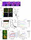
B cells acquire antigens from flexible membranes. (A) Sideview reconstruction of B220-stained primary B cells forming synapses with DiI-stained and anti-Igκ antigen-loaded (Ag) PMS or PLB. Arrowheads indicate the position of the substrate. Scalebars, 2 μm. (B) Image quantification of primary B cell antigen internalization (means±SEM, n=23-60 cells). (C) AFM force retraction curves of streptavidin-coated AFM tip and biotinylated antigens. Antigens were anti-Igκ for PLB, PM-PLB and PMS, and immune complexes of NIP antigen for DCs. Speed of retraction was 0.1μm/s. (D) Rupture distances and forces, mean±SD, n=31-109 retraction curves. (E) Colocalization of internalized antigen with DiI in primary B cells after internalization from the substrates. Means±SEM, n=12-21 cells. (F) B220-stained B1-8 primary B cell internalizing immune complexes of NIP antigen from a DC stained with DiI. Scalebar, 5 μm.
Fig. 2
Fig. 2
Primary B cells extract antigens by invaginating and pinching off the presenting membranes. (A) Changes in DiI intensity on antigen-loaded PMS after B cell spreading (white outline). (B) 3D localization of antigen. Top, DiI-stained PMS loaded with unlabeled antigen and antigen-Qdots before and after B cell spreading. Bottom, antigen-Qdots color-coded for their vertical position. Arrowhead shows a Qdot pulled-up by the B cell. (C) Vertical position of antigen-Qdots versus colocalized DiI fluorescence (n=10 cells, p for Pearson correlation test). (D) Antigen and DiI fluorescence in a single antigen microcluster. Arrow indicates internalization. (E) Lifetimes of invaginations (mean±SEM, n=7 cells). Inset, DiI fluorescence of a short-lived invagination. (F) Antigen fluorescence in invaginations grouped by lifetime. Dotted lines show fluorescence before and after invagination’s lifetime (mean±SEM, n=10 cells). *, p<0.01 in paired t-tests. (G) B cell expressing clathrin light chain-GFP spreading on antigen-loaded PMS. (H) Numbers and fluorescence of synaptic CCSs (mean±SEM, n=15-21 cells, *, p<0.05 in nonparametric tests for t>1 min against controls). (I) Percentage of invaginations colocalizing with CCSs (mean±SEM, n=12 cells). Black lines, colocalization with randomly scrambled CCSs. *, p<0.01 in nonparametric tests against scrambled controls. Scale bars, 5 μm.
Fig. 3
Fig. 3
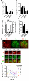
B cell antigen extraction requires myosin IIa contractility and clathrin-mediated endocytosis. (A) Antigen internalization (anti-Igμ) and (B) number of invaginations pulled from PMS by Ramos B cells expressing shRNA against AP2, dynamin2, Rock1, or myosin IIA. (C) Antigen internalization (anti-Igκ) from PMS (left y-axis) or solution (right y-axis) by primary B cells treated with inhibitors against dynamin2 (dynasore), Rock1 (Y-23632), or myosin IIA (blebbistatin). (D) Number of invaginations pulled from PMS by primary B cells treated with inhibitors. A-D, means±SEM, n=17-25 cells for internalization from PMS, n=7-15 cells for invagination numbers, n=3 experiments for soluble antigen internalization. *, p<0.01, **, p<0.05 in non-parametric tests against controls. F.u., fluorescence units. (E) Top, TIRF image of a primary B cell expressing myosin IIa regulatory light chain (RLC)-GFP (Myo) spread on DiD-labeled PMS loaded with antigen. Yellow squares show a region magnified below. Closed arrowheads show invagination, open arrowheads show myosin IIa structures. Scalebar, 1 μm. (F) Quantification of myosin IIa RLC fluorescence in invaginations grouped by lifetime (mean±SEM, n=15 cells). *, p<0.01 in paired t-tests.
Fig. 4
Fig. 4
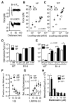
Forces generated by myosin IIa contractility promote affinity-dependent antigen internalization. (A) Force spectroscopy retraction curves showing rupture of bonds between NIP antigen and B1-8 Fab fragment (top) or B1-8 BCR on live primary B1-8 B cells (bottom). Speed of retraction was 0.1 μm/s. (B, C) Mean rupture forces (±SEM) measured by AFM force spectroscopy with NP (B) and NIP (C) binding to B1-8 Fab fragment on coverslips or to B1-8 BCR on B cells. Retraction speeds were 0.1-50 μm/s. N=41-185 retraction curves. (D, E) Internalization of antigens by B1-8 or wild type (WT) primary B cells from solution (D) or from PMS (E). (F) Lifetime of invaginations. (G) Antigen fluorescence associated with invaginations of indicated lifetime. (H) Internalization of antigens by B1-8 B cells treated with blebbistatin. D-H, means±SEM of individual cells, n=25-118. If error bars are not visible, they are smaller than the symbols. Asterisks show statistical significance in non-parametric tests at the indicated p-values.

References

    1. Carrasco YR, Batista FD. B cells acquire particulate antigen in a macrophage-rich area at the boundary between the follicle and the subcapsular sinus of the lymph node. Immunity. 2007;27:160–171. - PubMed
    1. Phan TG, et al. Immune complex relay by subcapsular sinus macrophages and noncognate B cells drives antibody affinity maturation. Nat. Immunol. 2009;10:786. - PMC - PubMed
    1. Junt T, et al. Subcapsular sinus macrophages in lymph nodes clear lymph-borne viruses and present them to antiviral B cells. Nature. 2007;450:110. - PubMed
    1. Qi H, Egen JG, Huang AYC, Germain RN. Extrafollicular activation of lymph node B cells by antigen-bearing dendritic cells. Science. 2006;312:1672–1676. - PubMed
    1. Gonzalez SF, et al. Capture of influenza by medullary dendritic cells via SIGN-R1 is essential for humoral immunity in draining lymph nodes. Nat. Immunol. 2010;11:427–434. - PMC - PubMed

Publication types