. 2013 Sep;8(4):998-1009.
doi: 10.1007/s11481-013-9471-7. Epub 2013 May 18.

Adenosine triphosphate released from HIV-infected macrophages regulates glutamatergic tone and dendritic spine density on neurons

Affiliations

Adenosine triphosphate released from HIV-infected macrophages regulates glutamatergic tone and dendritic spine density on neurons

Luis B Tovar-Y-Romo et al. J Neuroimmune Pharmacol. 2013 Sep.

Abstract

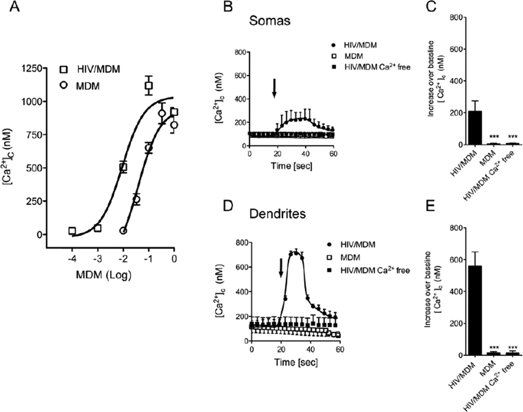
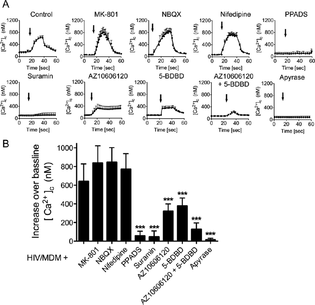
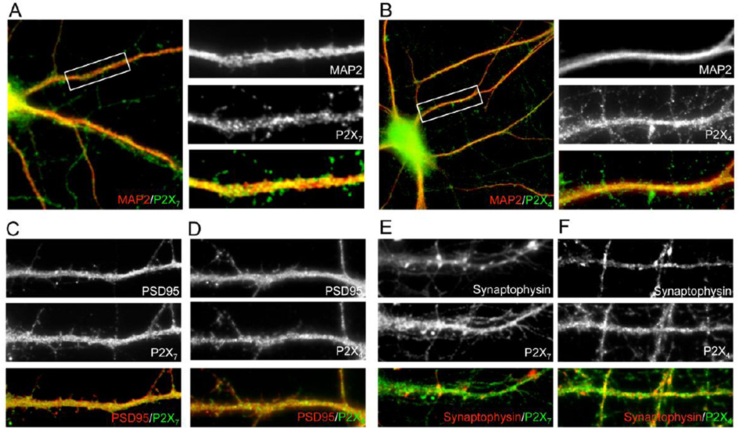
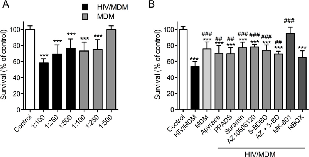
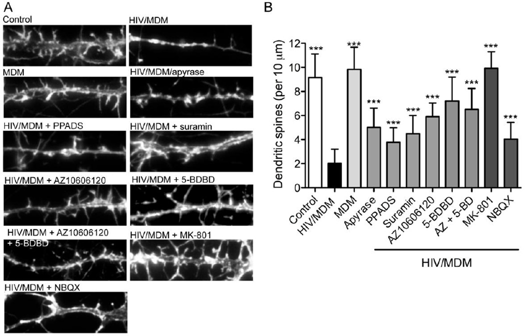
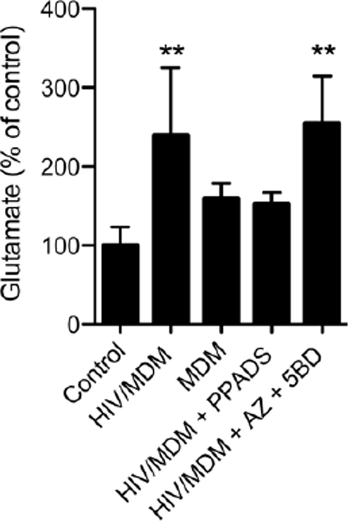
Despite wide spread use of combination antiretroviral therapy (cART) in developed countries, approximately half of HIV-infected patients will develop impairments in cognitive function. Accumulating evidence suggests that neuronal dysfunction can be precipitated by HIV-infection of macrophages by mechanisms that involve alterations in innate and adaptive immune responses. HIV-infection of macrophages is known to increase the release of soluble neurotoxins. However, the composition of products released from infected macrophages is complex and not fully known. In this study we provide evidence that ATP and other immuno-/neuromodulatory nucleotides are exported from HIV-infected macrophages and modify neuronal structure. Supernatants collected from HIV-infected macrophages (HIV/MDM) contained large amounts of ATP, ADP, AMP and small amounts of adenosine, in addition to glutamate. Dilutions of these supernatants that were sub-threshold for glutamate receptor activation evoked rapid calcium flux in neurons that were completely inhibited by the enzymatic degradation of ATP, or by blockade of calcium permeable purinergic receptors. Applications of these highly diluted HIV/MDM onto neuronal cultures increased the amount of extracellular glutamate by mechanisms dependent on purinergic receptor activation, and downregulated spine density on neurons by mechanisms dependent on purinergic and glutamate receptor activation. We conclude from these data that ATP released from HIV-infected macrophages downregulates dendritic spine density on neurons by a mechanism that involves purinergic receptor mediated modulation of glutamatergic tone. These data suggest that neuronal function may be depressed in HIV infected individuals by mechanisms that involve macrophage release of ATP that triggers secondary effects on glutamate handling.


Conflict of interest statement

COMPETING INTERESTS

The authors have no conflicts of interest or competing interests to disclose.

Figures

Figure 1
Figure 1. HIV/MDM supernatants stimulate extracellular calcium influx in cultured hippocampal neurons
MDM and HIV/MDM supernatants evoked dose-dependent increases of [Ca2+]c in primary hippocampal neurons. (A) Peak [Ca2+]c increases over baseline in neurons exposed to the indicated concentrations of supernatants from MDM and HIV/MDM. HIV/MDM evoked larger [Ca2+]c increases compared with MDM at all concentrations tested. Representative traces showing the time course and peak [Ca2+]c increases over baseline evoked by brief applications of supernatants from MDM, HIV-MDM, and HIV/MDM in calcium free buffer for neuronal somas (B–C) and dendrites (D–E). MDM and HIV/MDM supernatants were each used at 1:500 dilution (arrows indicate time of addition). Each data point represents the average ± SD of 10–12 somas or 25–30 dendrites derived from 7 separate experiments. ANOVA with Tukey’s post hoc comparisons. *** p < 0.001.
Figure 2
Figure 2. HIV/MDM activates calcium permeable purinergic receptors
HIV/MDM-evoked calcium flux was quantified in the presence of the indicated calcium channel antagonists. (A) Representative traces showing low dose HIV/MDM (1:500)-evoked [Ca2+]c responses in neuronal dendrites in the presence of vehicle (Control), or antagonists for NMDA (MK-801; 10 µM), AMPA (NBQX; 10 µM), L-type voltage operated calcium channels (Nifedipine; 10 µM), general blockers of calcium permeable P2X receptors (PPADS: 10 µM. Suramin; 100 µM), P2X7 (AZ10606120; 10 µM), P2X4 (5-BDBD; 10 µM), and HIV/MDM supernatants pre-incubated with apyrase (2U/10 µl). (B) Summary data showing peak amplitudes of [Ca2+]c responses for the indicated conditions. Data are the average ± SD of [Ca2+]c responses recorded from 30–35 dendrites in 7 independent experiments. ANOVA with Tukey’s post hoc comparisons. *** p < 0.001.
Figure 3
Figure 3. The purinergic receptors P2X7 and P2X4 are expressed at post-synaptic regions in hippocampal neurons
Representative images showing the expression of P2X7 and P2X4 in hippocampal neurons. Co-localization of P2X7(A) and P2X4(B) with the neuronal marker MAP-2. Images to the right of each primary image are enlarged to show the indicated portion of the dendrite. MAP-2 (red) and P2X4 or P2X7 (green) appear as yellow in the merged images when co-localized with MAP-2. P2X7(C) and P2X4(D)(green) co-localize with the postsynaptic marker PSD-9 (red). P2X7(E) and P2X4(F) (green) do not co-localize with the presynaptic marker synaptophysin (red).
Figure 4
Figure 4. Calcium permeable purinergic receptors regulate HIV/MDM-evoked excitotoxicity
Apoptotic nuclei with condensed or fragmented chromatin were determined by Hoechst 33342 DNA staining in neurons exposed to high-dose MDM or HIV/MDM supernatants (1:100 dilution) for 24 h. (A) Dose–dependent effect on neuronal survival upon exposure to MDM or HIV/MDM supernatants. (B) Neuronal apoptosis induced by HIV/MDM supernatants (1:100) was reduced by pre-treatment of HIV/MDM supernatant with apyrase (2 IU/10 µl HIV/MDM), and general antagonists of purinergic receptors (PPADS; 10 µM and Suramin; 100 µM), specific antagonists of P2X7 (AZ10606120; 10 µM), P2X4 (5-BDBD; 10 µM) and AMPA receptors (NBQX; 10 µM). Neuronal death was completely blocked by an antagonist to NMDA receptors (MK-801; 10 µM). Data are the average ± S.D. of 200 cells from three separate experiments per condition. ANOVA with Tukey’s post hoc comparisons. *** = p < 0.001, ** = p < 0.01 compared with control, and ### = p < 0.001, ## = p < 0.01 compared with HIV/MDM supernatant.
Figure 5
Figure 5. Purinergic and NMDA receptors mediate the downregulation of dendritic spines in response to low dose HIV/MDM supernatants
Neuronal dendrites were visualized by labeling F-actin with fluorescent phalloidin. (A) Representative images showing individual dendritic branches and associated spines in neurons treated with vehicle (Control), low dose HIV/MDM supernatants (1:250), MDM supernatants (1:250), and HIV/MDM supernatants in the presence of apyrase (2 IU/10 µl), or antagonists of purinergic receptors (PPADS; 10 µM and suramin; 100 µM), P2X7 (AZ10606120; 10 µM), P2X4 (5-BDBD; 10 µM), NMDA (MK801; 10 µM), and AMPA (NBQX; 10 µM). (B) Quantitative analysis of dendritic spine numbers. HIV/MDM supernatant-induced downregulation of dendritic spines was attenuated by degradation of ATP, purinergic, and AMPA receptor antagonists. NMDA-receptor blockade provided near complete protection from HIV/MDM induced downregulation of dendritic spine number. Quantitative data are the average ± S.D. of dendritic spine number per 10 µm. Data were obtained from a total of 10 different neurons in 3 independent experiments per condition. ANOVA with Tukey’s post hoc comparisons. *** = p < 0.001 compared with HIV/MDM sup.
Figure 6
Figure 6. ATP contained in HIV/MDM supernatants increases extracellular glutamate by a purinergic receptor mediated mechanism
Hippocampal neuronal cultures were subjected to 5 min incubations with HIV/MDM (1:100) in the presence of DL-TBOA (70 µM) to block glutamate transporters. HIV/MDM supernatants induced a 2.5-fold increase in the content of extracellular glutamate that was blocked by PPADS (10 µM), but not by co-administration of antagonists for P2×4 and P2X7 receptors (5-BDBD (5BD) + AZ10606120 (AZ); 10 µM each). Experiments were conducted in quadruplicate. Values are mean ± S.D. of glutamate normalized to controls. ANOVA with Tukey’s post hoc comparisons ** = p < 0.01 compared to control.

Similar articles

Cited by

References

    1. Ayna G, Krysko DV, Kaczmarek A, Petrovski G, Vandenabeele P, Fesus L. ATP release from dying autophagic cells and their phagocytosis are crucial for inflammasome activation in macrophages. PLoS One. 2012;7:e40069. - PMC - PubMed
    1. Bandaru VV, Patel N, Ewaleifoh O, Haughey NJ. A Failure to Normalize Biochemical and Metabolic Insults During Morphine Withdrawal Disrupts Synaptic Repair in Mice Transgenic for HIV-gp120. J Neuroimmune Pharmacol. 2011;6:640–649. - PMC - PubMed
    1. Brew BJ, Corbeil J, Pemberton L, Evans L, Saito K, Penny R, Cooper DA, Heyes MP. Quinolinic acid production is related to macrophage tropic isolates of HIV-1. J Neurovirol. 1995;1:369–374. - PubMed
    1. Buckner CM, Calderon TM, Willams DW, Belbin TJ, Berman JW. Characterization of monocyte maturation/differentiation that facilitates their transmigration across the blood-brain barrier and infection by HIV: implications for NeuroAIDS. Cell Immunol. 2011;267:109–123. - PMC - PubMed
    1. Chen W, Sulcove J, Frank I, Jaffer S, Ozdener H, Kolson DL. Development of a human neuronal cell model for human immunodeficiency virus (HIV)-infected macrophage-induced neurotoxicity: apoptosis induced by HIV type 1 primary isolates and evidence for involvement of the Bcl-2/Bcl-xL-sensitive intrinsic apoptosis pathway. J Virol. 2002;76:9407–9419. - PMC - PubMed

Publication types

LinkOut - more resources