. 2013 Jun 15;190(12):6340-50.
doi: 10.4049/jimmunol.1300397. Epub 2013 May 17.

Truncated form of TGF-βRII, but not its absence, induces memory CD8+ T cell expansion and lymphoproliferative disorder in mice

Affiliations

Truncated form of TGF-βRII, but not its absence, induces memory CD8+ T cell expansion and lymphoproliferative disorder in mice

Harumichi Ishigame et al. J Immunol. .

Abstract

Inflammatory and anti-inflammatory cytokines play an important role in the generation of effector and memory CD8(+) T cells. We used two different models, transgenic expression of truncated (dominant negative) form of TGF-βRII (dnTGFβRII) and Cre-mediated deletion of the floxed TGF-βRII to examine the role of TGF-β signaling in the formation, function, and homeostatic proliferation of memory CD8(+) T cells. Blocking TGF-β signaling in effector CD8(+) T cells using both of these models demonstrated a role for TGF-β in regulating the number of short-lived effector cells but did not alter memory CD8(+) T cell formation and their function upon Listeria monocytogenes infection in mice. Interestingly, however, a massive lymphoproliferative disorder and cellular transformation were observed in Ag-experienced and homeostatically generated memory CD8(+) T cells only in cells that express the dnTGFβRII and not in cells with a complete deletion of TGF-βRII. Furthermore, the development of transformed memory CD8(+) T cells expressing dnTGFβRII was IL-7- and IL-15-independent, and MHC class I was not required for their proliferation. We show that transgenic expression of the dnTGFβRII, rather than the absence of TGF-βRII-mediated signaling, is responsible for dysregulated expansion of memory CD8(+) T cells. This study uncovers a previously unrecognized dominant function of the dnTGFβRII in CD8(+) T cell proliferation and cellular transformation, which is caused by a mechanism that is different from the absence of TGF-β signaling. These results should be considered during both basic and translational studies where there is a desire to block TGF-β signaling in CD8(+) T cells.


Conflict of interest statement

Disclosures

The authors declare no competing financial interests.

Figures

FIGURE 1
FIGURE 1
Conditional deletion of Tgfbr2 in OTI cells results in increased number of SLECs, but does not lead to increased accumulation of total effector cells during acute Listeria infection (A) Naive OTI-WT and OTI-dnTGFβRII cells on different CD45 congenic backgrounds were adoptively co-transferred into naive CD11c-dnTGFβRII hosts, followed by infection with LM-OVA 1 day later. The percentage of OTI-WT and OTI-dnTGFβRII cells was monitored among the PBLs of infected mice over time. (B) The ratio of OTI-dnTGFβRII over OTI-WT cells in PBLs. (C) The percentage of OTI-WT and OTI-dnTGFβRII SLECs in the blood after LM-OVA infection at the indicated time point (left). Representative plots of the expression of CD127 and KLRG1 on blood OTI-WT and OTI-dnTGFβRII cells at day 7 p.i. (right). (D) The percentage of OTI-control (CD45.1.1) and OTI-CD4Cre-Tgfbr2f/f (CD45.1.2) cells among PBLs was monitored over time. (E) The ratio of OTI-CD4Cre-Tgfbr2f/f over OTI-control cells in PBLs. (F) The percentage of SLECs in the blood OTI-control and OTI-CD4Cre-Tgfbr2f/f cells after LM-OVA infection at the indicated time point. Representative plots of the expression of CD127 and KLRG1 in blood OTI-WT or OTI-CD4Cre-Tgfbr2f/f cells at day 5 p.i. are shown on the right. Each dot represents 1 mouse. NS, not significant. *** p<0.001, ** p<0.01, * p<0.05 versus control. Data are representative of at least 3 independent experiments.
FIGURE 2
FIGURE 2
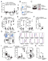
Transgenic expression of dnTGFβRII, but not conditional deletion of Tgfbr2, results in massive accumulation of late memory OTI cells after Listeria infection. (A) Naive OTI-WT or OTI-dnTGFβRII cells were adoptively transferred into CD11c-dnTGFβRII hosts, followed by infection with LM-OVA 1 day later. C57BL/6 mice receiving naive OTI-WT were also used as controls. The percentage of OTI-WT and OTI-dnTGFβRII after contraction was monitored in PBLs over time. (B) Representative dot plots of transferred OTI-WT (top) and OTI-dnTGFβRII (bottom) cells in the PBLs at day 307 p.i. (C) Gross morphology of spleen and LNs from infected hosts at 11 months p.i. (D–E) The percentage (D) and absolute number (E) of donor OTI cells in the indicated tissues at day 374–448 p.i. Results are combined from 4 independent experiments. (F) The percentage of host CD8+ T cells in the spleen of the same mice as in (D). (G) Representative dot plots of donor OTI (CD45.1+) and host CD8+ T cells (CD45.1) in the spleen at day 374 p.i (top). The expression of CD44 and CD62L in the spleen of host CD8+ T cells (bottom). (H) Expression pattern of indicated surface cell markers was analyzed on OTI-WT and OTI-dnTGFβRII cells isolated from spleen of LM-OVA-infected mice at day 340 p.i. (I) Naive OTI-control and OTI-GzmBCre-Tgfbr2f/f cells on different congenic backgrounds were adoptively co-transferred into naive C57BL/6 hosts, followed by infection with LM-OVA 1 day later. The percentage of OTI-control and OTI-GzmBCre-Tgfbr2f/f cells in PBLs was monitored over time at indicated time points. (J) The percentage of SLECs, MPECs and KLRG1+CD127+ (DP) cells among the OTI-control or OTI-GzmBCre-Tgfbr2f/f cells in the PBLs at 280 days p.i. Representative plots of the expression of CD127 and KLRG1 at day 280 p.i. are shown on the right. (K) The percentage of OTI-control and OTI-GzmBCre-Tgfbr2f/f cells in the spleen at day 438 p.i. NS, not significant versus OTI-WT. * p<0.05, ** p<0.01 *** p<0.001 versus OTI-WT. (A, B, G–K) Data are representative of 3 independent experiments. Each dot represents 1 mouse.
FIGURE 3
FIGURE 3
IL-15 is not required for accumulation of late memory OTI-dnTGFβRII cells. (A) C57BL/6 hosts (CD45.2.2) receiving OTI-WT cells (CD45.1.1) (left), Il15+/+ CD11c-dnTGFβRII (middle) or Il15−/− CD11c-dnTGFβRII (right) hosts (CD45.2.2) receiving both OTI-WT (CD45.1.1) and OTI-dnTGFβRII (CD45.1.2) cells, were infected with LM-OVA, and the percentage of donor cells in the PBLs was analyzed at day 225 p.i. Each dot represents 1 mouse. (B) Representative plots of transferred OTI cells in the PBLs shown in (A). (C) Naive OTI-dnTGFβRII cells were transferred into CD11c-dnTGFβRII mice, followed by LM-OVA infection 1 day later. On day 370 p.i., dysregulated memory OTI-dnTGFβRII cells were isolated and CFSE-labeled. CFSE-labeled late memory cells (2 × 10⁶) were transferred into naive Il15+/+ CD11c-dnTGFβRII or Il15−/− CD11c-dnTGFβRII mice. Animals were bled at indicated time points and CFSE dilution was measured. Data are representative of 2 independent experiments with similar results.
FIGURE 4
FIGURE 4
IL-7 is not required for accumulation of late memory OTI-dnTGFβRII cells. (A) Naive OTI-WT and OTI-dnTGFβRII cells were adoptively co-transferred into naive Il7+/+ CD11c-dnTGFβRII (left) or Il7−/− CD11c-dnTGFβRII (right) hosts, followed by infection with LM-OVA 1 day later. The percentage of OTI-WT and OTI-dnTGFβRII in PBLs was monitored over time. Each dot represents 1 mouse. (B) The percentage of OTI-WT and OTI-dnTGFβRII SLECs in the blood of the same mice as (A) after LM-OVA infection at the indicated time points. (C–D) C57BL/6 hosts (CD45.2.2) receiving OTI-WT (CD45.1.1) cells, Il7+/+ CD11c-dnTGFβRII or Il7−/− CD11c-dnTGFβRII hosts (CD45.2.2) receiving both OTI-WT (CD45.1.1) and OTI-dnTGFβRII (CD45.1.2) cells, were infected with LM-OVA, and the percentage of donor cells in the PBLs was monitored over time. (D) Representative dot plots of transferred OTI-WT and OTI-dnRII cells in the PBLs at day 223 p.i. from (C). (E) Naive OTI-dnTGFβRII cells were transferred into CD11c-dnTGFβRII mice, followed by LM-OVA infection 1 day later. On day 448 p.i., dysregulated memory OTI-dnTGFβRII cells were isolated and CFSE-labeled. CFSE-labeled late memory cells (5 × 10⁵) were transferred into naive Il7+/+ CD11c-dnTGFβRII or Il7−/− CD11c-dnTGFβRII mice. Animals were bled at indicated time points and CFSE dilution was measured. Each dot represents 1 mouse. * p<0.05, ** p<0.01, NS, not significant. Data are representative of 2 independent experiments.
FIGURE 5
FIGURE 5
Proliferation of dysregulated memory OTI-dnTGFβRII cells is MHC class I-independent. Naive OTI-dnTGFβRII cells were adoptively transferred into CD11c-dnTGFβRII hosts, followed by LM-OVA infection 1 day later. Dysregulated memory OTI-dnTGFβRII cells were isolated and 2 × 10⁶ cells were transferred into sublethally irradiated C57BL/6 and b2m−/− mice. (A) Representative plots of transferred OTI-dnTGFβRII cells in the LNs and spleen 30 days post transfer. Plot represents cells in the live lymphocyte gate. (B–C) The percentage (B) and absolute number (C) of host CD8+ T cells and transferred OTI-dnTGFβRII cell in the LNs and spleen 30 days post transfer. Each dot represents 1 mouse. Data are representative of 2 independent experiments.
FIGURE 6
FIGURE 6
OTI-dnTGFβRII cells, but not OTI-CD4Cre-Tgfbr2f/f cells, show dysregulated expansion under steady state condition. (A) The percentage of CD44hi memory-like OTI cells in the LNs of OTI-dnTGFβRII-Rag1−/− or OTI-WT-Rag1−/− mice under steady-state conditions are plotted against age. Representative plots at 20 weeks of age are shown at the bottom. Plots show cells gated on CD8α+TCRβ+ cells. (B) The percentage of CD44hi memory-like OTI cells in the LNs of OTI-control (CD4Cre-Tgfbr2f/+ or Tgfbr2f/f)-Rag1−/− or OTI-CD4Cre-Tgfbr2f/f-Rag1−/− mice under steady-state conditions are plotted against age. Representative plots at 22 weeks of age are shown at the bottom. Plots show cells gated on CD8α+TCRβ+ cells. (C) The absolute number of CD44hi memory-like OTI cells in the LNs and spleen of OTI-dnTGFβRII-Rag1−/− or OTI-WT-Rag1−/− mice under steady-state conditions are plotted against age. (D) The absolute number of CD44hi memory-like OTI cells in the LNs and spleen of OTI-control (CD4Cre-Tgfbr2f/+ or Tgfbr2f/f)-Rag1−/− or OTI-CD4Cre-Tgfbr2f/f-Rag1−/− mice under steady-state conditions are plotted against age. (E) Cell surface makers on the CD8α+TCRβ+ OTI cells from the LNs of indicated 7-week old Rag1−/− mice under steady-state conditions. Data are representative of 2 independent experiments. Each dot represents 1 mouse.
FIGURE 7
FIGURE 7
Memory OTI-dnTGFβRII cells develop lymphoma. (A) Karyotype analysis of late memory OTI-dnTGFβRII cells isolated form LM-OVA-infected mice at day 420 p.i. as described in Fig. 1. Arrow indicates trisomy of chromosome 15. (B) CD44lo naive or CD44hi memory-like OTI cells were isolated from OTI-dnTGFβRII-Rag1−/− or OTI-WT-Rag1−/− mice at 2–3 months of age (prior to cellular transformation), and the mRNA expression of Myc, Id2 and Cdkn1a was determined with real-time PCR. The RNA sample was pooled from 3–4 mice for each group. The mRNA expression level in the CD44lo OTI-WT cells is defined as 1. Data represent the means +/− SEM. (C) CD44hi memory-like OTI cells were isolated from OTI-dnTGFβRII-Rag1−/− or OTI-WT-Rag1−/− mice at 6 months of age (after cellular transformation), and the mRNA expression of Myc was determined with real-time PCR. The RNA sample was pooled from 3–4 mice for each group. The mRNA expression level in the OTI-WT cells is defined as 1. Data represent the means +/− SEM. (B–C) Data are representative of 2 independent experiments.
FIGURE 8
FIGURE 8
Transformed late memory OTI-dnTGFβRII cells have effector and CTL activity, but do not proliferate after recall response. Adoptive transfer and LM-OVA infection was performed as described in Fig. 2. (A) Intracellular cytokine staining of splenocytes isolated at day 380 p.i. and stimulated ex vivo with SIINFEKL peptide. Memory OTI-WT and transformed memory OTI-dnTGFβRII cells were identified based on staining with CD8α and the corresponding congenic markers. (B) In vivo CTL assay performed with memory OTI-WT and transformed memory OTI-dnTGFβRII cells isolated from the spleen of infected mice at day 380 p.i. Representative histograms are shown on the left. NS, not significant compared to OTI-WT. (C–D) Memory OTI-WT or transformed memory OTI-dnTGFβRII cells isolated from LM-OVA-infected mice at day 375 p.i. were CFSE-labeled, and stimulated with indicated mitogens (C) or cytokines (D). CFSE dilution was analyzed 2 days (C) or 4 days (D) after stimulation. (E) Memory OTI-WT or transformed memory OTI-dnTGFβRII cells isolated from LM-OVA-infected mice at day 461 p.i. (5 × 10⁴ cells) were transferred into naive CD11c-dnTGFβRII recipients that were subsequently infected with LM-OVA. The percentage of OTI-WT and OTI-dnTGFβRII in PBLs was monitored over time. NS, not significant versus OTI-WT. *** p<0.001 versus OTI-WT. Data are representative of 2 independent experiments.

References

    1. Cui W, Kaech SM. Generation of effector CD8+ T cells and their conversion to memory T cells. Immunol Rev. 2010;236:151–166. - PMC - PubMed
    1. Lefrancois L, Obar JJ. Once a killer, always a killer: from cytotoxic T cell to memory cell. Immunol Rev. 2010;235:206–218. - PMC - PubMed
    1. Sallusto F, Lanzavecchia A, Araki K, Ahmed R. From vaccines to memory and back. Immunity. 2010;33:451–463. - PMC - PubMed
    1. Zhang N, Bevan MJ. CD8+ T cells: foot soldiers of the immune system. Immunity. 2011;35:161–168. - PMC - PubMed
    1. Sprent J, Surh CD. Normal T cell homeostasis: the conversion of naive cells into memory-phenotype cells. Nat Immunol. 2011;131:478–484. - PMC - PubMed

Publication types

MeSH terms

Substances