. 2013 Jun 15;190(12):6329-39.
doi: 10.4049/jimmunol.1300100. Epub 2013 May 17.

IL-1α signaling initiates the inflammatory response to virulent Legionella pneumophila in vivo

Affiliations

IL-1α signaling initiates the inflammatory response to virulent Legionella pneumophila in vivo

Kevin C Barry et al. J Immunol. .

Abstract

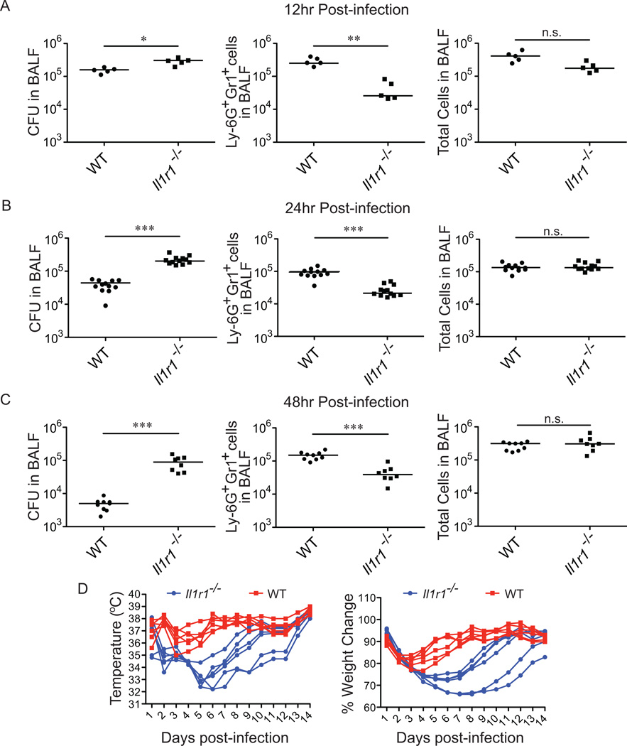
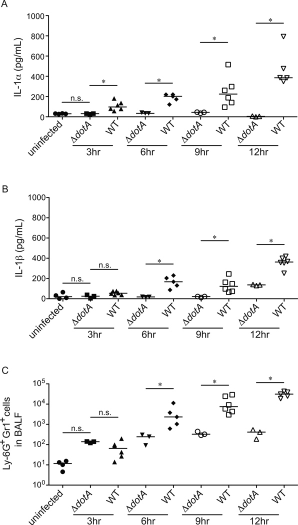
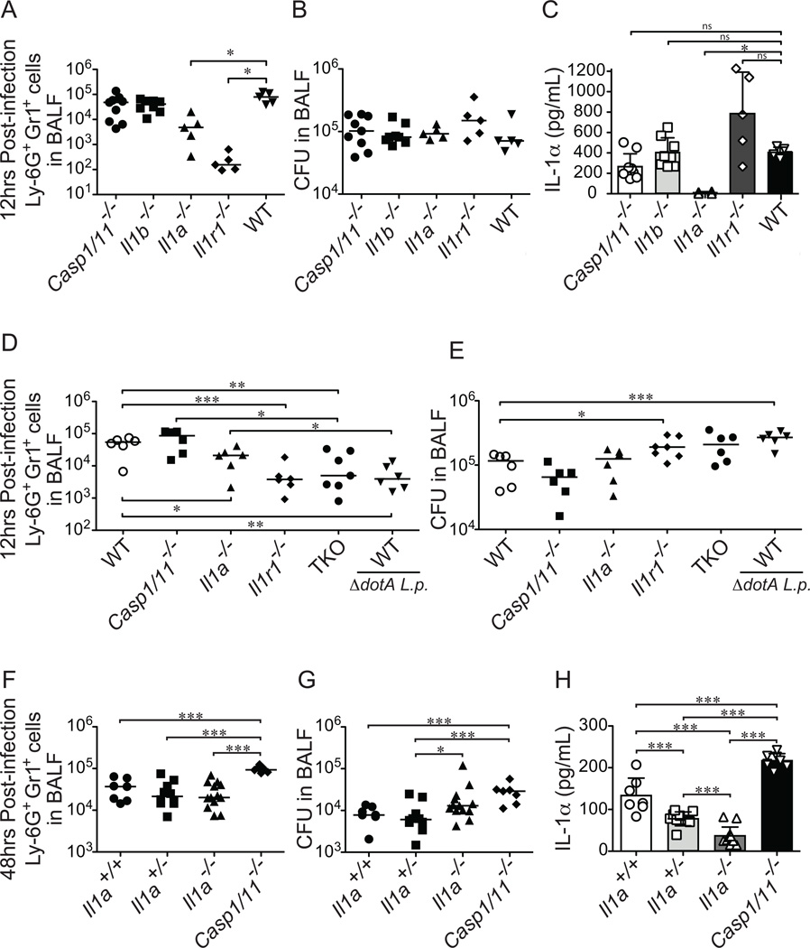
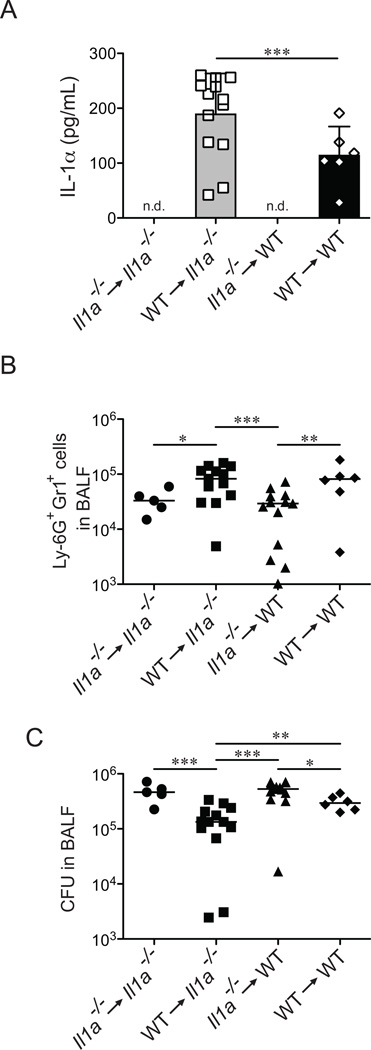
Legionella pneumophila is an intracellular bacterial pathogen that is the cause of a severe pneumonia in humans called Legionnaires' disease. A key feature of L. pneumophila pathogenesis is the rapid influx of neutrophils into the lungs, which occurs in response to signaling via the IL-1R. Two distinct cytokines, IL-1α and IL-1β, can stimulate the type I IL-1R. IL-1β is produced upon activation of cytosolic sensors called inflammasomes that detect L. pneumophila in vitro and in vivo. Surprisingly, we find no essential role for IL-1β in neutrophil recruitment to the lungs in response to L. pneumophila. Instead, we show that IL-1α is a critical initiator of neutrophil recruitment to the lungs of L. pneumophila-infected mice. We find that neutrophil recruitment in response to virulent L. pneumophila requires the production of IL-1α specifically by hematopoietic cells. In contrast to IL-1β, the innate signaling pathways that lead to the production of IL-1α in response to L. pneumophila remain poorly defined. In particular, although we confirm a role for inflammasomes for initiation of IL-1β signaling in vivo, we find no essential role for inflammasomes in production of IL-1α. Instead, we propose that a novel host pathway, perhaps involving inhibition of host protein synthesis, is responsible for IL-1α production in response to virulent L. pneumophila. Our results establish IL-1α as a critical initiator of the inflammatory response to L. pneumophila in vivo and point to an important role for IL-1α in providing an alternative to inflammasome-mediated immune responses in vivo.


Figures

Figure 1
Figure 1
The IL-1 Receptor Type I is essential for control of L. pneumophila infection. (A–C) IL-1R Type I-deficient mice were infected intranasally with 2×10⁶ L. pneumophila (LP01). Bronchoalveolar lavage (BAL) was performed at12 hours (A), 24 hours (B) and 48 hours (C) post-infection. Bacterial burden in the BAL fluid was determined by plating for colony forming units. The number of Ly-6G+Gr1+ cells was determined by flow cytometry and the total number of cells in the BAL fluid as determined by Guava ViaCount assay. (D) IL-1R-deficient (blue circles) and wild-type B6 (red squares) mice were infected with 2×10⁶ L. pneumophila (LP01) and monitored daily for temperature and weight change. Percent weight change is calculated to weight at day zero. Data are representative of two (D) or three (A, B, C) experiments. (Median in A-C). *, p<0.05. **, p<0.01. ***, p<0.005. (Statistical analysis: Mann-Whitney U test).
Figure 2
Figure 2
Dot/Icm T4SS-dependent IL-1α production precedes the recruitment of Ly-6G+Gr1+ cells to the lung. (A–C) B6 mice were infected intranasally with 2×10⁶ L. pneumophila (WT) or a mutant lacking a function type IV secretion system (ΔdotA). Bronchoalveolar lavage was performed at 3, 6, 9, or 12 hours post-infection. IL-1α (A) and IL-1β (B) levels were measured by ELISA. (C) The number of Ly-6G+Gr1+ positive cells in the BAL fluid was determined by flow cytometry. Data are representative of three experiments. (Median in A-C). *, p<0.05 (Statistical analysis: Mann-Whitney U test).
Figure 3
Figure 3
IL-1α is required for Ly-6G+Gr1+ cell recruitment to the lung in response to infection with L. pneumophila. (A–C) The indicated mouse strains were infected intranasally with 2×10⁶ L. pneumophila (LP01). At 12hrs post-infection bronchoalveolar lavage (BAL) fluid was collected. (A) Ly-6G+Gr1+ cells in the BAL fluid were enumerated by flow cytometry. (B) Bacterial burden in the lung was determined by plating BAL fluid for CFU. (C) IL-1α levels were measured by ELISA. (D–E) The indicated mouse strains were infected intranasally with 2×10⁶ L. pneumophila (LP01) or Dot/Icm T4SS-deficient L. pneumophila (LP01 ΔdotA) as noted. At 12 hours post-infection BAL fluid was harvested and Ly-6G+Gr1+ cell recruitment was measured by flow cytometry (D) and bacterial burden in the lung was measured by plating for CFU (E). (F–H). Il1a−/−Il1a+/−, and Il1a+/+ littermates were infected intranasally with 2×10⁶ L. pneumophila (LP01). Non-littermate Casp1/11−/− mice were also infected with LP01. Bronchoalveolar lavage fluid was collected 48hrs post-infection and Ly-6G+Gr1+ cells were quantified by flow cytometry (F). Bacterial burden was determined by plating for CFU (G). IL-1α levels in the BAL fluid were determined by ELISA (H). Data are representative of two (F–H) or three (A–E) experiments (Median in A, B, D-G. mean ± s.d. in C, H). The low level of apparent IL-1α protein produced in Il1a−/− mice at 48h post infection appears to be due to an unknown cross-reacting protein that produced a low signal on the ELISA assay. TKO, Il1a/Casp1/11−/− triple knockout mice. *, p<0.05. **, p<0.01. ***, p<0.005. (Statistical analysis: Mann-Whitney U test).
Figure 4
Figure 4
Hematopoietic cells are responsible for IL-1α production in response to L. pneumophila. (A–C) 6 week old Il1a−/− and congenically marked B6.SJL (CD45.1) mice were lethally irradiated and reconstituted with Il1a−/− (CD45.2) or B6.SJL bone marrow as indicated. After 12 weeks of recovery, chimeric mice were infected with L. pneumophila (LP01). Bronchoalveolar lavage (BAL) fluid was collected 12hrs post-infection. (A) IL-1α levels in BAL fluid were determined by ELISA. (B) Recruitment of Ly-6G+Gr1+ cells was determined by flow cytometry. (C) Bacterial burden in the lung was determined by plating BAL fluid for bacterial CFUs. Data are representative of two (A–C) experiments. (Mean ± s.d. in A. Median in B, C). n.d., not detectable. WT, B6.SJL. *, p<0.05. **, p<0.01. ***, p<0.005. (Statistical analysis: Mann-Whitney U test).
Figure 5
Figure 5
L. pneumophila mutants lacking bacterial effectors known to block translation have no defect in IL-1α production. (A) Wild-type B6 bone marrow derived macrophages were infected with the indicated strains of L. pneumophila (LP02) at a MOI of 1. 8hrs and 24hrs post-infection cell supernatants were collected and IL-1α levels were determined by ELISA. (B) Wild-type bone marrow derived macrophages were infected with the indicated strains of L. pneumophila (MOI=3) and at 6hrs (left panels) and 24hrs (right panels) post-infection cells were incubated with 35S-methionine for one hour followed by lysis in RIPA buffer. Gels were stained with coomassie blue to visualize equal loading (bottom panels) and global translation levels were determined by autoradiography (top panels). Intervening lanes on gel were removed for simplicity. Data are representative of two (B) or three (A) experiments. (Mean ± s.d. in A). n.d., not detectable. ns, not significant. (Statistical analysis: Mann-Whitney U test).
Figure 6
Figure 6
Translation inhibition in conjunction with TLR activation is sufficient to induce the production of IL-1α both in vitro and in vivo. (A–D) Wild-type B6 bone marrow derived macrophages were infected with the indicated strains of L. pneumophila (LP02) or treated with Pam3CSK4 (10µg/mL), Exotoxin A (50ng/µL) or both Pam3CSK4 and ExoA. Samples were collected 4, 8, or 24 hours post-treatment. (A) Cells were lysed with RIPA buffer and intracellular IL-1α levels were determined by ELISA. (B) Extracellular IL-1α levels were determined by performing ELISA on cell supernatants. (C) Il1a transcript levels were assayed by quantitative reverse transcriptase PCR. (D) Cell cytotoxicity was determined by measuring the release of Lactate Dehydrogenase into cell supernatants and values were normalized to an untreated control and a 100% lysis control where cells were treated with 1% TritonX-100 for 30 minutes. (E) Wild-type B6 mice were treated intranasally with Pam3CSK4 (10µg/mouse), Exotoxin A (2µg/mouse) or both in 20 µL of PBS. Bronchoalveolar lavage was performed 24hrs post-infection. IL-1α levels in BAL fluid were determined by ELISA. Data are representative of three (A–E) experiments (mean ± s.d. in A-E). Pam3, Pam3CSK4. ExoA, Exotoxin A. n.d., not detectable. ns, not significant.*, p<0.05. **,p<0.01. ***,p<0.001. (Statistical analysis: Unpaired T-Test (A–D), Mann-Whitney U Test (E)).

References

    1. Luo ZQ. Legionella secreted effectors and innate immune responses. Cellular microbiology. 2012;14:19–27. - PMC - PubMed
    1. Fontana MF, Vance RE. Two signal models in innate immunity. Immunol Rev. 2011;243:26–39. - PubMed
    1. Winn WC, Jr, Myerowitz RL. The pathology of the Legionella pneumonias. A review of 74 cases and the literature. Human pathology. 1981;12:401–422. - PubMed
    1. Trisolini R, Lazzari Agli L, Cancellieri A, Procaccio L, Candoli P, Alifano M, Patelli M. Bronchoalveolar lavage findings in severe community-acquired pneumonia due to Legionella pneumophila serogroup 1. Respir Med. 2004;98:1222–1226. - PubMed
    1. Yu H, Higa F, Koide M, Haranaga S, Yara S, Tateyama M, Li H, Fujita J. Lung abscess caused by Legionella species: implication of the immune status of hosts. Intern Med. 2009;48:1997–2002. - PubMed

Publication types

Substances