. 2014 Jul;73(7):1340-9.
doi: 10.1136/annrheumdis-2013-203301. Epub 2013 May 18.

Evidence-based detection of pulmonary arterial hypertension in systemic sclerosis: the DETECT study

Affiliations
Free PMC article

Evidence-based detection of pulmonary arterial hypertension in systemic sclerosis: the DETECT study

J Gerry Coghlan et al. Ann Rheum Dis. 2014 Jul.
Free PMC article

Abstract

Objective: Earlier detection of pulmonary arterial hypertension (PAH), a leading cause of death in systemic sclerosis (SSc), facilitates earlier treatment. The objective of this study was to develop the first evidence-based detection algorithm for PAH in SSc.

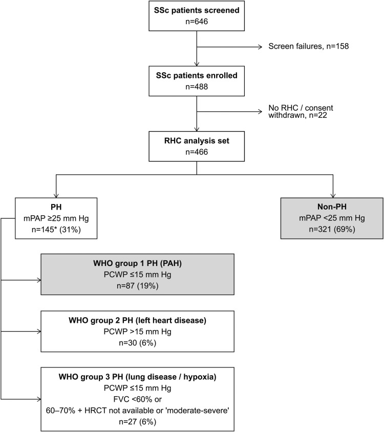
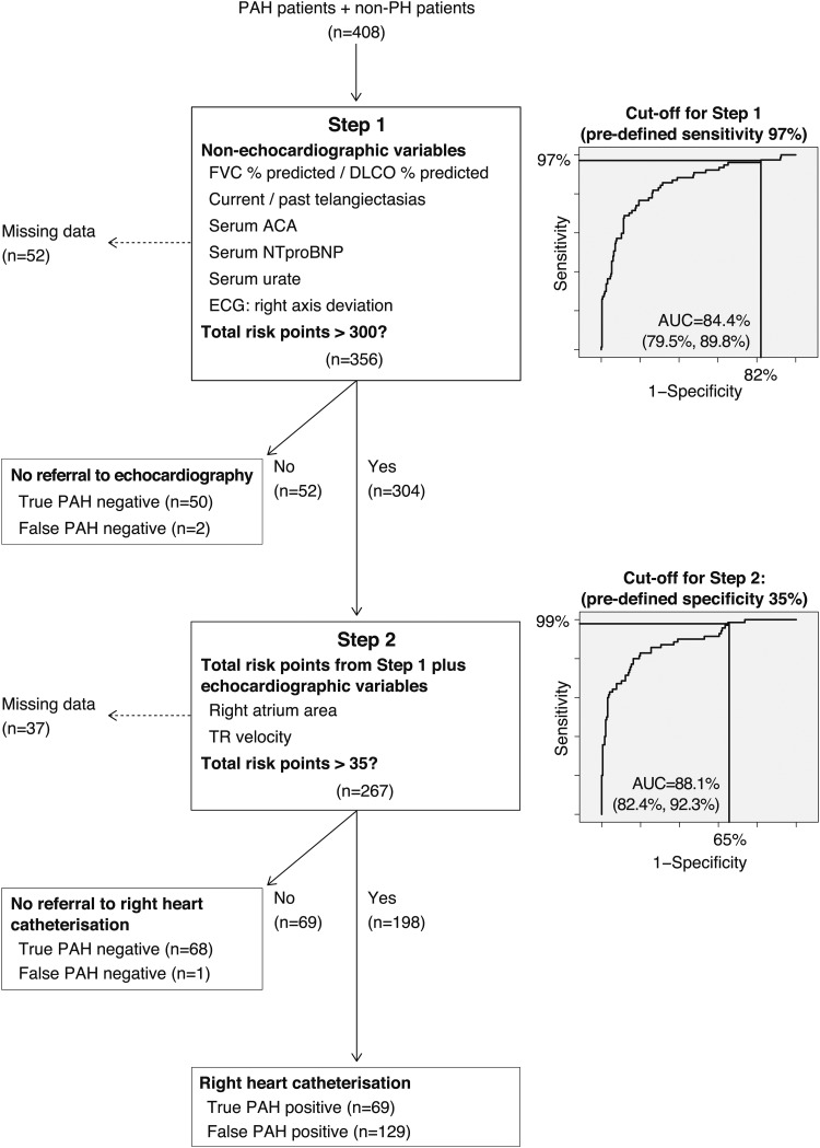
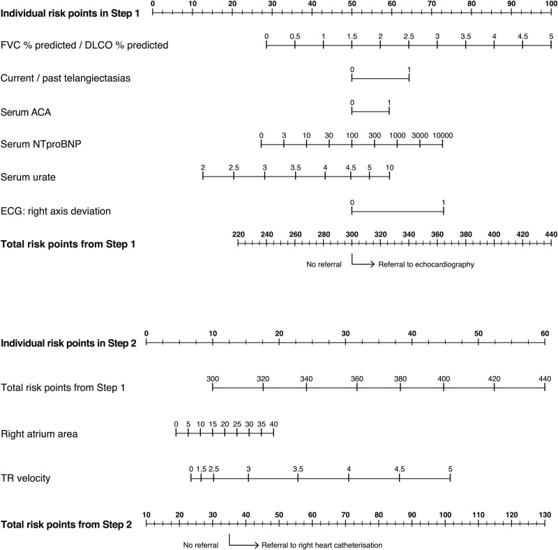
Methods: In this cross-sectional, international study conducted in 62 experienced centres from North America, Europe and Asia, adults with SSc at increased risk of PAH (SSc for >3 years and predicted pulmonary diffusing capacity for carbon monoxide <60%) underwent a broad panel of non-invasive assessments followed by diagnostic right heart catheterisation (RHC). Univariable and multivariable analyses selected the best discriminatory variables for identifying PAH. After assessment for clinical plausibility and feasibility, these were incorporated into a two-step, internally validated detection algorithm. Nomograms for clinical practice use were developed.

Results: Of 466 SSc patients at increased risk of PAH, 87 (19%) had RHC-confirmed PAH. PAH was mild (64% in WHO functional class I/II). Six simple assessments in Step 1 of the algorithm determined referral to echocardiography. In Step 2, the Step 1 prediction score and two echocardiographic variables determined referral to RHC. The DETECT algorithm recommended RHC in 62% of patients (referral rate) and missed 4% of PAH patients (false negatives). By comparison, applying European Society of Cardiology/European Respiratory Society guidelines to these patients, 29% of diagnoses were missed while requiring an RHC referral rate of 40%.

Conclusions: The novel, evidence-based DETECT algorithm for PAH detection in SSc is a sensitive, non-invasive tool which minimises missed diagnoses, identifies milder disease and addresses resource usage.

Keywords: Arterial Hypertension; Epidemiology; Systemic Sclerosis.


Figures

Figure 1
Figure 1
Patient disposition. The results reported here focus on the 408 SSc patients with PAH (n=87) and those without PH (n=321; grey boxes). *One patient could not be assigned to a PH group due to a missing PCWP value. FVC, forced vital capacity; HRCT, high-resolution CT; mPAP, mean pulmonary arterial pressure; PAH, pulmonary arterial hypertension; PH, pulmonary hypertension; PCWP, pulmonary capillary wedge pressure; RHC, right heart catheterisation; SSc, systemic sclerosis.
Figure 2
Figure 2
Two-step decision tree for detection of pulmonary arterial hypertension in systemic sclerosis patients: the DETECT algorithm. Of the 408 SSc patients (87 PAH and 321 non-PH) at risk for PAH (SSc of >3 years’ duration, DLCO <60% of predicted, FVC ≥40% of predicted), data from 319 patients (72 PAH and 247 non-PH) were used for construction of the algorithm. All patients underwent right heart catheterisation. Sensitivity and specificity of the two steps of the algorithm (and the corresponding risk point cut-offs) were selected by the Study Scientific Committee with the aim of minimising the number of missed PAH diagnoses. Step 1: A complete dataset was available for 356 patients. The combined discriminatory ability of the six selected non-echocardiographic variables expressed as the AUC of the ROC curve was 84.4% (95% CI 79.5% to 89.8%) showing good discriminatory performance and no statistically significant lack of fit (see online supplementary appendix 5). At Step 1, a predefined sensitivity cut-off of 97% (corresponding to >300 risk points, compare figure 3), determined no referral to echocardiography in 52 patients. Among these, 50 were true negatives (patients without PAH on right heart catheterisation) and two were false negatives (PAH confirmed on right heart catheterisation). Step 2: A complete dataset was available for 267 patients. The AUC of the ROC curve for the total risk points from Step 1, plus the two selected echocardiographic variables, was 88.1% (95% CI 82.4% to 92.3%). A predefined specificity cut-off of 35% (corresponding to >35 risk points, compare figure 3), determined no referral to right heart catheterisation in 69 patients. Among these, 68 were true negatives and one was a false negative. Right heart catheterisation in the remaining 198 patients yielded 69 true positives (PAH confirmed) and 129 false positives. Thus, overall, the algorithm missed 3 (4%) out of the 72 PAH patients who had sufficient data to be included in the analysis. Note that the algorithm uses cut-offs for the risk points of the two steps only but not for individual parameters. ACA, anticentromere antibody; AUC, area under the curve; DLCO, pulmonary diffusing capacity for carbon monoxide; FVC, forced vital capacity; NTproBNP, N-terminal probrain natriuretic peptide; PAH, pulmonary arterial hypertension; PH, pulmonary hypertension; ROC, receiver operating characteristic; SSc, systemic sclerosis; TR, tricuspid regurgitant jet.
Figure 3
Figure 3
Nomograms for practical application of the DETECT algorithm: determination of the likelihood of pulmonary arterial hypertension and cut-off points for decision to refer a patient to echocardiography (Step 1) and subsequent right heart catheterisation (Step 2). At Step 1 (top panel), risk points for each of the six non-echocardiographic variables are calculated by reading from ‘Individual risk points in Step 1’ and adding them up to obtain a total. If the ‘Total risk points from Step 1’ is >300 (corresponding to a sensitivity of 97% as selected by the Study Scientific Committee) the patient is referred to echocardiography. Similarly, at Step 2 (bottom panel), risk points for the carried forward ‘Total risk points from Step 1’ and the two echocardiographic variables are calculated by reading from the ‘Individual risk points in Step 2’. If the ‘Total risk points from Step 2’ is >35 (corresponding to a specificity of 35% as selected by the Study Scientific Committee) the patient is referred to right heart catheterisation. Alternatively, being less conservative (65% predefined specificity at Step 2), the patient would be referred to right heart catheterisation if ‘Total risk points from Step 2’ is >40 (compare table 3 for the performance of these two options). Note that all variables will always contribute risk points irrespective of the measured value; for example, a negative serum ACA will contribute 50 risk points. Exclusion of any single variable from the DETECT algorithm has only a small impact on model performance (see online supplementary appendix 9). If a single Step 1 variable is missing it should be assigned 50 risk points, with the exception of current/past telangiectasias which should be assigned 65 points. If a single Step 2 variable is missing it should be assigned 10 points. The nomograms cannot be reliably used if more than one variable out of the eight total variables is missing. ACA, anticentromere antibody; DLCO, pulmonary diffusing capacity for carbon monoxide; FVC, forced vital capacity; NTproBNP, N-terminal probrain natriuretic peptide; TR, tricuspid regurgitant jet.

References

    1. Galiè N, Hoeper MM, Humbert M, et al. Guidelines for the diagnosis and treatment of pulmonary hypertension: the Task Force for the Diagnosis and Treatment of Pulmonary Hypertension of the European Society of Cardiology (ESC) and the European Respiratory Society (ERS), endorsed by the International Society of Heart and Lung Transplantation (ISHLT). Eur Heart J 2009;30:2493–537 - PubMed
    1. McLaughlin VV, Archer SL, Badesch DB, et al. ACCF/AHA 2009 expert consensus document on pulmonary hypertension: a report of the American College of Cardiology Foundation Task Force on Expert Consensus Documents and the American Heart Association: developed in collaboration with the American College of Chest Physicians, American Thoracic Society, Inc., and the Pulmonary Hypertension Association. Circulation 2009;119:2250–94 - PubMed
    1. Steen VD, Medsger TA. Changes in causes of death in systemic sclerosis, 1972–2002. Ann Rheum Dis 2007;66:940–4 - PMC - PubMed
    1. Tyndall AJ, Bannet B, Vonk M, et al. Causes and risk factors for death in systemic sclerosis: a study from the EULAR Scleroderma Trial and Research (EUSTAR) database. Ann Rheum Dis 2010;69:1809–15 - PubMed
    1. Hachulla E, Carpentier P, Gressin V, et al. Risk factors for death and the 3-year survival of patients with systemic sclerosis: the French ItinérAIR-Sclérodermie study. Rheumatology (Oxford) 2009;48:304–8 - PMC - PubMed

Publication types

MeSH terms