. 2013 May 14;19(18):2781-92.
doi: 10.3748/wjg.v19.i18.2781.

MAWBP and MAWD inhibit proliferation and invasion in gastric cancer

Affiliations

MAWBP and MAWD inhibit proliferation and invasion in gastric cancer

Dong-Mei Li et al. World J Gastroenterol. .

Abstract

Aim: To investigate role of putative mitogen-activated protein kinase activator with WD40 repeats (MAWD)/MAWD binding protein (MAWBP) in gastric cancer (GC).

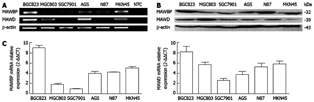
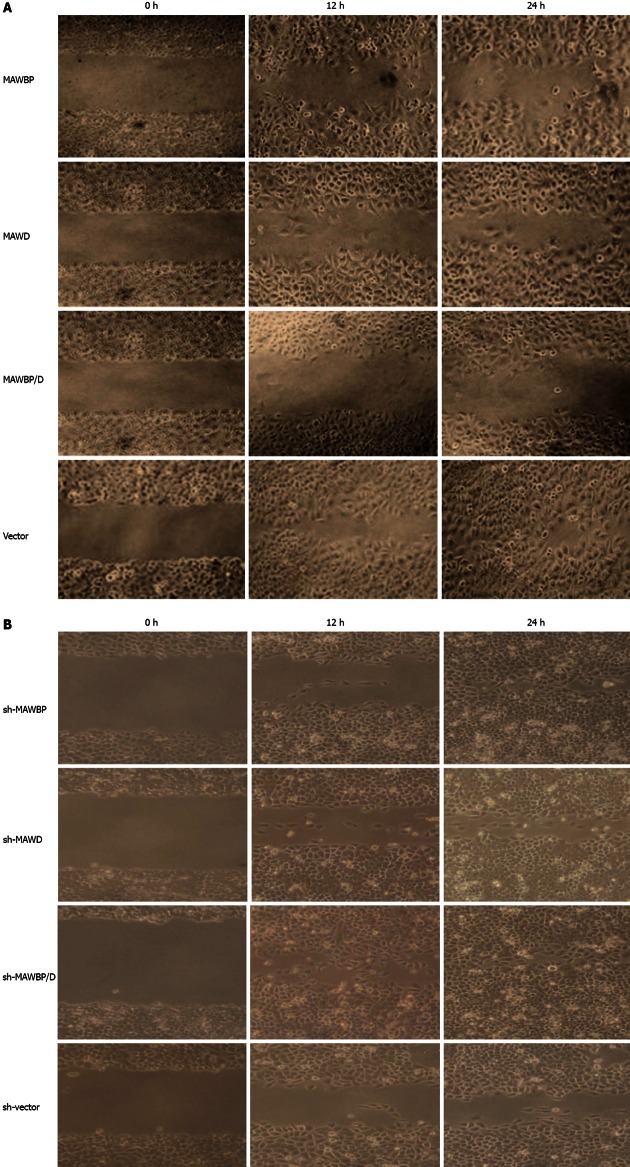
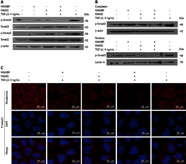
Methods: MAWBP and MAWD mRNA expression level was examined by real-time reverse transcriptase-polymerase chain reaction and semi-quantitative polymerase chain reaction in six GC cell lines. Western blotting was used to examine the protein expression levels. We developed GC cells that stably overexpressed MAWBP and MAWD, and downregulated expression by RNA interference assay. Proliferation and migration of these GC cells were analyzed by 3-(4,5-dimethyl-2-thiazolyl)-2,5-diphenyl tetrazolium bromide (MTT), soft agar, tumorigenicity, migration and transwell assays. The effect of expression of MAWBP and MAWD on transforming growth factor (TGF)-β1-induced epithelial-mesenchymal transition (EMT) was examined by transfection of MAWBP and MAWD into GC cells. We detected the levels of EMT markers E-cadherin, N-cadherin and Snail in GC cells overexpressing MAWBP and MAWD by Western blotting. The effect of MAWBP and MAWD on TGF-β signal was detected by analysis of phosphorylation level and nuclear translocation of Smad3 using Western blotting and immunofluorescence.

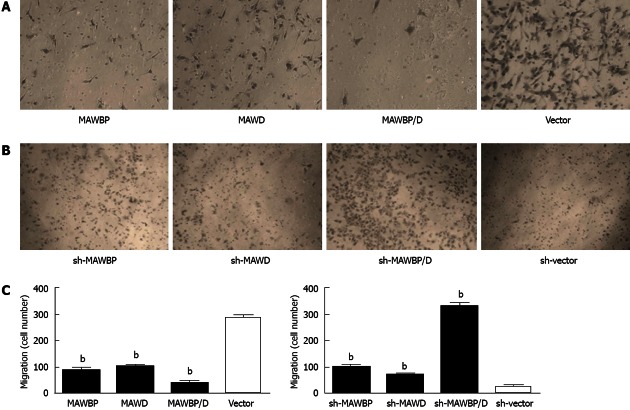
Results: Among the GC cell lines, expression of endogenous MAWBP and MAWD was lowest in SGC7901 cells and highest in BGC823 cells. MAWBP and MAWD were stably overexpressed in SGC7901 cells and knocked down in BGC823 cells. MAWBP and MAWD inhibited GC cell proliferation in vitro and in vivo. MTT assay showed that overexpression of MAWBP and MAWD suppressed growth of SGC7901 cells (P < 0.001), while knockdown of these genes promoted growth of BGC823 cells (P < 0.001). Soft agar colony formation experiments showed that overexpression of MAWBP and MAWD alone or together reduced colony formation compared with vector group in SGC7901 (86.25 ± 8.43, 12.75 ± 4.49, 30 ± 6.41 vs 336.75 ± 22.55, P < 0.001), and knocked-down MAWBP and MAWD demonstrated opposite effects (131.25 ± 16.54, 88.75 ± 11.12, 341.75 ± 22.23 vs 30.25 ± 8.07, P < 0.001). Tumorigenicity experiments revealed that overexpressed MAWBP and MAWD inhibited GC cell proliferation in vivo (P < 0.001). MAWBP and MAWD also inhibited GC cell invasion. Transwell assay showed that the number of traverse cells of MAWBP, MAWD and coexpression group were more than that in vector group (84 ± 16.57, 98.33 ± 9.8, 29 ± 16.39 vs 298 ± 11.86, P < 0.001). Coexpression of MAWBP and MAWD significantly decreased the cells traversing the matrix membrane. Conversely, knocked-down MAWBP and MAWD correspondingly promoted invasion of GC cells (100.67 ± 14.57, 72.66 ± 8.51, 330.67 ± 20.55 vs 27 ± 11.53, P < 0.001). More importantly, coexpression of MAWBP and MAWD promoted EMT. Cells that coexpressed MAWBP and MAWD displayed a pebble-like shape and tight cell-cell adhesion, while vector cells showed a classical mesenchymal phenotype. Western blotting showed that expression of E-cadherin was increased, and expression of N-cadherin and Snail was decreased when cells coexpressed MAWBP and MAWD and were treated with TGF-β1. Nuclear translocation of p-Smad3 was reduced by attenuating its phosphorylation.

Conclusion: Coexpression of MAWBP and MAWD inhibited EMT, and EMT-aided malignant cell progression was suppressed.

Keywords: Epithelial-mesenchymal transition; Gastric cancer; Invasion; Mitogen-activated protein kinase activator with WD40 repeats; Mitogen-activated protein kinase activator with WD40 repeats binding protein; Transforming growth factor-β.


Figures

Figure 1
Figure 1
Analysis of expression of mitogen-activated protein kinase activator with WD40 repeats binding protein and mitogen-activated protein kinase activator with WD40 repeats in six gastric cancer cell lines. A: mRNA expression for mitogen-activated protein kinase activator with WD40 repeats (MAWD)/MAWD binding protein (MAWBP) in BGC823, MGC803, SGC7901, AGS, N87 and MKN45 six gastric cancer cell lines was detected by reverse transcription-polymerase chain reaction (PCR). There was a low level of endogenous MAWBP and MAWD mRNA in SGC7901 cells, and a high level in BGC823 cells; B: Expression of MAWBP and MAWD proteins was detected by Western blotting. The expression level was lower in SGC7901 cells than in the other cell lines. Expression of MAWBP and MAWD was higher in BGC823 cells than in the other cells; C: mRNA expression for MAWBP and MAWD was detected by real-time PCR. There were lower levels of endogenous MAWD and MAWBP mRNA in SGC7901 cells, and higher levels in BGC823 cells. NTC: No template control.
Figure 2
Figure 2
Effect of mitogen-activated protein kinase activator with WD40 repeats binding protein and mitogen-activated protein kinase activator with WD40 repeats on proliferation and tumorigenicity of gastric cancer cells. A: 3-(4,5-Dimethyl-2-thiazolyl)-2,5-diphenyl tetrazolium bromide assay showed that growth of SGC7901 cells overexpressing mitogen-activated protein kinase activator with WD40 repeats (MAWD)/MAWD binding protein (MAWBP) was markedly inhibited; B: In BGC823 cells with inhibition of expression of MAWBP and MAWD, cell growth was inversely increased; C: In soft agar assay, colonies of cells with overexpression of MAWBP, MAWD and MAWBP/D were reduced in number and size compared with cells transfected with vectors alone (original magnification of clones: ×100); D: Knockdown group demonstrated the opposite effects. There was an increase in the number and size of sh-MAWBP, sh-MAWD, sh-MAWBP/D cells compared with cells transfected with vectors alone; E: Nude mouse xenografts. Tumors induced by MAWBP, MAWD and MAWBP/D cotransfected cells were smaller than those of the vector group; F: Xenograft weight. The average tumor weight were calculated from five nude mice in every group. Data are presented as the mean ± SD from three independent experiments. bP < 0.01 vs vector group.
Figure 3
Figure 3
Effect of mitogen-activated protein kinase activator with WD40 repeats binding protein and mitogen-activated protein kinase activator with WD40 repeats on migration of gastric cancer cells. A: Wound healing assay showed that migration of vector-transfected cells was faster than that of cells overexpressing mitogen-activated protein kinase activator with WD40 repeats (MAWD)/MAWD binding protein (MAWBP). The scratch gap in vector group was almost closed at 24 h. The migration of MAWBP/D cotransfected cells was slowest; B: Cells with combined downregulation of MAWBP and MAWD expression migrated faster than the other cells. Migration of sh-vector cells was slowest (original magnification, ×100).
Figure 4
Figure 4
Effect of mitogen-activated protein kinase activator with WD40 repeats binding protein and mitogen-activated protein kinase activator with WD40 repeats on invasive ability of gastric cancer cells. A: Transwell assay showed that invasive ability of mitogen-activated protein kinase activator (MAWD) with WD40 repeats binding protein (MAWBP)/D cotransfected cells was weakest, with the lowest number of cells to cross the matrix membranes. Vector-transfected cells migrated farthest; B: Knockdown of MAWBP and MAWD increased the invasive ability of gastric cancer (GC) cells (original magnification: ×100); C: Number of cells that traverse the matrix membrane in the different groups. Data are presented as the mean ± SD from three independent experiments. bP < 0.01 vs vector group.
Figure 5
Figure 5
Effect of mitogen-activated protein kinase activator with WD40 repeats binding protein and mitogen-activated protein kinase activator with WD40 repeats on expression of biomarkers specific for epithelial-mesenchymal transition induced by transforming growth factor-β1. A: According to the expression of transforming growth factor (TGF)-β downstream reporter gene plasminogen activator inhibitor (PAI)-1, the optimum TGF-β1 concentration and time were confirmed as 4 ng/mL for 24 h; B: SGC7901 cells overexpressing mitogen-activated protein kinase activator with WD40 repeats (MAWD)/MAWD binding protein (MAWBP) were stimulated by 4 ng/mL TGF-β1 for 24 h. Cells that overexpressed both MAWBP and MAWD displayed a pebble-like shape, while vector cells showed a classical mesenchymal phenotype (original magnification, ×400); C: Expression of E-cadherin was strongest in the MAWBP/D cotransfection group and weakest in the vector group, using Western blotting. Snail and N-cadherin were inversely associated with E-cadherin expression.
Figure 6
Figure 6
Combination of mitogen-activated protein kinase activator with WD40 repeats binding protein and mitogen-activated protein kinase activator with WD40 repeats inhibited the transforming growth factor-β pathway. A: The level of p-Smad3 was lowest in the mitogen-activated protein kinase activator (MAWD) with WD40 repeats binding protein (MAWBP)/D cotransfected group and highest in the vector group. p-Smad2 was also lower in the MAWBP/D group; B, C: Nuclear translocation capability of p-Smad3 in cotransfected cells was weakest that means the activity of transforming growth factor (TGF)-β pathway in co-expression group was inhibited. DAPI: 4’,6-diamidino-2-phenylindole.

Similar articles

Cited by

References

    1. Parkin DM, Bray F, Ferlay J, Pisani P. Global cancer statistics, 2002. CA Cancer J Clin. 2005;55:74–108. - PubMed
    1. Becker K, Langer R, Reim D, Novotny A, Meyer zum Buschenfelde C, Engel J, Friess H, Hofler H. Significance of histopathological tumor regression after neoadjuvant chemotherapy in gastric adenocarcinomas: a summary of 480 cases. Ann Surg. 2011;253:934–939. - PubMed
    1. Hass HG, Smith U, Jäger C, Schäffer M, Wellhäuber U, Hehr T, Markmann HU, Nehls O, Denzlinger C. Signet ring cell carcinoma of the stomach is significantly associated with poor prognosis and diffuse gastric cancer (Lauren’s): single-center experience of 160 cases. Onkologie. 2011;34:682–686. - PubMed
    1. Leal MF, Calcagno DQ, Borges da Costa Jde F, Silva TC, Khayat AS, Chen ES, Assumpção PP, de Arruda Cardoso Smith M, Burbano RR. MYC, TP53, and chromosome 17 copy-number alterations in multiple gastric cancer cell lines and in their parental primary tumors. J Biomed Biotechnol. 2011;2011:631268. - PMC - PubMed
    1. Cidon EU, Centeno RG, Lagarto EG, Peral JI. HER-2 Evaluation in a Specific Gastric Cancer Population with the Highest Rate of Mortality in Spain. J Oncol. 2011;2011:391564. - PMC - PubMed

Publication types

MeSH terms