. 2013 Apr 16:19:927-34.
Print 2013.

The protective effect of antioxidants on orbital fibroblasts from patients with Graves' ophthalmopathy in response to oxidative stress

Affiliations

The protective effect of antioxidants on orbital fibroblasts from patients with Graves' ophthalmopathy in response to oxidative stress

Chieh-Chih Tsai et al. Mol Vis. .

Abstract

Purpose: To investigate the biphasic effects of hydrogen peroxide (H2O2) on the orbital fibroblasts of patients with Graves' ophthalmopathy (GO) and the relation to antioxidants and proinflammatory cytokines.

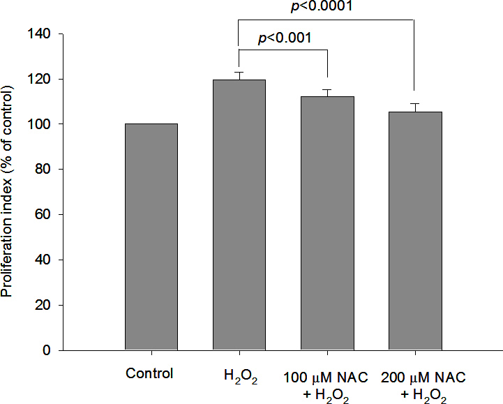
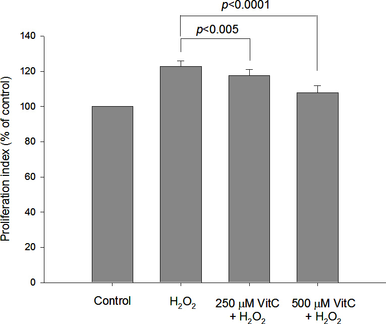
Methods: Proliferation of cultured orbital fibroblasts from patients with GO and normal controls was evaluated in response to various concentrations of H2O2. The effect of low concentrations of H2O2 (6.25 μM) on the cellular proliferation and induction of intracellular proinflammatory cytokines, and reactive oxygen species of orbital fibroblasts were assessed. Protective effects of N-acetylcysteine and vitamin C on GO fibroblasts in response to 6.25 μM H2O2 stimulation were also investigated.

Results: When the GO fibroblasts were exposed to H2O2 at a concentration of 50 μM or above, significant cytotoxicity was observed. In contrast, lower concentrations of H2O2 (3.125-25 μM) increased the survival of GO fibroblasts with the peak cellular proliferation at 6.25 μM H2O2. However, this biphasic effect of H2O2 on the viability of orbital fibroblasts was not found in normal controls. In addition, 6.25 μM H2O2 led to significant elevation of the levels of transforming growth factor, beta 1, interleukin-1β, and superoxide anion in GO fibroblasts, but no significant change in the normal controls. Pretreatment with N-acetylcysteine or vitamin C reversed the enhanced proliferation capacity and the induction of transforming growth factor, beta 1, interleukin-1β and superoxide anion of GO fibroblasts in response to 6.25 μM H2O2.

Conclusions: These findings revealed the biphasic effect of H2O2 on cellular proliferation of GO orbital fibroblasts. Importantly, a low level of H2O2 can stimulate proliferation of GO orbital fibroblasts and induce the production of proinflammatory cytokines, which can be inhibited by pretreatment with antioxidants. This provides a theoretical basis for the rational use of antioxidant in treating GO at an early stage.


Figures

Figure 1
Figure 1
Comparison of the effects of hydrogen peroxide at various concentrations on the viability of orbital fibroblasts between patients with Graves’ ophthalmopathy (GO) and normal controls. We treated orbital fibroblasts from patients with GO (n=7) and age-matched normal subjects (n=5) with various concentrations of hydrogen peroxide (H2O2) for 24 h. The cell proliferation rate was examined with the AlamarBlue assay as methods described and normalized to each control not exposed to H2O2. The mean values of cell proliferation in H2O2-treated orbital fibroblasts are shown in the histogram. Treatment with a low concentration of H2O2 (<25 μM) in GO orbital fibroblasts significantly induced the cell proliferation, but the effect was not observed in normal subjects. The cytotoxicity of H2O2 was observed in GO orbital fibroblasts above 50 μM and in normal subjects above 100 μM. The data are presented as mean ± standard deviation of the results from three independent experiments. (Significant increase when ** p<0.01 and *p<0.05; significant decrease when ##p<0.01 and #p<0.05.)
Figure 2
Figure 2
Protective effect of N-acetylcysteine against 6.25 μM hydrogen peroxide–induced proliferation of orbital fibroblasts from patients with Graves’ ophthalmopathy. After pretreatment of Graves’ ophthalmopathy (GO) orbital fibroblasts (n=7) with 100 μM or 200 μM N-acetylcysteine (NAC) for 1 h, followed by the addition of 6.25 μM hydrogen peroxide (H2O2) for 24 h, the cell proliferation rate was examined with the AlamarBlue assay. The data were normalized to each control that was not exposed to H2O2, and the mean values of cell proliferation are shown in the histogram. The pretreatment of NAC at 100 μM and 200 μM in GO orbital fibroblasts significantly abolished H2O2-induced cell proliferation. The data are presented as mean ± standard deviation of the results from three independent experiments. (p<0.001 and p<0.0001 represented significant decrease.)
Figure 3
Figure 3
Protective effect of vitamin C against 6.25 μM hydrogen peroxide–induced proliferation of orbital fibroblasts from patients with Graves’ ophthalmopathy. After pretreatment of Graves’ ophthalmopathy (GO) orbital fibroblasts (n=7) with 250 μM or 500 μM vitamin C (VitC) for 1 h, followed by the addition of 6.25 μM hydrogen peroxide (H2O2) for 24 h, the cell proliferation rate was examined with the AlamarBlue assay. The data were normalized to each control not exposed to H2O2, and the mean values of cell proliferation from GO orbital fibroblasts are shown in the histogram. The pretreatment of VitC at 250 μM and 500 μM in GO orbital fibroblasts significantly inhibited H2O2-induced cell proliferation. The data are presented as mean ± standard deviation of the results from three independent experiments. (p<0.005 and p<0.0001 represented significant decrease.)
Figure 4
Figure 4
Protective effect of N-acetylcysteine and vitamin C against hydrogen peroxide–induced expression of intracellular levels of interleukin-1β and transforming growth factor, beta 1 in Graves’ ophthalmopathy orbital fibroblasts. After pretreatment of Graves’ ophthalmopathy (GO) orbital fibroblasts (n=7) with 200 μM N-acetylcysteine (NAC) or 500 μM vitamin C (VitC) for 1 h, followed by the addition of 6.25 μM hydrogen peroxide (H2O2) for 24 h, the release of the intracellular levels of interleukin-1β (IL-1β) and transforming growth factor, beta 1 (TGF-β1) was determined with an enzyme-linked immunosorbent assay kit. No H2O2 treatment in GO orbital fibroblasts was represented as the control (basal level). The mean values of IL-1β and TGF-β1 from the GO orbital fibroblasts are shown in the histogram (A and B, respectively). The H2O2-induced release of the IL-1β and TGF-β1 levels in the GO orbital fibroblasts was significantly abolished by pretreatment with 200 μM NAC or 500 μM VitC, respectively. The data are presented as mean ± standard deviation of the results from three independent experiments. (p<0.05, p<0.005, and p<0.001 represented significant decrease.)
Figure 5
Figure 5
Protective effect of N-acetylcysteine and vitamin C against hydrogen peroxide–induced elevation of superoxide anion production in Graves’ ophthalmopathy orbital fibroblasts. After pretreatment of Graves’ ophthalmopathy (GO) orbital fibroblasts (n=7) with 200 μM N-acetylcysteine (NAC) or 500 μM vitamin C (VitC) for 1 h, followed by the addition of 6.25 μM hydrogen peroxide (H2O2) for 24 h, the intracellular levels of the superoxide anions were determined with dihydroethidine staining with flow cytometry. The mean values of the superoxide anions from the GO orbital fibroblasts are shown in the histogram. The H2O2-induced intracellular levels of the superoxide anions in the GO orbital fibroblasts were significantly abolished by pretreatment with 200 μM NAC or 500 μM VitC. The data are presented as mean ± standard deviation of the results from three independent experiments. (p<0.0005, and p<0. 01 versus the indicated group.)

Similar articles

Cited by

References

    1. Douglas RS, Gupta S. The pathophysiology of thyroid eye disease: implications for immunotherapy. Curr Opin Ophthalmol. 2011;22:385–90. - PMC - PubMed
    1. Kazim M, Goldberg RA, Smith TJ. Insights into the pathogenesis of thyroid associated orbitopathy: evolving rationale for therapy. Arch Ophthalmol. 2002;120:380–6. - PubMed
    1. Naik VM, Naik MN, Goldberg RA, Smith TJ, Douglas RS. Immunopathogenesis of thyroid eye disease: emerging paradigms. Surv Ophthalmol. 2010;55:215–26. - PMC - PubMed
    1. Smith TJ, Tsai CC, Shih MJ, Tsui S, Chen B, Han R, Naik V, King CS, Press C, Kamat S, Goldberg RA, Phipps RP, Douglas RS, Gianoukakis AG. Unique attributes of orbital fibroblasts and global alterations in IGF-1 receptor signaling could explain thyroid-associated ophthalmopathy. Thyroid. 2008;18:983–8. - PMC - PubMed
    1. Bartalena L, Tanda ML, Piantanida E, Lai A. Oxidative stress and Graves' ophthalmopathy: in vitro studies and therapeutic implications. Biofactors. 2003;19:155–63. - PubMed

Publication types

MeSH terms

LinkOut - more resources