

Induction of spermatogenic synchrony by retinoic acid in neonatal mice
- PMID: 23687613
- PMCID: PMC3644044
- DOI: 10.4161/spmg.23180
Induction of spermatogenic synchrony by retinoic acid in neonatal mice
Abstract
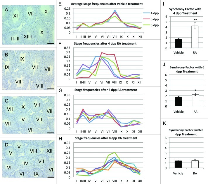
Retinoic acid (RA) is required for the successful differentiation and meiotic entry of germ cells in the murine testis. The availability of RA to undifferentiated germ cells begins in a variable, uneven pattern during the first few days after birth and establishes the asynchronous pattern of germ cell differentiation in adulthood. It has been shown that synchronous spermatogenesis can be induced in 2 d postpartum mice, but not in adult mice, by treating vitamin A sufficient males with RA. In this study, neonatal males were treated at different ages with a single dose of RA and spermatogenesis was examined after recovery to adulthood. The failure of exogenous RA to alter asynchrony correlates with the appearance of meiotic preleptotene spermatocytes within the seminiferous epithelium.
Keywords: asynchronous spermatogenesis; differentiation; preleptotene spermatocytes; retinoic acid; spermatogenic wave; spermatogonia; synchronous spermatogenesis.
Figures
Similar articles
-
Expression of stimulated by retinoic acid gene 8 (Stra8) in spermatogenic cells induced by retinoic acid: an in vivo study in vitamin A-sufficient postnatal murine testes.Biol Reprod. 2008 Jul;79(1):35-42. doi: 10.1095/biolreprod.107.066795. Epub 2008 Mar 5. Biol Reprod. 2008. PMID: 18322276 Free PMC article.
-
Potential roles of gonadotropins to control pulsatile retinoic acid signaling during spermatogenesis.Med Hypotheses. 2015 Sep;85(3):303-4. doi: 10.1016/j.mehy.2015.05.021. Epub 2015 Jun 3. Med Hypotheses. 2015. PMID: 26141633
-
Retinoic acid is able to reinitiate spermatogenesis in vitamin A-deficient rats and high replicate doses support the full development of spermatogenic cells.Endocrinology. 1991 Feb;128(2):697-704. doi: 10.1210/endo-128-2-697. Endocrinology. 1991. PMID: 1989855
-
Cycles, waves, and pulses: Retinoic acid and the organization of spermatogenesis.Andrology. 2020 Jul;8(4):892-897. doi: 10.1111/andr.12722. Epub 2019 Nov 20. Andrology. 2020. PMID: 31670467 Free PMC article. Review.
-
Surfing the wave, cycle, life history, and genes/proteins expressed by testicular germ cells. Part 1: background to spermatogenesis, spermatogonia, and spermatocytes.Microsc Res Tech. 2010 Apr;73(4):241-78. doi: 10.1002/jemt.20783. Microsc Res Tech. 2010. PMID: 19941293 Review.
Cited by
-
Spermatogenesis: The Commitment to Meiosis.Physiol Rev. 2016 Jan;96(1):1-17. doi: 10.1152/physrev.00013.2015. Physiol Rev. 2016. PMID: 26537427 Free PMC article. Review.
-
Retinoic acid induces multiple hallmarks of the prospermatogonia-to-spermatogonia transition in the neonatal mouse.Biol Reprod. 2014 Mar 27;90(3):64. doi: 10.1095/biolreprod.113.114645. Print 2014 Mar. Biol Reprod. 2014. PMID: 24478393 Free PMC article.
-
Beyond stem cells: Commitment of progenitor cells to meiosis.Stem Cell Res. 2018 Mar;27:169-171. doi: 10.1016/j.scr.2018.01.032. Epub 2018 Jan 31. Stem Cell Res. 2018. PMID: 29415862 Free PMC article.
-
What has single-cell RNA-seq taught us about mammalian spermatogenesis?Biol Reprod. 2019 Sep 1;101(3):617-634. doi: 10.1093/biolre/ioz088. Biol Reprod. 2019. PMID: 31077285 Free PMC article. Review.
-
Retinoic acid and meiosis induction in adult versus embryonic gonads of medaka.Sci Rep. 2016 Sep 28;6:34281. doi: 10.1038/srep34281. Sci Rep. 2016. PMID: 27677591 Free PMC article.
References
-
- Russell L. Histologial and histopathological evaluation of the testis. Clearwater, FL: Cache River Press; 1990.
-
- McCARTHY PTC, Cerecedo LR. Vitamin A deficiency in the mouse. J Nutr. 1952;46:361–76. - PubMed
Grants and funding
LinkOut - more resources
Full Text Sources
Other Literature Sources