

Review
doi: 10.1186/1477-7819-11-102.
Botryoid Wilms' tumor: a case report and review of the literature
Affiliations
- PMID: 23687909
- PMCID: PMC3661349
- DOI: 10.1186/1477-7819-11-102
Item in Clipboard
Review
Botryoid Wilms' tumor: a case report and review of the literature
World J Surg Oncol.
.
Abstract
Here, we report a new case of botryoid Wilms' tumor, a 4-year-old boy, who was referred to us with a chief complaint of dysuria and gross hematuria. The computed tomography and radical nephroureterectomy showed that a botryoid sarcoma-like appearance occupied the right renal pelvis and extended into the bladder. Histologic examination further confirmed this case was a mixed type of Wilms' tumor. In a word, we demonstrated a rare case of botryoid Wilms' tumor, which extended from the renal pelvis into the ureter and bladder, then some degenerative and necrotic tissue with calcification discharged from urethra. Postoperative adjuvant chemotherapy was executed. At 24-month follow-up, there was no evidence of recurrence.
Figures

Non-contrasted CT and Contrasted enhanced CT. (A) Non-contrast CT illustrated enlarged inhomogeneous right kidney consisting of multiple water-density masses, which filled the pelvicalyceal system. (B) Contrasted-enhanced CT showed that the masses at the same level were enhanced slightly and inhomogeneously.

Nephrogenic phase sagittal and coronal CT. (A,B) Sagittal CT showed the botryoid tumor mass extended from the renal pelvis into the ureter and the bladder. (C,D) Coronal CT illustrated a botryoid sarcoma-like appearance.

Lesion characteristics under microscope. (A) Microscopically, tumor extended into the pelvis by transitional cell of the urothelium. (B) The tissue discharged from urethra illustrated degenerative and necrotic tissue with calcification.
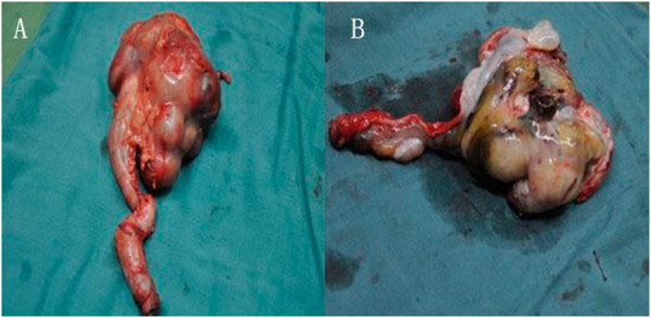
Resected specimen. (A) Botryoid sarcoma-like appearance, occupied the right renal pelvicaliceal system, renal pelvis and ureter. (B) Grayish polypoid mass with coagulation necrosis.
References
-
- Wicklund RA, Tank ES. Polypoid renal pelvic lesions in children. J Urol. 1980;11:943–944. - PubMed
-
- Chiba T, Ohashi E. Wilms’ tumor extending into the dilated renal pelvis as a mold. J Urol. 1980;11:130–131. - PubMed
-
- Weinberg AG, Currarino G, Hurt GEJR. Botryoid Wilms’ tumor of the renal pelvis. Arch Path Lab Med. 1984;11:147–148. - PubMed
-
- Johnson KM, Horvath LJ, Gaisie G, Mesrobian HG, Koepke JF, Askin FB. Wilms tumor occurring as a botryoid renal pelvicalycealmass. Radiology. 1987;11:385–386. - PubMed
Publication types
MeSH terms
LinkOut - more resources
Full Text Sources
Other Literature Sources
Medical
