

Effects of fiber orientation on the frictional properties and damage of regenerative articular cartilage surfaces
- PMID: 23688110
- PMCID: PMC3761558
- DOI: 10.1089/ten.TEA.2012.0580
Effects of fiber orientation on the frictional properties and damage of regenerative articular cartilage surfaces
Abstract
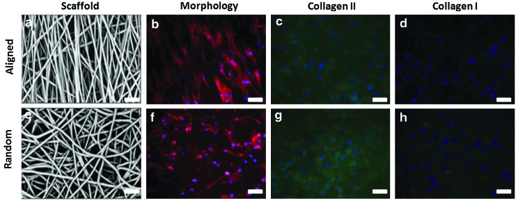
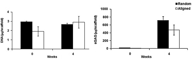
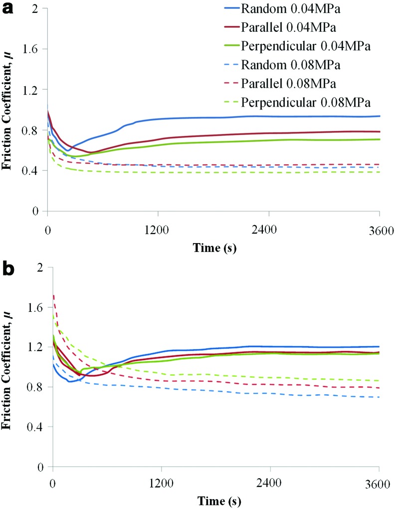
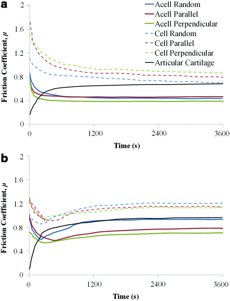
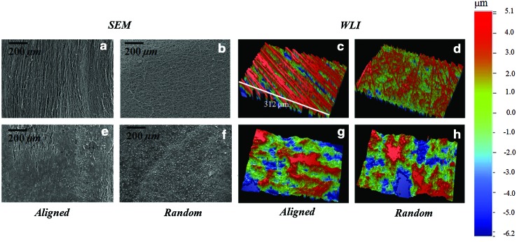
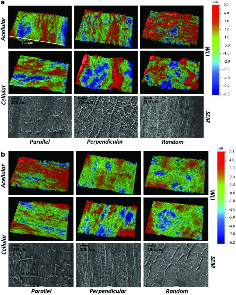
Articular cartilage provides a low-friction, wear-resistant surface for diarthrodial joints. Due to overloading and overuse, articular cartilage is known to undergo significant wear and degeneration potentially resulting in osteoarthritis (OA). Regenerative medicine strategies offer a promising solution for the treatment of articular cartilage defects and potentially localized early OA. Such strategies rely on the development of materials to restore some aspects of cartilage. In this study, microfibrous poly(ɛ-caprolactone) scaffolds of varying fiber orientations (random and aligned) were cultured with bovine chondrocytes for 4 weeks in vitro, and the mechanical and frictional properties were evaluated. Mechanical properties were quantified using unconfined compression and tensile testing techniques. Frictional properties were investigated at physiological compressive strains occurring in native articular cartilage. Scaffolds were sheared along the fiber direction, perpendicular to the fiber direction and in random orientation. The evolution of damage as a result of shear was evaluated via white light interferometry and scanning electron microscopy. As expected, the fiber orientation strongly affected the tensile properties as well as the compressive modulus of the scaffolds. Fiber orientation did not significantly affect the equilibrium frictional coefficient, but it was, however, a key factor in dictating the evolution of surface damage on the surface. Scaffolds shear tested perpendicular to the fiber orientation displayed the highest surface damage. Our results suggest that the fiber orientation of the scaffold implanted in the joint could strongly affect its resistance to damage due to shear. Scaffold fiber orientation should thus be carefully considered when using microfibrous scaffolds.
Figures
Similar articles
-
In vitro chondrocyte behavior on porous biodegradable poly(e-caprolactone)/polyglycolic acid scaffolds for articular chondrocyte adhesion and proliferation.J Biomater Sci Polym Ed. 2015;26(7):401-19. doi: 10.1080/09205063.2015.1015864. Epub 2015 Mar 12. J Biomater Sci Polym Ed. 2015. PMID: 25671317
-
Anisotropic fibrous scaffolds for articular cartilage regeneration.Tissue Eng Part A. 2012 Oct;18(19-20):2073-83. doi: 10.1089/ten.TEA.2011.0606. Epub 2012 Aug 3. Tissue Eng Part A. 2012. PMID: 22655795 Free PMC article.
-
Functional properties of cell-seeded three-dimensionally woven poly(epsilon-caprolactone) scaffolds for cartilage tissue engineering.Tissue Eng Part A. 2010 Apr;16(4):1291-301. doi: 10.1089/ten.TEA.2009.0480. Tissue Eng Part A. 2010. PMID: 19903085 Free PMC article.
-
The role of computational models in the search for the mechanical behavior and damage mechanisms of articular cartilage.Med Eng Phys. 2005 Dec;27(10):810-26. doi: 10.1016/j.medengphy.2005.03.004. Med Eng Phys. 2005. PMID: 16287601 Review.
-
Toward low-friction and high-adhesion solutions: Emerging strategies for nanofibrous scaffolds in articular cartilage engineering.Biomater Adv. 2025 Apr;169:214129. doi: 10.1016/j.bioadv.2024.214129. Epub 2024 Nov 30. Biomater Adv. 2025. PMID: 39642717 Review.
Cited by
-
Experimental Study on the Mechanical Properties of Porcine Cartilage with Microdefect under Rolling Load.J Healthc Eng. 2017;2017:2306160. doi: 10.1155/2017/2306160. Epub 2017 Jun 12. J Healthc Eng. 2017. PMID: 29065577 Free PMC article.
-
A Non-woven Path: Electrospun Poly(lactic acid) Scaffolds for Kidney Tissue Engineering.Tissue Eng Regen Med. 2018 Feb 14;15(3):301-310. doi: 10.1007/s13770-017-0107-5. eCollection 2018 Jun. Tissue Eng Regen Med. 2018. PMID: 30603555 Free PMC article.
-
Hydrogels for Cartilage Regeneration, from Polysaccharides to Hybrids.Polymers (Basel). 2017 Dec 4;9(12):671. doi: 10.3390/polym9120671. Polymers (Basel). 2017. PMID: 30965974 Free PMC article. Review.
-
Combinatorial scaffold morphologies for zonal articular cartilage engineering.Acta Biomater. 2014 May;10(5):2065-75. doi: 10.1016/j.actbio.2013.12.030. Epub 2013 Dec 25. Acta Biomater. 2014. PMID: 24370641 Free PMC article.
-
Electrospun cartilage-derived matrix scaffolds for cartilage tissue engineering.J Biomed Mater Res A. 2014 Nov;102(11):3998-4008. doi: 10.1002/jbm.a.35068. Epub 2014 Jan 9. J Biomed Mater Res A. 2014. PMID: 24375991 Free PMC article.
References
-
- Buckwalter J.A. Martin J.A. Osteoarthritis. Adv Drug Deliv Rev. 2006;58:150. - PubMed
-
- Brittberg M. Cell carriers as the next generation of cell therapy for cartilage repair: a review of the matrix-induced autologous chondrocyte implantation procedure. Am J Sports Med. 2010;38:1259. - PubMed
-
- Nuernberger S. Cyran N. Albrecht C. Redl H. Vecsei V. Marlovits S. The influence of scaffold architecture on chondrocyte distribution and behavior in matrix-associated chondrocyte transplantation grafts. Biomaterials. 2011;32:1032. - PubMed
-
- Behrens P. Ehlers E.M. Kochermann K.U. Rohwedel J. Russlies M. Plotz W. New therapy procedure for localized cartilage defects. Encouraging results with autologous chondrocyte implantation. MMW Fortschr Med. 1999;141:49. - PubMed
Publication types
MeSH terms
Grants and funding
LinkOut - more resources
Full Text Sources
Other Literature Sources
Research Materials