. 2013 May 20:13:246.
doi: 10.1186/1471-2407-13-246.

Expression of follicle-stimulating hormone receptor by the vascular endothelium in tumor metastases

Affiliations

Expression of follicle-stimulating hormone receptor by the vascular endothelium in tumor metastases

Ahsan Siraj et al. BMC Cancer. .

Abstract

Background: The Follicle Stimulating Hormone receptor (FSHR) is expressed by the vascular endothelium in a wide range of human tumors. It was not determined however if FSHR is present in metastases which are responsible for the terminal illness.

Methods: We used immunohistochemistry based on a highly FSHR-specific monoclonal antibody to detect FSHR in cancer metastases from 6 major tumor types (lung, breast, prostate, colon, kidney, and leiomyosarcoma ) to 6 frequent locations (bone, liver, lymph node, brain, lung, and pleura) of 209 patients.

Results: In 166 patients examined (79%), FSHR was expressed by blood vessels associated with metastatic tissue. FSHR-positive vessels were present in the interior of the tumors and some few millimeters outside, in the normally appearing tissue. In the interior of the metastases, the density of the FSHR-positive vessels was constant up to 7 mm, the maximum depth available in the analyzed sections. No significant differences were noticed between the density of FSHR-positive vessels inside vs. outside tumors for metastases from lung, breast, colon, and kidney cancers. In contrast, for prostate cancer metastases, the density of FSHR-positive vessels was about 3-fold higher at the exterior of the tumor compared to the interior. Among brain metastases, the density of FSHR-positive vessels was highest in lung and kidney cancer, and lowest in prostate and colon cancer. In metastases of breast cancer to the lung pleura, the percentage of blood vessels expressing FSHR was positively correlated with the progesterone receptor level, but not with either HER-2 or estrogen receptors. In normal tissues corresponding to the host organs for the analyzed metastases, obtained from patients not known to have cancer, FSHR staining was absent, with the exception of approx. 1% of the vessels in non tumoral temporal lobe epilepsy samples.

Conclusion: FSHR is expressed by the endothelium of blood vessels in the majority of metastatic tumors.


Figures
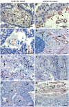
Figure 1
Figure 1
Expression of FSHR by microvascular endothelial cells in brain metastases of four major cancers. (A,B) lung cancer; (C,D) breast cancer; (E,F) kidney cancer; (G,H) prostate cancer. Immunohistochemical analysis was performed on paraffin-embedded sections of human metastatic tissues using the anti-FSHR monoclonal antibody 323, followed by a secondary goat anti-mouse Ig antibody coupled to peroxidase, visualized by the red-brown peroxidase-reaction product. Sections were also stained with hematoxylin. Arrows point to the blood vessels. The scale bar represents 20 μm in all panels.
Figure 2
Figure 2
FSHR-expression in metastases to liver of breast cancer (A) and colon cancer (B). Immunohistochemical analysis was performed as for the Figure 1. Arrows point to the blood vessels. bc, breast cancer; cc, colon cancer. Panels C and D show FSHR expression in the normal appearing tissue surrounding metastasis of colon cancer to liver. Endothelial cells of the arterioles and capillaries (arrowheads) derived from the hepatic artery (HA) express FSHR (C). The majority of the branches of the portal vein (PV) do not express FSHR. No staining is visible in the central vein (CV) and its associated sinusoids (*) (D). The scale bar represents 20 μm in all panels.
Figure 3
Figure 3
FSHR-expression in metastases of breast cancer to pleura, and of prostate cancer to lymph node and bone. Immunohistochemistry was performed as for the preceding figures. Arrows point to blood vessels. Panel A shows specimens obtained from patients with breast cancer localized to pleura. Panels C and E show prostate cancer that metastased to lymph node (C) and to bone (E). No staining for FSHR is observed in blood vessels of normal tissues in pleura (B), lymph node (D), and bone (F). The scale bar represents 20 μm in all panels.
Figure 4
Figure 4
Density of FSHR-positive vessels in metastases. A) Metastases of prostate cancer to brain, lymph nodes, and bone. B) Metastases to brain of tumors from prostate, breast, kidney, colon, and lung. White and black bars represent vessel densities inside and outside tumors, respectively. Error bars: standard error of the mean.

References

    1. Sprengel R, Braun T, Nikolics K, Segaloff DL, Seeburg PH. The testicular receptor for follicle-stimulating hormone: structure and functional expression of cloned cDNA. Mol Endocrinol. 1990;4:525–530. doi: 10.1210/mend-4-4-525. - DOI - PubMed
    1. Simoni M, Gromoll J, Nieschlag E. The follicle stimulating hormone receptor: biochemistry, molecular biology, physiology, and pathophysiology. Endocr Rev. 1997;18:739–773. doi: 10.1210/er.18.6.739. - DOI - PubMed
    1. Vannier B, Loosfelt H, Meduri G, Pichon C, Milgrom E. Anti-human FSH receptor monoclonal antibodies: immunochemical and immunocytochemical characterization of the receptor. Biochemistry. 1996;35:1358–1366. doi: 10.1021/bi952290f. - DOI - PubMed
    1. Vu Hai MT, Lescop P, Loosfelt H, Ghinea N. Receptor-mediated transcytosis of follicle stimulating hormone through the rat testicular microvasculature. Biol Cell. 2004;96:133–144. doi: 10.1016/j.biolcel.2003.11.008. - DOI - PubMed
    1. Radu A, Pichon C, Camparo P, Antoine M, Allory Y, Couvelard A, Fromont G, Vu Hai MT, Ghinea N. Expression of follicle-stimulating hormone receptor in tumor blood vessels. N Engl J Med. 2010;363:1621–1630. doi: 10.1056/NEJMoa1001283. - DOI - PubMed

Publication types

MeSH terms