. 2013 Jun 4;85(11):5555-61.
doi: 10.1021/ac400761e. Epub 2013 May 20.

Glycomic analysis using glycoprotein immobilization for glycan extraction

Affiliations

Glycomic analysis using glycoprotein immobilization for glycan extraction

Shuang Yang et al. Anal Chem. .

Abstract

Glycosylation is one of the most common protein modifications and is involved in many functions of glycoproteins. Investigating aberrant protein glycosylation associated with diseases is useful in improving disease diagnostics. Due to the nontemplate nature of glycan biosynthesis, the glycans attached to glycoproteins are enormously complex; thus, a method for comprehensive analysis of glycans from biological or clinical samples is needed. Here, we describe a novel method for glycomic analysis using glycoprotein immobilization for glycan extraction (GIG). Proteins or peptides from complex samples were first immobilized on solid support, and other nonconjugated molecules were removed. Glycans were enzymatically or chemically modified on solid phase before releasing from glycoproteins/glycopeptides for mass spectrometry analysis. The method was applied to the glycomic analysis of both N- and O-glycans.


Figures

Figure 1
Figure 1
Schematic diagram of glycan capture and release using glycoprotein immobilization for glycan extraction (GIG) method.
Figure 2
Figure 2
Coupling of protein onto the solid support. Protein, ribonuclease B (RNase B, 400 µg), was mixed with 200 µL of Aminolink slurry after denatured in 100°C for 10 min. Protein concentration was determined after conjugation protein to beads for 0, 0.5, 1, 2, 4, 6, 24 hrs. Over 95% binding efficiency was observed after 4 hrs incubation in pH 10.0 buffer.
Figure 3
Figure 3
Analysis of glycans from SGP using GIG and modification of glycan on solid support. The mass spectra for the glycans of SGP without any modification (i), desialylation by neuraminidase treatment (ii), and sialic acid stabilization (iii). Three glycans (1663.1 (1 Na+), 1976.1 (2 Na+) and 2289.1(3 Na+) were observed before sialic acid modification (i); only one glycan (1663.1) was detected after neuraminidase treatment (ii); only one glycan (2424.3) was detected after complete sialic acid derivatization (iii).
Figure 4
Figure 4
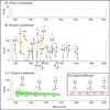
N-Glycan analysis from human serum using GIG method in low mass range (A) and high mass range (B).
Figure 4
Figure 4
N-Glycan analysis from human serum using GIG method in low mass range (A) and high mass range (B).
Figure 5
Figure 5
O-Glycan analysis from mucin using GIG method after incubation in ammonium hydroxide for 4 hrs (A), 24 hrs (B), and 72 hrs (C). N-glycan analysis from mucin using GIG by PNGase F (D).

References

    1. Arnold JN, Wormald MR, Sim RB, Rudd PM, Dwek RA. Annu. Rev. Immunol. 2007;25:21–50. - PubMed
    1. Peracaula R, Barrabes S, Sarrats A, Rudd PM, de Llorens R. Dis. Markers. 2008;25:207–218. - PMC - PubMed
    1. Marth JD, Grewal PK. Nat. Rev. Immunol. 2008;8:874–887. - PMC - PubMed
    1. Lowe JB. Cell. 2001;104:809–812. - PubMed
    1. Isenberg DA. Clin. Exp. Rheumatol. 1995;13:17–20. - PubMed

Publication types

MeSH terms