. 2013 Jul;132(1):205-13.
doi: 10.1016/j.jaci.2013.03.048. Epub 2013 May 17.

Lung type 2 innate lymphoid cells express cysteinyl leukotriene receptor 1, which regulates TH2 cytokine production

Affiliations

Lung type 2 innate lymphoid cells express cysteinyl leukotriene receptor 1, which regulates TH2 cytokine production

Taylor A Doherty et al. J Allergy Clin Immunol. 2013 Jul.

Abstract

Background: Cysteinyl leukotrienes (CysLTs) contribute to asthma pathogenesis, in part through cysteinyl leukotriene receptor 1 (CysLT1R). Recently discovered lineage-negative type 2 innate lymphoid cells (ILC2s) potently produce IL-5 and IL-13.

Objectives: We hypothesized that lung ILC2s might be activated by leukotrienes through CysLT1R.

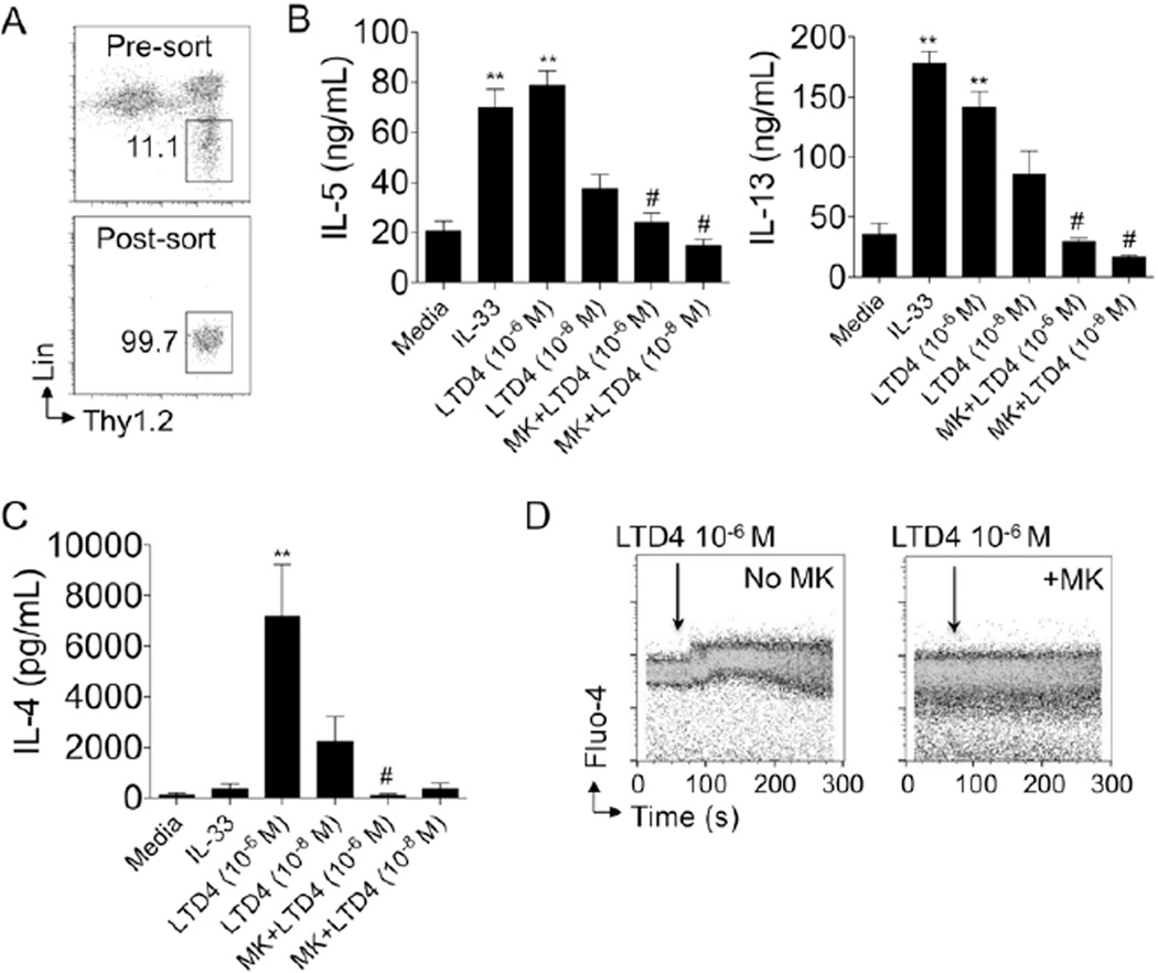
Methods: ILC2s (Thy1.2(+) lineage-negative lymphocytes) and CysLT1R were detected in the lungs of wild-type, signal transducer and activator of transcription 6-deficient (STAT6(-/-)), and recombination-activating gene 2-deficient (RAG2(-/-)) mice by means of flow cytometry. T(H)2 cytokine levels were measured in purified lung ILC2s stimulated with leukotriene D₄ (LTD₄) in the presence or absence of the CysLT1R antagonist montelukast. Calcium influx was measured by using Fluo-4 intensity. Intranasal leukotriene C₄, D₄, and E₄ were administered to naive mice, and levels of ILC2 IL-5 production were determined. Finally, LTD₄ was coadministered with Alternaria species repetitively to RAG2(-/-) mice (with ILC2s) and IL-7 receptor-deficient mice (lack ILC2s), and total ILC2 numbers, proliferation (Ki-67(+)), and bronchoalveolar lavage fluid eosinophil numbers were measured.

Results: CysLT1R was expressed on lung ILC2s from wild-type, RAG2(-/-), and STAT6(-/-) naive and Alternaria species-challenged mice. In vitro LTD₄ induced ILC2s to rapidly generate high levels of IL-5 and IL-13 within 6 hours of stimulation. Interestingly, LTD4, but not IL-33, induced high levels of IL-4 by ILC2s. LTD₄ administered in vivo rapidly induced ILC2 IL-5 production that was significantly reduced by montelukast before treatment. Finally, LTD₄ potentiated Alternaria species-induced eosinophilia, as well as ILC2 accumulation and proliferation.

Conclusions: We present novel data that CysLT1R is expressed on ILC2s and LTD₄ potently induces CysLT1R-dependent ILC2 production of IL-4, IL-5, and IL-13. Additionally, LTD₄ potentiates Alternaria species-induced eosinophilia and ILC2 proliferation and accumulation.


Figures

Figure 1
Figure 1. CysLT1R is expressed on lung and bone marrow type 2 innate lymphoid cells
Single cell suspensions from lungs of naïve mice were stained with CD45, lineage (CD3, CD4, CD5, CD8, CD19, Gr-1, CD11b, CD11c, B220, Ter-119, NK1.1, FcεR1, TCRβ, and TCRγδ), and Thy1.2. ILC2 gated on CD45+ lineage-negative Thy1.2+ lymphocytes (A, left). Cells were then stained with a C-terminus antibody for CysLTR1 with and without blocking peptide using permeabilized methods (A, right). (B) ILC2 from naïve WT mice were stained with an antibody to the extracellular portion of CysLT1R (RB34) using non-permeabilized methods. (C) CysLT1R mRNA levels from FACS purifed lung ILC2, CD4+T1/ST2+, and CD4+T1/ST2- cells from WT mice receiving 4 challenges with Alternaria. Data shown from triplicate samples, p < 0.05, Mann-whitney. (D) Bone marrow was taken from naïve WT mice and processed for FACS analysis of ILC2 CysLT1R expression using the RB34 antibody. (E) Lung ILC2 CysLT1R expression from WT and STAT6−/− mice and (F) from RAG2−/− mice after staining with the RB34 antibody. Plots representative of at least two independent experiments, control antibody staining in solid grey.
Figure 2
Figure 2. ILC2 expression of CysLT1R is stable after allergen challenges and independent of STAT6 or RAG2
WT, STAT6−/−, and RAG2−/− mice were administered a single intranasal challenge with Alternaria allergen once and lung cells obtained at 24 hours and 3 days or given 4 challenges over 10 days. Single suspensions of lung were stained for ILC2 and surface CysLT1R using the RB34 antibody or control antibody (grey). Histograms gated on lung ILC2 (CD45+ Lineage-negative Thy1.2+ lymphocytes). (A) WT and STAT6−/− ILC2 CysLT1R expression in Alternaria-challenged mice at 24 hours, 3 days, or 10 days. (B) WT and RAG2−/− ILC2 CysLT1R expression in Alternaria-challenged mice at 24 hours, 3 days, or 10 days. Plots representative of two independent experiments at each time point, control antibody staining in solid grey.
Figure 3
Figure 3. Leukotriene D4 induces ILC2 Th2 cytokine production through CysLT1R
(A) Lung ILC2 were expanded in-vivo after 3 Alternaria challenges followed by FACS sorting and resting for 40 hours prior to in-vitro studies. Pre- (top) and post-sort (bottom) ILC2% shown. (B) IL-5, IL-13 and (C) IL-4 levels determined by ELISA from supernatants of purified ILC2 were stimulated with IL-33, LTD4 (10−6 M and 10−8 M) for 6 hours with and without 1 µM montelukast pre-treatment for 2 hours. (D) ILC2 Fluo-4 intensity over time in seconds after LTD4 10−6 M was added at 60 seconds without montelukast (left) and after pre-treatment with montelukast (right). Data in A & D are representative of two independent experiments and B & C are combined from triplicate wells of each condition of two independent experiments. MK = montelukast. ** P < 0.005 compared to media, # p < 0.01 compared to the same dose of LTD4 alone, Mann-whitney.
Figure 4
Figure 4. STAT6 dependent CysLTs are induced by Alternaria and promote ILC2 IL-5 production in vivo
(A) BAL CysLTs measured from naïve WT and STAT6−/− mice 12 hours after a single intranasal challenge with 100 mg of Alternaria. 9 mice/group Alternaria challenged, 3 mice per group PBS, Mann-whitney,**P < 0.005. (B) WT naïve mice received a single intranasal challenge with LTC4, LTD4, LTE4, or PBS and some mice were administered three doses of intragastric montelukast before challenges. Lung cells obtained three hours after challenge were cultured overnight with protein transport inhibitor and stained for ILC2 intracellular IL-5. Lung ILC2 (left) were gated and intracellular IL-5 from mice receiving LTC4, LTD4 and LTD4 with (bottom row) and without montelukast (top row). Control antibody staining and ILC2 IL-5 from mice receiving PBS challenge as shown. MK = montelukast. Data shown is representative of 2–3 independent experiments.
Figure 5
Figure 5. LTD4 induces accumulation and proliferation of lung ILC2 and potentiates Alternaria-induced eosinophilia
RAG2−/− and IL-7R−/− mice received 4 intranasal challenges with Alternaria alone or with LTD4. (A) Representative FACS plots of ILC2 % from pooled lungs of naïve RAG2−/− (left) and IL-7R−/− mice (middle left) and RAG2−/− mice after 4 challenges with Alternaria alone (middle right) or with LTD4 (right). (B) Total ILC2 per mouse (left), Ki-67+ ILC2 (middle) and percent Ki-67% ILC2 in mice receiving Alternaria alone or with LTD4 (right). (C) Total IL-5+ lung ILC2 (top) from RAG2−/− mice and BAL eosinophils (bottom) from RAG2−/− and IL-7R−/− mice receiving 4 intranasal challenges with Alternaria alone or with LTD4. Total ILC2, Ki-67, and IL-5 ILC2/mouse are from two independent experiments with pooled lungs from 2 mice per group (t-test, *P < 0.05, **P < 0.005). BAL eosinophils from 8 mice per group (Mann-whitney,*P < 0.05).

Comment in

References

    1. Moro K, Yamada T, Tanabe M, Takeuchi T, Ikawa T, Kawamoto H, et al. Innate production of T(H)2 cytokines by adipose tissue-associated c-Kit(+)Sca-1(+) lymphoid cells. Nature. 2010;463:540–544. - PubMed
    1. Neill DR, Wong SH, Bellosi A, Flynn RJ, Daly M, Langford TK, et al. Nuocytes represent a new innate effector leukocyte that mediates type-2 immunity. Nature. 2010;464:1367–1370. - PMC - PubMed
    1. Price AE, Liang HE, Sullivan BM, Reinhardt RL, Eisley CJ, Erle DJ, et al. Systemically dispersed innate IL-13-expressing cells in type 2 immunity. Proc Natl Acad Sci U S A. 2010;107:11489–11494. - PMC - PubMed
    1. Spits H, Artis D, Colonna M, Diefenbach A, Di Santo JP, Eberl G, et al. Innate lymphoid cells--a proposal for uniform nomenclature. Nat Rev Immunol. 2013;13:145–149. - PubMed
    1. Brickshawana A, Shapiro VS, Kita H, Pease LR. Lineage-Sca1+c-Kit-CD25+ Cells Are IL-33-Responsive Type 2 Innate Cells in the Mouse Bone Marrow. J Immunol. 2011;187:5795–5804. - PMC - PubMed

Publication types