

TNF-α and LPA promote synergistic expression of COX-2 in human colonic myofibroblasts: role of LPA-mediated transactivation of upregulated EGFR
- PMID: 23688423
- PMCID: PMC3663734
- DOI: 10.1186/1471-230X-13-90
TNF-α and LPA promote synergistic expression of COX-2 in human colonic myofibroblasts: role of LPA-mediated transactivation of upregulated EGFR
Abstract
Background: Enhanced EGF receptor (EGFR) signaling is a hallmark of many human cancers, though the role of enhanced EGFR signaling within the surrounding tumor stroma has not been well studied. The myofibroblast is an important stromal cell that demonstrates enhanced EGFR expression in the setting of inflammation, though the functional relevance is not known. We recently reported that TNF-α and the G protein-coupled receptor (GPCR) agonist lysophosphatidic acid (LPA) lead to synergistic cyclo-oxygenase-2 (COX-2) expression, an enzyme strongly associated with the development of colitis-associated cancer. Here, we investigate whether EGFR signaling plays a role in the synergistic COX-2 expression induced by LPA and TNF-α.
Methods: 18Co cells, a model of human colonic myofibroblasts, were grown to confluence on 35 × 10 mm cell culture dishes and were used from passages 10-14. 18Co cells were treated with TNF-α (8.3 ng/ml) and LPA (10 μM). EGFR and COX-2 protein expression, Y1068 phosphorylation, and p42/44 MAPK phosphorylation were assessed by Western Blot, in the presence and absence of various inhibitors.
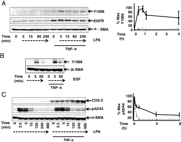
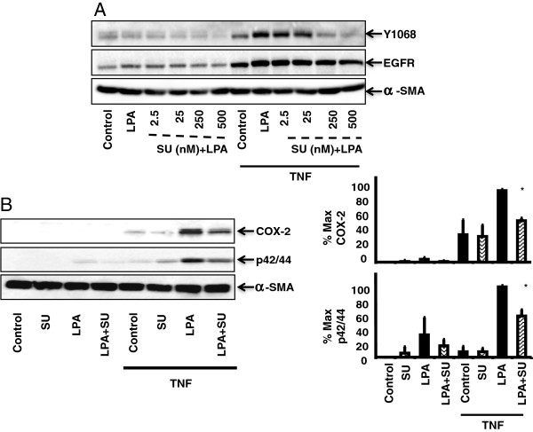
Results: Exposure of 18Co cells to either TNF-α or LPA alone had no effect on EGFR autophosphorylation at Y1068. However, chronic exposure to TNF-α led to upregulation of EGFR in association with sustained LPA-mediated EGFR phosphorylation at Y1068. TNF-α and LPA also led to sustained p42/44 MAPK phosphorylation and synergistic COX-2 expression, effects that were partially inhibited by the EGFR tyrosine kinase inhibitor AG1478. p42/44 MAPK phosphorylation and COX-2 expression were inhibited to the same degree by the MMP inhibitors GM6001 and BB-94, suggesting that LPA-mediated EGFR transactivation involved MMP-mediated release of EGFR ligands from the cell surface. The Src inhibitor SU6556 inhibited TNF-α/LPA-mediated EGFR phosphorylation at Y1068, p42/44 MAPK phosphorylation, and COX-2 expression in a dose-dependent fashion, suggesting an upstream role of Src in the transactivation of EGFR.
Conclusion: Synergistic COX-2 expression induced by TNF-α and LPA involves Src/MMP-mediated transactivation of EGFR and downstream p42/44 MAPK activation in human colonic myofibroblasts. Enhanced EGFR expression induced by TNF-α promotes GPCR-mediated EGFR transactivation in colonic myofibroblasts, providing an important mechanism for stromal COX-2 over-expression that may predispose to the development of colitis-associated cancer.
Figures


Similar articles
-
TNF-α potentiates lysophosphatidic acid-induced COX-2 expression via PKD in human colonic myofibroblasts.Am J Physiol Gastrointest Liver Physiol. 2011 Apr;300(4):G637-46. doi: 10.1152/ajpgi.00381.2010. Epub 2011 Feb 3. Am J Physiol Gastrointest Liver Physiol. 2011. PMID: 21292998 Free PMC article.
-
TNF-α induces upregulation of EGFR expression and signaling in human colonic myofibroblasts.Am J Physiol Gastrointest Liver Physiol. 2012 Apr 15;302(8):G805-14. doi: 10.1152/ajpgi.00522.2011. Epub 2012 Feb 2. Am J Physiol Gastrointest Liver Physiol. 2012. PMID: 22301110 Free PMC article.
-
Angiogenin regulates PKD activation and COX-2 expression induced by TNF-α and bradykinin in the colonic myofibroblast.Biochem Biophys Res Commun. 2020 May 14;525(4):870-876. doi: 10.1016/j.bbrc.2020.02.169. Epub 2020 Mar 12. Biochem Biophys Res Commun. 2020. PMID: 32171525 Free PMC article.
-
Transactivation of EGFR by G protein-coupled receptor in the pathophysiology of intimal hyperplasia.Curr Vasc Pharmacol. 2014 Mar;12(2):190-201. doi: 10.2174/1570161112666140226123745. Curr Vasc Pharmacol. 2014. PMID: 24568153 Review.
-
GPCR-ErbB transactivation pathways and clinical implications.Cell Signal. 2021 Oct;86:110092. doi: 10.1016/j.cellsig.2021.110092. Epub 2021 Jul 22. Cell Signal. 2021. PMID: 34303814 Review.
Cited by
-
Rho-kinase expression in Hirschsprung's disease.Pediatr Surg Int. 2015 Nov;31(11):1077-85. doi: 10.1007/s00383-015-3762-4. Epub 2015 Aug 15. Pediatr Surg Int. 2015. PMID: 26276426
-
Primary Myofibroblasts Maintain Short-Term Viability following Submucosal Injection in Syngeneic, Immune-Competent Mice Utilizing Murine Colonoscopy.PLoS One. 2015 May 27;10(5):e0127258. doi: 10.1371/journal.pone.0127258. eCollection 2015. PLoS One. 2015. PMID: 26016485 Free PMC article.
-
Free-fatty acid receptor-4 (FFA4) modulates ROS generation and COX-2 expression via the C-terminal β-arrestin phosphosensor in Raw 264.7 macrophages.Biochem Pharmacol. 2017 Dec 15;146:139-150. doi: 10.1016/j.bcp.2017.09.008. Epub 2017 Sep 21. Biochem Pharmacol. 2017. PMID: 28943238 Free PMC article.
-
Assessment of the correlation between oxidative stress and expression of MMP-2, TIMP-1 and COX-2 in human aortic smooth muscle cells.Arch Med Sci Atheroscler Dis. 2021 Sep 20;6:e158-e165. doi: 10.5114/amsad.2021.109255. eCollection 2021. Arch Med Sci Atheroscler Dis. 2021. PMID: 34703944 Free PMC article.
-
Natural history of hepatic metastases from colorectal cancer--pathobiological pathways with clinical significance.World J Gastroenterol. 2014 Apr 14;20(14):3719-37. doi: 10.3748/wjg.v20.i14.3719. World J Gastroenterol. 2014. PMID: 24744570 Free PMC article. Review.
References
-
- Powell DW, Mifflin RC, Valentich JD, Crowe SE, Saada JI, West AB. Myofibroblasts. I. Paracrine cells important in health and disease. Am J Physiol. 1999;277(1 Pt 1):C1–C9. - PubMed
Publication types
MeSH terms
Substances
Grants and funding
LinkOut - more resources
Full Text Sources
Other Literature Sources
Research Materials
Miscellaneous
