

A tailored ML-EM algorithm for reconstruction of truncated projection data using few view angles
- PMID: 23689102
- PMCID: PMC3745016
- DOI: 10.1088/0031-9155/58/12/N157
A tailored ML-EM algorithm for reconstruction of truncated projection data using few view angles
Abstract
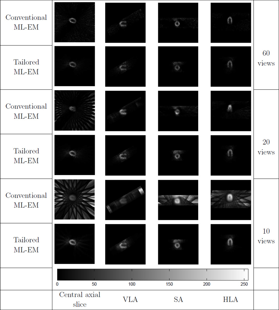
Dedicated cardiac single photon emission computed tomography (SPECT) systems have the advantage of high speed and sensitivity at no loss, or even a gain, in resolution. The potential drawbacks of these dedicated systems are data truncation by the small field of view (FOV) and the lack of view angles. Serious artifacts, including streaks outside the FOV and distortion in the FOV, are introduced to the reconstruction when using the traditional emission data maximum-likelihood expectation-maximization (ML-EM) algorithm to reconstruct images from the truncated data with a small number of views. In this note, we propose a tailored ML-EM algorithm to suppress the artifacts caused by data truncation and insufficient angular sampling by reducing the image updating step sizes for the pixels outside the FOV. As a consequence, the convergence speed for the pixels outside the FOV is decelerated. We applied the proposed algorithm to truncated analytical data, Monte Carlo simulation data and real emission data with different numbers of views. The computer simulation results show that the tailored ML-EM algorithm outperforms the conventional ML-EM algorithm in terms of streak artifacts and distortion suppression for reconstruction from truncated projection data with a small number of views.
Figures


Similar articles
-
Small field-of-view dedicated cardiac SPECT systems: impact of projection truncation.Eur J Nucl Med Mol Imaging. 2010 Mar;37(3):528-36. doi: 10.1007/s00259-009-1223-9. Epub 2009 Sep 1. Eur J Nucl Med Mol Imaging. 2010. PMID: 19722106 Free PMC article.
-
Effective noise-suppressed and artifact-reduced reconstruction of SPECT data using a preconditioned alternating projection algorithm.Med Phys. 2015 Aug;42(8):4872-87. doi: 10.1118/1.4926846. Med Phys. 2015. PMID: 26233214 Free PMC article.
-
Few-view single photon emission computed tomography (SPECT) reconstruction based on a blurred piecewise constant object model.Phys Med Biol. 2013 Aug 21;58(16):5629-52. doi: 10.1088/0031-9155/58/16/5629. Epub 2013 Jul 29. Phys Med Biol. 2013. PMID: 23892823 Free PMC article.
-
Small field-of-view cardiac SPECT can be implemented on hybrid SPECT/CT platforms where data acquisition and reconstruction are guided by CT.Nucl Med Commun. 2009 Sep;30(9):718-26. doi: 10.1097/MNM.0b013e32832eabec. Nucl Med Commun. 2009. PMID: 19617862
-
Application of geometric shape-based CT field-of-view extension algorithms in an all-digital positron emission tomography/computed tomography system.Med Phys. 2024 Feb;51(2):1034-1046. doi: 10.1002/mp.16888. Epub 2023 Dec 16. Med Phys. 2024. PMID: 38103259
Cited by
-
Extreme Few-View Tomography without Training Data.Biomed J Sci Tech Res. 2024;55(2):46779-46884. doi: 10.26717/bjstr.2024.55.008672. Epub 2024 Feb 23. Biomed J Sci Tech Res. 2024. PMID: 38883320 Free PMC article.
-
The impact of system matrix dimension on small FOV SPECT reconstruction with truncated projections.Med Phys. 2016 Jan;43(1):213. doi: 10.1118/1.4938098. Med Phys. 2016. PMID: 26745914 Free PMC article.
-
Development of a Solvability Map.Med Res Arch. 2023;10(11):10.18103/mra.v10i11.3312. doi: 10.18103/mra.v10i11.3312. Epub 2022 Nov 28. Med Res Arch. 2023. PMID: 37063930 Free PMC article.
-
Design and development of the DE-SPECT system: a clinical SPECT system for broadband multi-isotope imaging of peripheral vascular disease.Phys Med Biol. 2024 Jun 11;69(12):125016. doi: 10.1088/1361-6560/ad5266. Phys Med Biol. 2024. PMID: 38815617 Free PMC article.
-
Segmented slant hole collimator for stationary cardiac SPECT: Monte Carlo simulations.Med Phys. 2015 Sep;42(9):5426-34. doi: 10.1118/1.4928484. Med Phys. 2015. PMID: 26328991 Free PMC article.
References
-
- Funk T, Kirch DL, Koss JE, Botvinick E, Hasegawa BH. A novel approach to multipinhole SPECT for myocardial perfusion imaging. Journal of Nuclear Medicine. 2006;47(4):595–602. - PubMed
-
- Steele PP, Kirch DL, Koss JE. Comparison of simultaneous dual-isotope multipinhole SPECT with rotational SPECT in a group of patients with coronary artery disease. Journal of Nuclear Medicine. 2008;49(7):1080–1089. - PubMed
-
- Esteves FP, Raggi P, Folks RD, Keidar Z, Wells Askew J, Rispler S, OConnor MK, Verdes L, Garcia EV. Novel solid-state-detector dedicated cardiac camera for fast myocardial perfusion imaging: multicenter comparison with standard dual detector cameras. Journal of Nuclear Cardiology. 2009;16(6):927–934. - PMC - PubMed
-
- Natterer F. The mathematics of computerized tomography. volume 32. Society for Industrial Mathematics; 2001.
Publication types
MeSH terms
Grants and funding
LinkOut - more resources
Full Text Sources
Other Literature Sources
Research Materials