. 2013 Jul 5;288(27):20064-75.
doi: 10.1074/jbc.M113.481556. Epub 2013 May 20.

Analysis of polymorphic residues reveals distinct enzymatic and cytotoxic activities of the Streptococcus pyogenes NAD+ glycohydrolase

Affiliations

Analysis of polymorphic residues reveals distinct enzymatic and cytotoxic activities of the Streptococcus pyogenes NAD+ glycohydrolase

Sukantha Chandrasekaran et al. J Biol Chem. .

Abstract

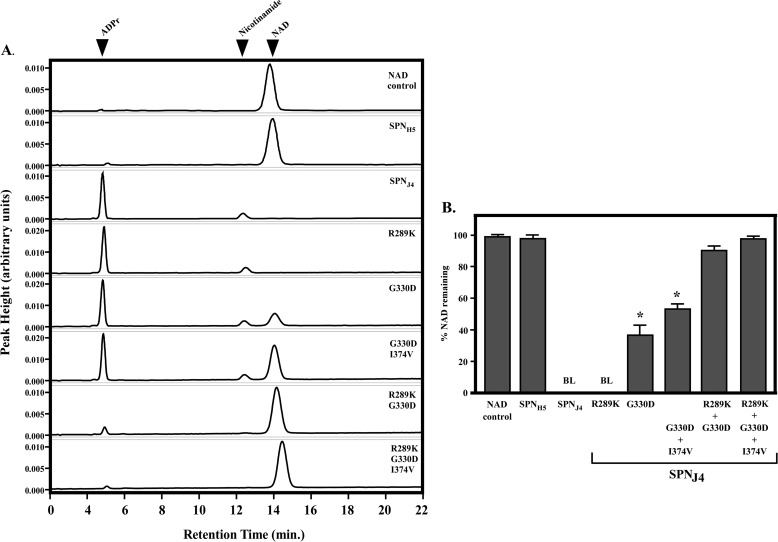
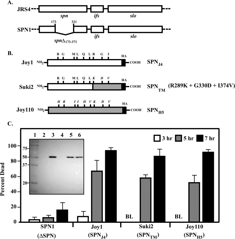
The Streptococcus pyogenes NAD(+) glycohydrolase (SPN) is secreted from the bacterial cell and translocated into the host cell cytosol where it contributes to cell death. Recent studies suggest that SPN is evolving and has diverged into NAD(+) glycohydrolase-inactive variants that correlate with tissue tropism. However, the role of SPN in both cytotoxicity and niche selection are unknown. To gain insight into the forces driving the adaptation of SPN, a detailed comparison of representative glycohydrolase activity-proficient and -deficient variants was conducted. Of a total 454 amino acids, the activity-deficient variants differed at only nine highly conserved positions. Exchanging residues between variants revealed that no one single residue could account for the inability of the deficient variants to cleave the glycosidic bond of β-NAD(+) into nicotinamide and ADP-ribose; rather, reciprocal changes at 3 specific residues were required to both abolish activity of the proficient version and restore full activity to the deficient variant. Changing any combination of 1 or 2 residues resulted in intermediate activity. However, a change to any 1 residue resulted in a significant decrease in enzyme efficiency. A similar pattern involving multiple residues was observed for comparison with a second highly conserved activity-deficient variant class. Remarkably, despite differences in glycohydrolase activity, all versions of SPN were equally cytotoxic to cultured epithelial cells. These data indicate that the glycohydrolase activity of SPN may not be the only contribution the toxin has to the pathogenesis of S. pyogenes and that both versions of SPN play an important role during infection.

Keywords: Allelic Variation; Bacteria; Bacterial Pathogenesis; Bacterial Toxins; Cell Death; Cytotoxicity; Enzymes; Group A Streptococcus; NAD+ Glycohydrolase.


Figures

FIGURE 1.
FIGURE 1.
Distribution of polymorphic residues between two representative SPN variants. The top of the figure shows the domain structure of SPN from a strain (JRS4) with an active NADase activity (SPNJ4). The signal sequence (SS) is not part of the mature protein. Indicated above the figure are the locations of the various polymorphic residues when compared with SPN from a strain (HSC5) representative of the major class deficient for NAD+ glycohydrolase activity (SPNH5). The asterisks below show the location of two glutamate residues implicated in catalysis. The bottom of the figure lists the identities of the polymorphic residues at each position. Also indicated is whether the gene encoding the spn allele is associated with an intact (full-length) or degraded (truncated) gene encoding IFS, the endogenous inhibitor of SPN.
FIGURE 2.
FIGURE 2.
Three residues are required to restore NADase activity to SPNH5. Polymorphic residues in SPNJ4 were changed to the corresponding residue in SPNH5, and those in SPNH5 were changed to their counterparts in SPNJ4 as indicated by the gray and black bars, respectively, for comparison with the unmodified proteins (Unmod). The genes encoding the mutations were expressed from a plasmid (supplemental Table S2) introduced into a derivative of JRS4 (SPN1) with a deletion of its resident gene. NADase activities were determined from cell-free culture supernatants as described under “Experimental Procedures” and are presented relative to SPNJ4. “BL” indicates that activity was below the limit of detection (<0.5%). A double asterisk indicates significantly less NADase activity when compared with unmodified SPNJ4 (p < 0.01). Data shown are the mean and S.E. (error bars) derived from at least three independent experiments.
FIGURE 3.
FIGURE 3.
SPNJ4 single and double swap proteins have NADase activity. The various polymorphic residues from SPNH5 were introduced into SPNJ4 as indicated in the figure. The NADase activity of purified proteins was then analyzed using an HPLC-based assay. A, representative HPLC chromatograms showing the products of enzymatic cleavage of β-NAD+ following a 1-h reaction at 37 °C. Also evaluated was a reaction mixture lacking protein (NAD control). Identities of the various products, including β-NAD+ (NAD), nicotinamide, and ADP-ribose (ADPr) are indicated at the top of the figure. B, relative activity of each of the proteins shown in A quantitated as the percentage of the β-NAD+ substrate remaining uncleaved following a 1-h reaction. BL indicates that values were below the limit of detection (<0.05%), and an asterisk indicates that significantly more substrate was consumed when compared with SPNH5 (p < 0.05). Data presented are the mean and S.E. (error bars) derived from at least three independent experiments.
FIGURE 4.
FIGURE 4.
Cluster 4 swaps have low but detectable NADase activity. A, residues polymorphic between the Bayesian cluster 2 (SPNJ4) and cluster 4 proteins are shown. An asterisk indicates that the cluster 4 residue corresponds to the residue in the cluster 3 NADase activity-deficient (SPNH5) protein (compare with Fig. 1A). B, polymorphic residues from NADase activity-deficient cluster 4 were introduced into SPNJ4 as indicated and analyzed as described in Fig. 3. Shown are representative HPLC chromatograms indicating the products of enzymatic cleavage of β-NAD+ following a 1-h reaction. The β-NAD+ control is a reaction that does not include enzyme. C, relative activity of selected proteins following the extended incubation periods shown in the figure. Data presented are the mean and S.E. (error bars) derived from at least three independent experiments.
FIGURE 5.
FIGURE 5.
NADase-deficient SPN remains cytotoxic to A549 cells. A, the chromosomal spn loci of JRS4 and an in-frame deletion mutant of JRS4 (SPN1; Ref. 4) are shown. Adjacent to the gene encoding SPNJ4 (spn), ifs encodes immunity factor for SPN, and slo encodes streptolysin O. B, strain Joy1 is a derivative of JRS4 expressing SPNJ4 with an HA epitope tag (black box) from the native spn locus. Derived from Joy1 are strains Suki2 and Joy110, which express the chimeric SPN proteins listed in parentheses at the right of the figure. Regions and residues derived from SPNH5 are shown by shaded boxes and in italics, respectively. C, the strains expressing the proteins listed in parentheses were used to infect A549 cells, and cytotoxicity was evaluated at the indicated times by fluorescence microscopy following LIVE/DEAD staining. SPN1 does not express SPN protein (ΔSPN). Refer to supplemental Table S3 for detailed construction of these strains. Data presented represent the mean and S.E. (error bars) derived from at least three independent experiments. BL, below the limit of detection (<0.3%). The inset shows an overlay of a protein ladder with the Western blot analysis of A549 cytosolic fractions prepared after 5 h of infection developed with an HA epitope antiserum. Lane 1, molecular weight standards; lane 2, uninfected; lane 3, Joy1 (SPNJ4); lane 4, SPN1 (ΔSPN); lane 5, Suki2 (SPNTM); lane 6, Joy110 (SPNH5). The molecular weights of selected standards are indicated at the left of the inset.
FIGURE 6.
FIGURE 6.
Catalytic biglutamic acid residues are not required for cytotoxicity. The biglutamic acid residues involved in catalysis (see Fig. 1) were changed to glycines in SPN proteins SPNJ4, SPNTM (R289K/G330D/I374V), and SPNH5 at the chromosomal spn locus in strain JRS4 to generate strains SPNJ4-GG, SPNTM-GG, and SPNH5-GG, respectively. Refer to supplemental Table S3 for detailed construction of these strains. The strains expressing the proteins listed in parentheses shown in the figure were used to infected A549 cells, and cytotoxicity was monitored at the indicated times as described for Fig. 5. SPN1 does not express SPN protein (ΔSPN). The data presented show the mean and S.E. (error bars) derived from at least three independent experiments. The inset shows an overlay of a protein ladder with the Western blot analysis of the cytosolic fraction of A549 cells following a 5-h infection using an HA epitope antiserum. Lane 1, molecular weight standards; lane 2, uninfected; lane 3, Joy1 (SPNJ4); lane 4, SPN1 (ΔSPN); lane 5, Suki6 (SPNJ4-GG); lane 6, Suki7 (SPNTM-GG); lane 7, Suki8 (SPNH5-GG). The molecular weights of selected standards are indicated at the left of the inset.
FIGURE 7.
FIGURE 7.
Polymorphic residues that influence catalysis are located in the substrate binding pocket of SPN. A, orthogonal view of the enzymatic domain of SPN (residues 191–451) modeled in complex with β-NAD+. The model was developed from a structural alignment with cholera toxin bound to β-NAD+ (Protein Data Bank code 2A5F) followed by superimposition of β-NAD+ into the binding pocket of SPN. Residues of the ADP-ribosyl turn-turn motif, catalytic residues, and relevant polymorphic resides are labeled and are highlighted in red, and β-NAD+ is shown in blue. B, Gly-330 forms a hydrogen bond with Gln-216 to form one wall of the binding pocket. C, proximity of Arg-289 to adjacent residues Asp-286 and Lys-288, which form a surface that may position β-NAD+ in the binding pocket. D, Ile-374 is in proximity to catalytic residue Glu-391 but lies buried in an adjacent hydrophobic pocket that includes Phe-200 and Tyr-243.

Similar articles

Cited by

References

    1. Cunningham M. W. (2000) Pathogenesis of group A streptococcal infections. Clin. Microbiol. Rev. 13, 470–511 - PMC - PubMed
    1. Riddle D. J., Bessen D. E., Caparon M. G. (2010) Variation in Streptococcus pyogenes NAD+ glycohydrolase is associated with tissue tropism. J. Bacteriol. 192, 3735–3746 - PMC - PubMed
    1. Meehl M. A., Pinkner J. S., Anderson P. J., Hultgren S. J., Caparon M. G. (2005) A novel endogenous inhibitor of the secreted streptococcal NAD-glycohydrolase. PLoS Pathog. 1, e35. - PMC - PubMed
    1. Madden J. C., Ruiz N., Caparon M. (2001) Cytolysin-mediated translocation (CMT): a functional equivalent of type III secretion in Gram-positive bacteria. Cell 104, 143–152 - PubMed
    1. Ghosh J., Caparon M. G. (2006) Specificity of Streptococcus pyogenes NAD+ glycohydrolase in cytolysin-mediated translocation. Mol. Microbiol. 62, 1203–1214 - PubMed

Publication types

MeSH terms