Review
. 2014 Mar;71(5):745-70.
doi: 10.1007/s00018-013-1355-8. Epub 2013 May 21.

Bacterial serine proteases secreted by the autotransporter pathway: classification, specificity, and role in virulence

Affiliations
Review

Bacterial serine proteases secreted by the autotransporter pathway: classification, specificity, and role in virulence

Fernando Ruiz-Perez et al. Cell Mol Life Sci. 2014 Mar.

Abstract

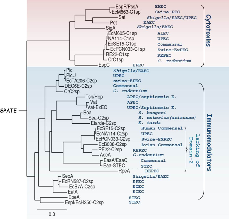
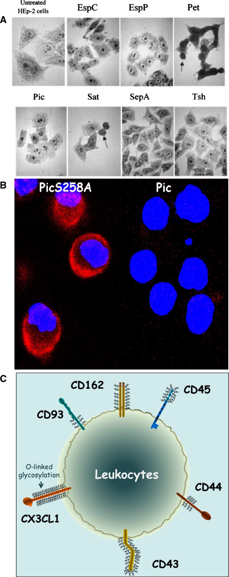
Serine proteases exist in eukaryotic and prokaryotic organisms and have emerged during evolution as the most abundant and functionally diverse group. In Gram-negative bacteria, there is a growing family of high molecular weight serine proteases secreted to the external milieu by a fascinating and widely employed bacterial secretion mechanism, known as the autotransporter pathway. They were initially found in Neisseria, Shigella, and pathogenic Escherichia coli, but have now also been identified in Citrobacter rodentium, Salmonella, and Edwardsiella species. Here, we focus on proteins belonging to the serine protease autotransporter of Enterobacteriaceae (SPATEs) family. Recent findings regarding the predilection of serine proteases to host intracellular or extracellular protein-substrates involved in numerous biological functions, such as those implicated in cytoskeleton stability, autophagy or innate and adaptive immunity, have helped provide a better understanding of SPATEs' contributions in pathogenesis. Here, we discuss their classification, substrate specificity, and potential roles in pathogenesis.


Figures

Fig. 1
Fig. 1
General structure of the serine protease autotransporter from Enterobacteriaceae (SPATE). Autotransporters comprise three functionally different domains: the signal peptide, which targets the autotransporter to the inner membrane SecYEG translocon; the N-terminal passenger domain (also called α-domain), which encodes the biological function of the AT-molecule; and the pore-forming C-terminal translocator domain (also known as the β-domain). Here, the crystal structure of a prototype SPATE protease; EspP passenger and translocator domains are depicted (PBD ID:3SZE and PBD ID:3SLT). The passenger domain (orange) shows the characteristic serine protease motif GDSGS on SPATE proteins along with the residues involved in the formation of the catalytic triad (His, Asp and Ser). The passenger domain is incised between the two Asn residues (violet) bridging passenger and translocator domains inside the pore during the passenger secretion. Residues in the catalytic triad and cleavage site between passenger and translocator domains are illustrated in colored letters and spheres
Fig. 2
Fig. 2
Prevailing model of AT biogenesis. The AT molecule is targeted to the periplasmic space by the Sec apparatus. Once in the periplasm, the AT intermediate is stabilized by periplasmic chaperones such as Skp, SurAm and/or DegP or, when a chaperone shortage exists, perhaps by FkpA. This step is believed to prevent non-productive aggregation, premature folding and/or to maintain the species in a partially folded “translocation-competent state”. Recent data suggest that the Bam complex catalyzes both the integration of the β-domain into the OM, and the translocation of the passenger domain across the OM in a C- to N-terminal direction, followed by intra-barrel cleavage and disengagement of the passenger domain (in the case of SPATEs)
Fig. 3
Fig. 3
Stereo ribbon diagrams showing the overall structure of the passenger domain of class 1 and class-2 SPATEs. EspP(3SZE) and Hbp(1WXR) PDB annotations were used to model prototype structures of class-1 and class-2 SPATE proteins, respectively. By using the SWISS-MODEL [162], Jmol (http://www.jmol.org/) and SignalP 4.1 servers [163], along with the two SPATE PDB annotations, the hypothetical structure of AdcA; a class-2 SPATE lacking of domain-2 was modeled (QMEAN4 score 0.639, z score:−2.02). Helices and strands are colored red for the protease domain (Domain-1; residue S1–N256 in EspP, G1–N256 in Hbp and S1-D253 in AdcA). Domain-2 is shown in violet; predicted as a chitinase-like domain in Hbp (residues A481–N556), but not present in EspP and AdcA. Domain-3, which forms a helix–turn–helix motif facing domain-1 is shown in orange (residues G512–E575 in EspP, residues G607-E644 in HbP, and G532-E569 in AdcA). Domain-4 is shown in yellow (residues T615–G645 in EspP, S684-S714 in Hbp, and N609-S639 in AdcA). The β-strands in the parallel β-helix, classical in most AT proteins, are colored in gray for EspP and Hbp, but white for the hypothetical AdcA structure. Bars underneath the structures illustrate positions of globular regions in the mature passenger domain
Fig. 4
Fig. 4
Domain-2 and -3 among SPATE autotransporters proteins. Alignment of the aminoacid sequence of 28 SPATE passenger domains with Clustal-Omega [164] revealed differences in domain-2 and -3 between SPATE classes. Class-1 SPATEs (pink) lack domain-2, while it is present in only a number of class-2 SPATEs (blue). Domain-3 in class-1 SPATEs is larger than its equivalent in class-2 SPATEs and includes a potential disulphide bond (two cysteines), which is missing in class-2 SPATEs. Identical residues are shaded in black, similar residues are shaded in gray, conserved cysteines(C) in domain-3 class-1 SPATEs are shaded in blue. Aminoacid sequence of Tsh/Hbp domain-2(A481-N556), and the aminoacid sequences of domain-3 for EspP(G512-E575), AdcA(G532-E569), and Tsh/Hbp(G607-E644) are shown in blue and correspond to the globular regions illustrated in Fig 3
Fig. 5
Fig. 5
Sequence relationships of SPATE proteins. Phylogenetic analysis of the aminoacidic sequence of the SPATE passenger domain reveals two distinctive classes of SPATE proteins: denominated class-1, cytotoxic; and class-2, lectin-like immunomodulators. Class-2 spates also includes a cluster of SPATEs found mostly in animal pathogens, which lack the classic domain-2 (see also Figs. 3, 4). Within both classes, clusters of allelic variants are also appreciated. For instance, allelic variants for EspP, Cr-C1sp, Pic, Tsh/Hbp, Boa, EcSE15-C2sp, EaaA/EaaC, SepA, and EpeA are observed. Bacterial species from which SPATE sequences were originated are shown on the right side. Phylogenetic analysis was performed as follows: alignment of sequences was built with Clustal-Omega [164] and cured by Gblocks [165]. Phylogenetic tree was constructed with PhyML/bootstrapping [166] procedure and visualized with TreeDyn [167]
Fig. 6
Fig. 6
Effect of Class-1 and Class-2 SPATEs on host cells. a Effects of SPATEs on HEp-2 cell monolayers are shown by oil immersion light microscopy of Giemsa-stained HEp-2 cells after treatment with SPATE proteins at 500 nM for 5 h. Rounding of cells is seen with Pet and Sat (arrows). Obvious cytotoxic effects of the less active EspP and EspC on cells are only seen with higher protein concentrations and exposure times (see text). Copyright© 2002, American Society for Microbiology [56]. b. Pic produced from Shigella flexneri and pathogenic E. coli degrades a broad range of leukocyte glycoproteins. To visualize degradation of O-linked glycoproteins by Pic, whole blood leukocytes were treated with Pic and the protease defective PicS258A for 30 min, stained for DNA (blue) and PSGL-1(red), and analyzed by confocal microscopy. Complete degradation of PSGL-1 can be observed on the Pic-treated leukocyte population. Only degradation of PSGL-1 is shown, but the array of Pic targets included CD43, CD44, CD45, CD93, and CX3CL1; illustrated in (c)

References

    1. Pallen MJ, Chaudhuri RR, Henderson IR. Genomic analysis of secretion systems. Curr Opin Microbiol. 2003;6(5):519–527. - PubMed
    1. Henderson IR, Navarro-Garcia F, Nataro JP. The great escape: structure and function of the autotransporter proteins. Trends Microbiol. 1998;6(9):370–378. - PubMed
    1. Jacob-Dubuisson F, Locht C, Antoine R. Two-partner secretion in Gram-negative bacteria: a thrifty, specific pathway for large virulence proteins. Mol Microbiol. 2001;40(2):306–313. - PubMed
    1. Linke D, et al. Trimeric autotransporter adhesins: variable structure, common function. Trends Microbiol. 2006;14(6):264–270. - PubMed
    1. Salacha R, et al. The Pseudomonas aeruginosa patatin-like protein PlpD is the archetype of a novel Type V secretion system. Environ Microbiol. 2010;12(6):1498–1512. - PubMed

Publication types

MeSH terms

Substances

LinkOut - more resources