. 2013 Jul;21(7):1306-15.
doi: 10.1038/mt.2013.86. Epub 2013 May 21.

Therapeutic response in feline sandhoff disease despite immunity to intracranial gene therapy

Affiliations

Therapeutic response in feline sandhoff disease despite immunity to intracranial gene therapy

Allison M Bradbury et al. Mol Ther. 2013 Jul.

Abstract

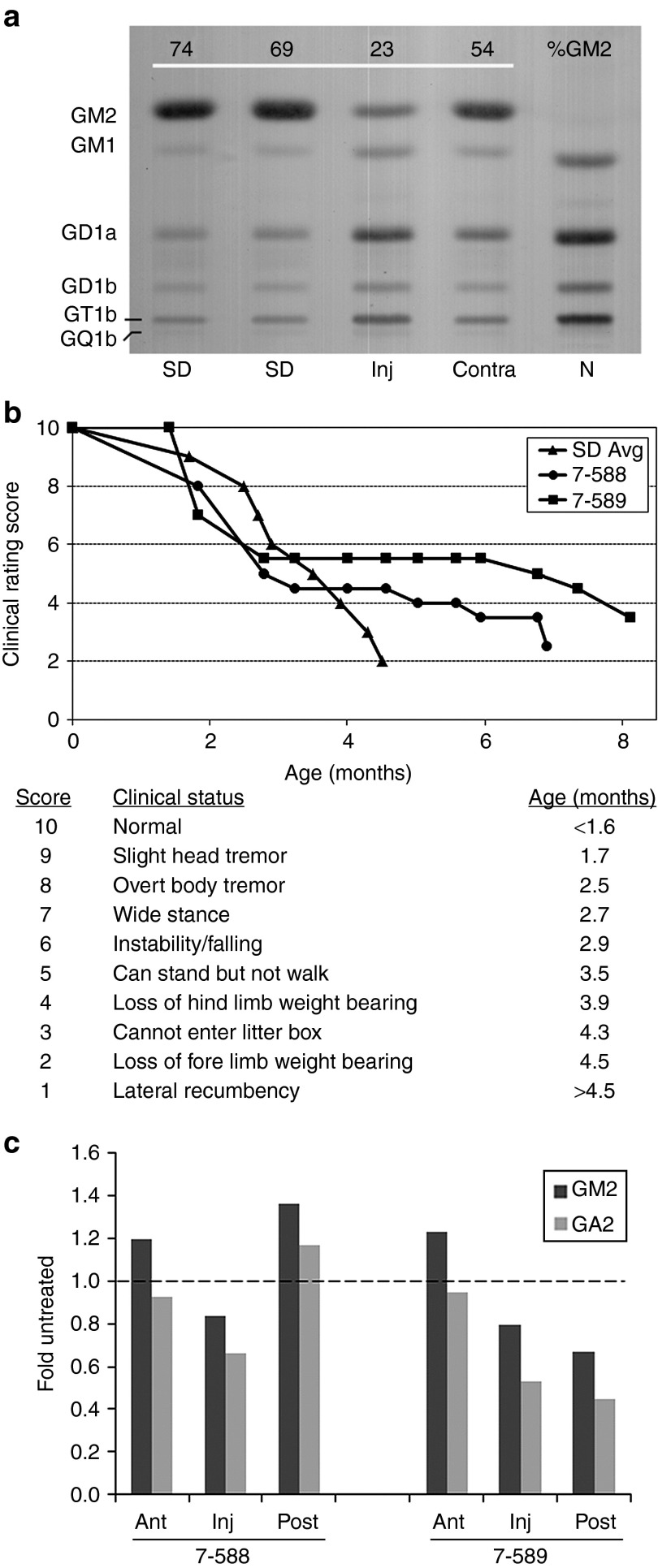
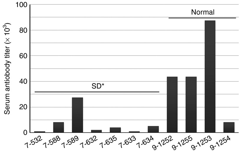
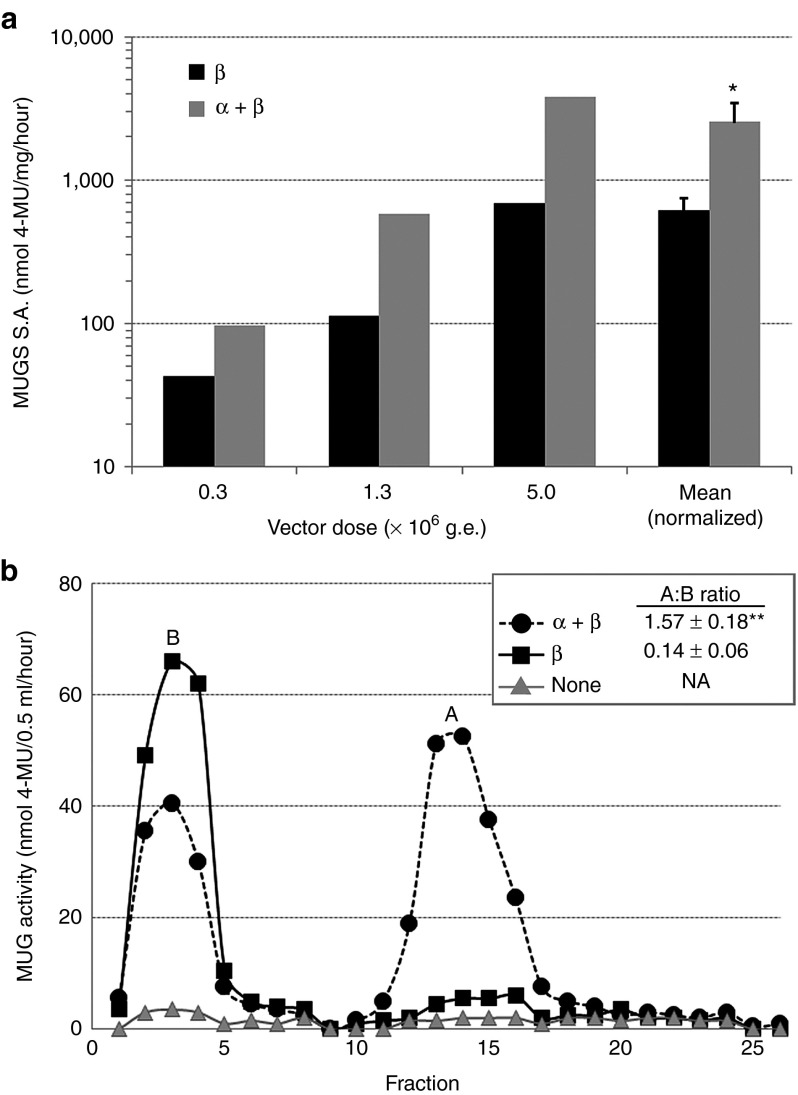
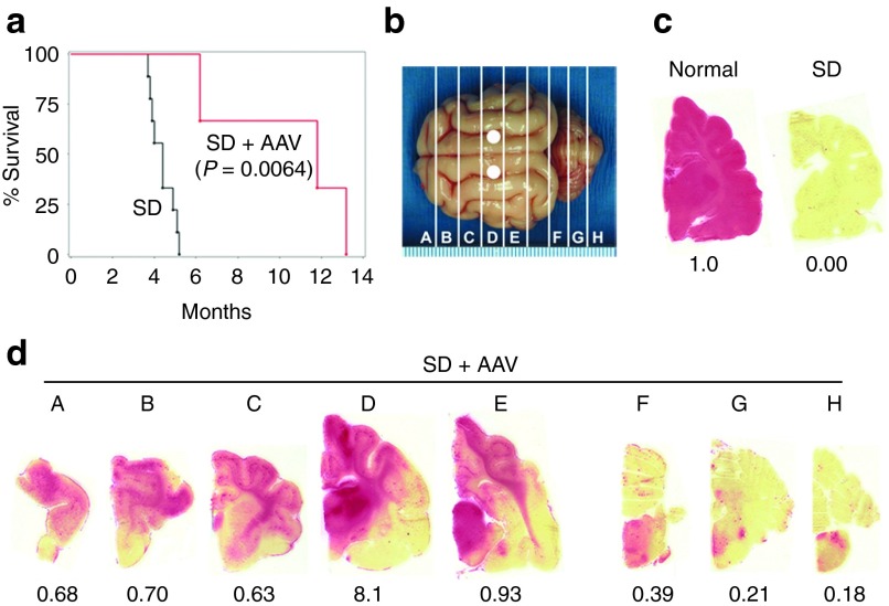
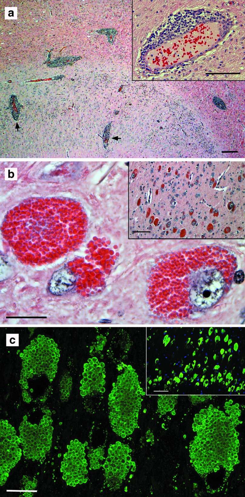
Salutary responses to adeno-associated viral (AAV) gene therapy have been reported in the mouse model of Sandhoff disease (SD), a neurodegenerative lysosomal storage disease caused by deficiency of β-N-acetylhexosaminidase (Hex). While untreated mice reach the humane endpoint by 4.1 months of age, mice treated by a single intracranial injection of vectors expressing human hexosaminidase may live a normal life span of 2 years. When treated with the same therapeutic vectors used in mice, two cats with SD lived to 7.0 and 8.2 months of age, compared with an untreated life span of 4.5 ± 0.5 months (n = 11). Because a pronounced humoral immune response to both the AAV1 vectors and human hexosaminidase was documented, feline cDNAs for the hexosaminidase α- and β-subunits were cloned into AAVrh8 vectors. Cats treated with vectors expressing feline hexosaminidase produced enzymatic activity >75-fold normal at the brain injection site with little evidence of an immune infiltrate. Affected cats treated with feline-specific vectors by bilateral injection of the thalamus lived to 10.4 ± 3.7 months of age (n = 3), or 2.3 times as long as untreated cats. These studies support the therapeutic potential of AAV vectors for SD and underscore the importance of species-specific cDNAs for translational research.


Figures

Figure 1
Figure 1
Therapeutic benefit of human Hex expressed from an AAV1 vector in Sandhoff disease (SD) cats. (a) Cat 7-532 was injected in the right thalamus with a 1:2 ratio of AAV1-CAG-hHEXA/B-Tat-WPRE (A:B = 1 × 1011 g.e.:2 × 1011 g.e., Table 1). Tissue was collected 6 weeks post-injection, and GM2 storage was analyzed by high-performance thin-layer chromatography. Shown are samples from the injected thalamus (Inj), the contralateral thalamus (Contra), and corresponding sections from untreated SD and normal (N) cats. GM2 levels were quantitated densitometrically and expressed as a percentage of total ganglioside on the plate (%GM2). Gangliosides were identified by comparison to known standards and labeled accordingly. The absence of a prominent GM2 band in the normal control lane caused a slight migration shift for GM1 and other bands. (b) Two SD cats treated for long-term follow-up (7-588 and 7-589) had delayed disease progression and increased life span (7.0 and 8.2 months, respectively). Before intracranial injection, cat 7-589 was treated by intravenous injection of AAV8 vectors with a liver-targeted promoter. Disease progression was scored according to a clinical rating scale developed for untreated SD cats, with average age of symptom acquisition compiled from nine separate animals (SD Avg). (c) Total brain lipids were analyzed as in a from the injection site (Inj) and the adjacent anterior (Ant) and posterior (Post) blocks from AAV-treated cats (7-588 and 7-589) at humane endpoint. GM2 (dark bar) and GA2 (light bar) levels were compared with corresponding blocks of an untreated SD cat at its humane endpoint (4.3 months of age). Data for AAV-treated cats is expressed as “fold untreated”, with the untreated level indicated by a dashed line. AAV, adeno-associated virus; g.e., genome equivalents.
Figure 2
Figure 2
Serum antibody titers to human HexA in AAV-treated cats. Sandhoff disease (SD) or normal cats treated by intracranial injection of AAV vectors expressing human Hex were assayed at experimental or humane endpoint for serum antibodies to human HexA. AAV-treated SD cats had significantly lower titers compared with AAV-treated normal cats (*P = 0.011 with the inclusion of 7-589, or P = 0.009 with omission of 7-589, which was pre-treated by intravenous injection before intracranial injection). Average titers of at least two assays are shown. AAV, adeno-associated virus.
Figure 3
Figure 3
HexA activity after expression of feline Hex α- and β-subunits. Immortalized skin fibroblasts from a Sandhoff disease (SD) cat were transduced with AAVrh8 vectors expressing the feline Hex subunits: β-subunit alone (β), or α- and β-subunits combined (α + β). The vector dose per subunit remained constant, and three doses were tested: 0.3, 1.3 or 5.0 × 106 g.e./cell. (a) Specific activity (S.A.) was calculated for the MUGS (HexA preferred) substrate at each dose and plotted on a logarithmic scale. After normalization to vector dose, specific activities were averaged (mean-normalized) and found to be 4.2-fold higher after α + β coexpression than after β-subunit expression alone (*P = 0.043). (b) DEAE cellulose anion exchange chromatography was used to separate HexA (A) and HexB (B) isozymes. Fractions of 0.5 ml eluates were collected using a 0–400 mmol/l NaCl gradient, and MUG substrate (cleaved by all Hex isozymes) was used to measure enzyme activity (nmol 4-MU/0.5 ml/hour). The total activity per isozyme was calculated by summing activities from fractions 1–8 (B) or 10–20 (A) and ratios of A:B were calculated for α + β (closed circles) or β (closed squares)(n = 3 each). MUG activity was measured in untreated cells as a control (none, gray triangles). A:B ratios were statistically higher in cells treated with α + β (1.57) than with β alone (0.14) (**P = 0.002). HexS was not detected in any sample but generally elutes after fraction 20. DEAE, diethylaminoethyl; g.e., genome equivalents; MUG, 4-MU-N-acetyl-β-D-glucosaminide; MUGS, 4-methylumbelliferyl 6-sulfo-2-acetamido-2-deoxy-β-D-glucopyranoside; NA, not applicable.
Figure 4
Figure 4
Therapeutic benefit in Sandhoff disease (SD) cats after intrathalamic injection of AAVrh8 vectors expressing feline Hex. SD cats (n = 3) were followed to the humane endpoint after bilateral thalamic injection of AAVrh8-fHEX vectors (1:1 ratio of α:β total dose = 3.2 × 1012 g.e.). (a) Mean survival of treated SD cats (SD + AAV) was 10.4 months, a statistically significant increase compared with nine untreated cats (SD; P = 0.0064, log-rank test). (b) Injection sites (white circles) and 0.6 cm coronal brain blocks (A–H) are shown. (c) Naphthol staining for Hex activity (red) is shown for untreated normal and SD cat brains. The fold normal Hex activity (measured by MUGS substrate) is listed below each brain block. (d) Naphthol staining for one representative AAV-treated SD cat (SD + AAV, 7-856) demonstrates Hex activity throughout the cerebrum (A–E) with minor activity in brainstem and cerebellum (F–H). MUGS activity ranged from 0.18- to 8.1-fold normal. Letter designations correspond to b. AAV, adeno-associated virus; g.e., genome equivalents; MUGS, 4-methylumbelliferyl 6-sulfo-2-acetamido-2-deoxy-β-D-glucopyranoside.
Figure 5
Figure 5
Histological findings after AAV injection in the normal and Sandhoff disease (SD) cat brain. (a) Perivascular cuffing (black arrows) was apparent in the brains of normal cats expressing human Hex from AAV1 vectors, with greatest concentrations near the thalamic injection site. One representative cat (9-1254) is shown. Bar = 200 μm (inset = 100 μm). (b) Strongly eosinophilic neurons were observed in discrete areas of the brain (inset), corresponding to areas of highest Hex activity. At high magnification, granular to botryoid intracellular inclusions were apparent. Bar = 20 μm (inset = 100 μm). The region of the dorsal thalamic nuclei from cat 7-708 is shown. (c) Immunofluorescence with a monoclonal antibody to the feline β-subunit demonstrated that botryoid inclusions contained high levels of Hex (green). Dorsal thalamic nuclei of cat 7-708 were stained. Inset was taken with a standard fluorescence microscope with DAPI nuclear counterstain (bar = 100 μm). Main panel is a merged stack of 20 layers (6 μm total) from a confocal microscope (bar = 20 μm). AAV, adeno-associated virus; DAPI, 4′,6-diamidino-2-phenylindole.

References

    1. Gravel RA, Clarke JTR, Kaback MM, Mahuran D, Sandhoff K, Suzuki K.1995The GM2 gangliosidoses. Scriver CR, Beaudet AL, Sly WS.Valle D.eds). The Metabolic and Molecular Bases of Inherited Disease7th edn., vol. 2McGraw-Hill, Inc.New York; pp. 2839–2879.
    1. Tay W. Symmetrical changes in the region of the yellow spot in each eye of an infant. Tran Ophthalmol Soc UK. 1881;1:155–157.
    1. Arfi A, Bourgoin C, Basso L, Emiliani C, Tancini B, Chigorno V, et al. Bicistronic lentiviral vector corrects beta-hexosaminidase deficiency in transduced and cross-corrected human Sandhoff fibroblasts. Neurobiol Dis. 2005;20:583–593. - PubMed
    1. Guidotti JE, Mignon A, Haase G, Caillaud C, McDonell N, Kahn A, et al. Adenoviral gene therapy of the Tay-Sachs disease in hexosaminidase A-deficient knock-out mice. Hum Mol Genet. 1999;8:831–838. - PubMed
    1. Itakura T, Kuroki A, Ishibashi Y, Tsuji D, Kawashita E, Higashine Y, et al. Inefficiency in GM2 ganglioside elimination by human lysosomal beta-hexosaminidase beta-subunit gene transfer to fibroblastic cell line derived from Sandhoff disease model mice. Biol Pharm Bull. 2006;29:1564–1569. - PubMed

Publication types

Substances