. 2013 Jul;21(7):1324-34.
doi: 10.1038/mt.2013.88. Epub 2013 May 21.

Selective forelimb impairment in rats expressing a pathological TDP-43 25 kDa C-terminal fragment to mimic amyotrophic lateral sclerosis

Affiliations

Selective forelimb impairment in rats expressing a pathological TDP-43 25 kDa C-terminal fragment to mimic amyotrophic lateral sclerosis

Robert D Dayton et al. Mol Ther. 2013 Jul.

Abstract

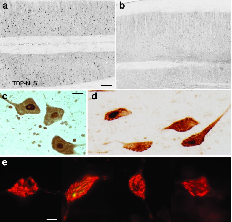
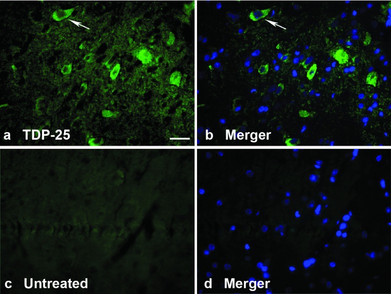
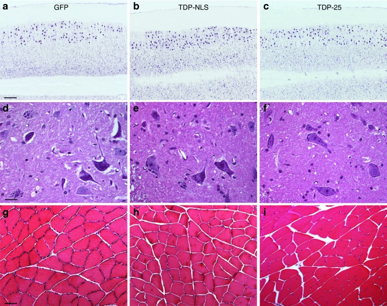
Pathological inclusions containing transactive response DNA-binding protein 43 kDa (TDP-43) are common in several neurodegenerative diseases including amyotrophic lateral sclerosis (ALS). TDP-43 normally localizes predominantly to the nucleus, but during disease progression, it mislocalizes to the cytoplasm. We expressed TDP-43 in rats by an adeno-associated virus (AAV9) gene transfer method that transduces neurons throughout the central nervous system (CNS). To mimic the aberrant cytoplasmic TDP-43 found in disease, we expressed a form of TDP-43 with mutations in the nuclear localization signal sequence (TDP-NLS). The TDP-NLS was detected in both the cytoplasm and the nucleus of transduced neurons. Unlike wild-type TDP-43, expression of TDP-NLS did not induce mortality. However, the TDP-NLS induced disease-relevant motor impairments over 24 weeks. We compared the TDP-NLS to a 25 kDa C-terminal proaggregatory fragment of TDP-43 (TDP-25). The clinical phenotype of forelimb impairment was pronounced with the TDP-25 form, supporting a role of this C-terminal fragment in pathogenesis. The results advance previous rodent models by inducing cytoplasmic expression of TDP-43 in the spinal cord, and the non-lethal phenotype enabled long-term study. Approaching a more relevant disease state in an animal model that more closely mimics underlying mechanisms in human disease could unlock our ability to develop therapeutics.


Figures

Figure 1
Figure 1
Green fluorescent protein (GFP) expression derived from intravenous administration of an adeno-associated virus (AAV9) vector. (a) Biophotonic imaging. For each tissue shown, there is an age-matched blank control rat and a GFP rat. In the central nervous system (CNS), there is intense and uniform GFP expression in the spinal cord, with apparently less GFP in the brain. With this promoter system, there is strong expression in the muscle, heart, and liver (as shown in fh). (b) Imaging of a GFP and control rat's spinal cord and brain by a small hand-held ultraviolet (UV) lamp. (c) View of intact dissected spinal cord from fluorescent microscope. (d,e) Lumbar spinal cord showing expression in large motor neurons in the ventral horn. ac,f,g, 24 weeks; d,e,h, 12 weeks. Bars: in c = 536 μm; in d = 134 μm; in e = 84 μm; in f = 268 μm, in fh same magnification.
Figure 2
Figure 2
Longitudinal studies of green fluorescent protein (GFP), TDP-NLS, and TDP-25 rats over 24 weeks. One TDP-43 construct bears mutations in the nuclear localization sequence (TDP-NLS) and the other is a 25 kDa C-terminal fragment (TDP-25). (ac) Weight gain appeared normal in the GFP group, but there were subtle effects on weight gain in the TDP-NLS and TDP-25 rats (see Results). (d) Both TDP-NLS and TDP-25 rats were impaired for the rotarod. (e) Total locomotor activity was affected in TDP-NLS and TDP-25 rats progressively over time. (f) In contrast, the rearing behavior was selectively affected in TDP-25 rats. (g,h) Observations of limb impairments. All of the TDP-NLS rats were impaired in their hindlimbs by 5 weeks, though forelimb impairment was more frequent in TDP-25 rats (7/13) versus TDP-NLS (2/16). NLS, nuclear localization signal.
Figure 3
Figure 3
Limb dysfunction after gene delivery of two forms of the transactive response DNA-binding protein 43 kDa (TDP-43). (a) Loss of proper hindlimb function in a TDP-NLS rat on the rotarod task. (bd) When briefly hanging an animal upside down, the green fluorescent protein (GFP) rat shows the normal extension response, but the TDP-NLS and TDP-25 rats are impaired. (e,f) Abnormal gait in a TDP-25 rat with the hindlimb reaching across the tail (arrows). Forelimb defects were more frequently observed in TDP-25 rats. (g) Normal rearing in a GFP rat with both forepaws touching the wall. (hk) In contrast, TDP-25 rats would attempt to rear using one or neither forepaw. Aberrant extension and spasticity of one of the forepaws/digits shown in j (arrow). In a, 4 weeks; in bk, 12 weeks. NLS, nuclear localization signal.
Figure 4
Figure 4
TDP-NLS and TDP-25 transgene expression. (a) RNA was extracted from lumbar spinal cord and probed for a sequence specific to the C-terminus of human TDP-43 mRNA. Four green fluorescent protein (GFP) rats (G1-4) were blank for the human TDP-43 sequence, and four TDP-NLS rats (N1-4) expressed the target band. The target sequence is also within the TDP-25 sequence and was detected in a TDP-25 rat (25). Different molecular weight (MW) markers were used in the two panels. (b) Western blot for TDP-43 with a non-species–specific antibody. There was a consistent one- to twofold upregulation in the TDP-NLS group in the cervical (C) and lumbar (L, arrow) spinal cord for 12 weeks. (c) The TDP-25 fragment could be selectively expressed and detected using a phospho-specific TDP-43 antibody (from M.A.G.). An immunoblot for tubulin below confirms similar protein loading. GFP and TDP-NLS, 12 weeks; TDP-25, 6 weeks. NLS, nuclear localization signal; RT-PCR, reverse transcription-PCR.
Figure 5
Figure 5
Human-specific TDP-43 expression in the spinal cord with the TDP-NLS AAV9. (a) Widespread expression of cytoplasmic TDP-43 immunoreactivity was successfully achieved, demonstrated on this horizontal section of the ventral horn of the lumbar region at a 24-week interval. (b) Background staining from a matching green fluorescent protein control sample stained with the human TDP-43–specific antibody. (c,d) Twelve- and 24-week samples of spinal motor neurons expressing the human TDP-43 in the cytoplasm. There was a trend towards a more granular staining pattern at the later interval. (e) Examples of hippocampal neurons in the brain expressing the human TDP-43, after a stereotaxic injection of the TDP-NLS AAV9, 2 weeks earlier (immunofluorescence). Some of the transduced neurons had punctuate and dystrophic staining patterns consistent with aggregation. Bars: in a = 335 μm, in a,b = same magnification; in c = 20 μm, in c,d = same magnification; in e = 9 μm. AAV, adeno-associated virus; NLS, nuclear localization signal.
Figure 6
Figure 6
Cytoplasmic TDP-25 expression. (a) Specific immunoreactivity for a phospho-TDP-43 antibody (green) in the cytoplasm of neurons in the lumbar spinal cord in a TDP-25 rat at 6 weeks. (b) Merger with a nuclear counterstain (DAPI, blue) demonstrates that the phospho-TDP-43 immunoreactivity is not expressed in the nucleus, arrows. (c,d) Background staining with this antibody from an untreated control imaged under matching conditions to a,b. Bars: in a = 34 μm, ad = same magnification. DAPI, 4′,6-diamidino-2-phenylindole.
Figure 7
Figure 7
Lack of neuronal loss or severe muscle atrophy at 24 weeks. (ac) Horizontal sections of green fluorescent protein (GFP), TDP-NLS, and TDP-25 samples of the ventral part of the lumbar spinal cord stained for Nissl substance. The large motor neurons are preserved in the TDP-NLS and TDP-25 groups. (df) Ventral horn including motor neurons from each group stained with hematoxylin and eosin (H&E). (gi) Gastrocnemius muscle from each group stained with H&E. The sample from the TDP-NLS group suggested slight angulation of muscle fibers. Bars: in a = 335 μm, ac = same magnification; in d = 21 μm, df = same magnification; in g = 67 μm, gi = same magnification. NLS, nuclear localization signal.

References

    1. Neumann M, Sampathu DM, Kwong LK, Truax AC, Micsenyi MC, Chou TT, et al. Ubiquitinated TDP-43 in frontotemporal lobar degeneration and amyotrophic lateral sclerosis. Science. 2006;314:130–133. - PubMed
    1. Mackenzie IR, Bigio EH, Ince PG, Geser F, Neumann M, Cairns NJ, et al. Pathological TDP-43 distinguishes sporadic amyotrophic lateral sclerosis from amyotrophic lateral sclerosis with SOD1 mutations. Ann Neurol. 2007;61:427–434. - PubMed
    1. Arai T, Hasegawa M, Akiyama H, Ikeda K, Nonaka T, Mori H, et al. TDP-43 is a component of ubiquitin-positive tau-negative inclusions in frontotemporal lobar degeneration and amyotrophic lateral sclerosis. Biochem Biophys Res Commun. 2006;351:602–611. - PubMed
    1. Arai T, Mackenzie IR, Hasegawa M, Nonoka T, Niizato K, Tsuchiya K, et al. Phosphorylated TDP-43 in Alzheimer's disease and dementia with Lewy bodies. Acta Neuropathol. 2009;117:125–136. - PubMed
    1. Igaz LM, Kwong LK, Xu Y, Truax AC, Uryu K, Neumann M, et al. Enrichment of C-terminal fragments in TAR DNA-binding protein-43 cytoplasmic inclusions in brain but not in spinal cord of frontotemporal lobar degeneration and amyotrophic lateral sclerosis. Am J Pathol. 2008;173:182–194. - PMC - PubMed

Publication types

Substances