. 2013 Jul;353(1):99-115.
doi: 10.1007/s00441-013-1645-3. Epub 2013 May 21.

Transmembrane protein PERP is a component of tessellate junctions and of other junctional and non-junctional plasma membrane regions in diverse epithelial and epithelium-derived cells

Affiliations

Transmembrane protein PERP is a component of tessellate junctions and of other junctional and non-junctional plasma membrane regions in diverse epithelial and epithelium-derived cells

Werner W Franke et al. Cell Tissue Res. 2013 Jul.

Abstract

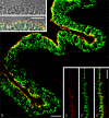
Protein PERP (p53 apoptosis effector related to PMP-22) is a small (21.4 kDa) transmembrane polypeptide with an amino acid sequence indicative of a tetraspanin character. It is enriched in the plasma membrane and apparently contributes to cell-cell contacts. Hitherto, it has been reported to be exclusively a component of desmosomes of some stratified epithelia. However, by using a series of newly generated mono- and polyclonal antibodies, we show that protein PERP is not only present in all kinds of stratified epithelia but also occurs in simple, columnar, complex and transitional epithelia, in various types of squamous metaplasia and epithelium-derived tumors, in diverse epithelium-derived cell cultures and in myocardial tissue. Immunofluorescence and immunoelectron microscopy allow us to localize PERP predominantly in small intradesmosomal locations and in variously sized, junction-like peri- and interdesmosomal regions ("tessellate junctions"), mostly in mosaic or amalgamated combinations with other molecules believed, to date, to be exclusive components of tight and adherens junctions. In the heart, PERP is a major component of the composite junctions of the intercalated disks connecting cardiomyocytes. Finally, protein PERP is a cobblestone-like general component of special plasma membrane regions such as the bile canaliculi of liver and subapical-to-lateral zones of diverse columnar epithelia and upper urothelial cell layers. We discuss possible organizational and architectonic functions of protein PERP and its potential value as an immunohistochemical diagnostic marker.


Figures

Fig. 1
Fig. 1
Immunoblot demonstration of the SDS-polyacrylamide gel electrophoresis (SDS-PAGE)-separated plasma membrane protein PERP (p53 apoptosis effector related to PMP-22) in mammalian epithelial and carcinoma cells and in cardiac tissue by using mouse monoclonal antibodies (mAbs) m8.2.9 (lane 1) or m26.3 (lane 2) with cultured human keratinocytes of line HaCat (lanes 1, 2) or bovine muzzle epithelium (lane 3; reaction of a 1:1 volume mixture of both murine mAbs). Lanes 4–16 show reactions with guinea pig polyclonal antibodies (pAbs) against PERP serum (gpPERP-4A) on SDS-PAGE-separated polypeptides of bovine muzzle (lane 4) or human tongue (lane 5) epithelium, human heart (lane 6) and liver (lane 7) tissue, human hepatic (lane 8) and cholangiocellular carcinoma (lane 9) tissue, bovine liver tissue (lane 10), various human cultured cells of the hepatocellular carcinoma lines Hep3b (lane 11), HuH-7 (lane 12) and PLC (lane 13) and cytoskeletal fractions from cultured cells of the human hepatocellular carcinoma line HepG2 (lane 14) or the human colon carcinoma lines CaCo2 (lane 15) and HT29 (lane 16). Relative molecular weight values (in kDa) of reference polypeptides analyzed in a parallel lane are given left. Note that all these cells and tissues and the cytoskeletal fractions show the specific reaction with the PERP polypeptide with an electrophoretic mobility corresponding to 23 kDa. The weak band with an electrophoretic mobility corresponding to an approximate molecular weight of 68 kDa in lane 4 might represent a homo- or heteromeric complex of PERP, which has not been completely separated under these specific solubilization conditions. Exposure times of the specific blots to the film were 10 min (lanes 1–4) and 15 min (lanes 5–16)
Fig. 2
Fig. 2
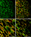
Double-label confocal laser-scanning immunofluorescence microscopy of a cryostat section through a lateral portion of bovine tongue mucosa after reaction with murine mAb m26.3 (a) against protein PERP and rabbit pAbs to β-catenin (a’), followed by reaction with the specific secondary Abs. Cryostat section treated with formaldehyde for 8 min, followed by treatment with saponin-containing buffer. a Primary murine Ab against PERP (red). a’ Primary rabbit Abs against β-catenin (green). a’’ Double-label (merged colors). Note the dominance of the membrane-membrane contact lines (yellow merged color). Bar 50 μm
Fig. 3
Fig. 3
Double-label confocal laser scanning immunofluorescence micrograph of an oblique grazing section of bovine tongue mucosa (frozen tissue section as in Fig. 2) and guinea pig pAbs against desmoplakin (a, green) or a murine mAb against protein PERP (a’, red; mAb 26.3). a Note the desmoplakin reactions, i.e., distinct dot-like desmosomes. a’–a’’’ Merged color reaction with intensely immunostained punctate desmosomes (a–a’’’, green dots) in direct comparison with the red-stained PERP-positive structures (a’–a’’’). a’’, a’’’ Higher magnifications of a’. Note that, in many places, desmosomal and non-desmosomal structures are clearly distinguishable, whereas in other places, they cannot be individually resolved but appear in the yellow merged color. Note also that in may places tiny yellow or red dots appear at the margins or even within green desmosomes. Bars 20 μm
Fig. 4
Fig. 4
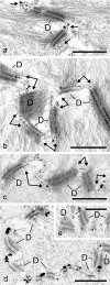
Immunoelectron microscopy of frozen bovine tongue mucosa epithelium, treated with Abs to protein PERP (ac), to occludin (d), or to claudin-4 (e) and reacted with immunogold and a tertiary enhancement silver reaction. Note that the silver-enhanced antigen-gold grains decorating PERP are predominantly located in the regions between the desmosomes (D), i.e., in the interdesmosomal space (arrows in a–c), not infrequently in direct contact with desmosomal margins. In such interdesmosomal regions of stratified epithelia, various proteins typical for tight junctions (TJs) and for adherens junctions (AJs) are also localized (tessellate junctions). Bars 500 nm
Fig. 5
Fig. 5
Confocal laser-scanning double-label immunofluorescence microscopy of cryostat sections through snap-frozen bovine liver after reactions with guinea pig pAbs to desmoplakin (green) and murine mAbs (mAb 8.2.9) against protein PERP (red). Note the typical arrays of dot-like desmosomes (green) along the bile canalicular plasma membrane structures of the hepatocytes (a) and the extended PERP-positive reactions (red) near some of these desmosomes and along the canalicular surface membrane (a’, a’’). Note, in particular, the difference between the distinct dotted (green) desmosomal structures and the continuous (b, c) or interrupted (d) canalicular plasma membrane reaction of PERP Abs (red). Bars 20 μm (a), 5 μm (b–d)
Fig. 6
Fig. 6
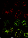
Confocal laser-scanning double-label immunofluorescence microscopy of a cryostat cross-section through snap-frozen bovine intestinal epithelium, after 10 min treatment with acetone and 5 min with buffer containing 0.2% Triton X-100, followed by a reaction with murine mAbs (26.2.22) against protein PERP (a red) and guinea pig pAbs against desmoplakin (a’, green; only the hybrid double-color picture is shown here in which the coincidence of both colors appears in yellow). Note the enrichment of both desmosomes and PERP-containing structures in the subapical cell-cell junction-containing region (red in a, yellow in a’') but also that some of the desmosomes at the basolateral sides appear as yellow dots indicative of the close vicinity of the two proteins, whereas many strictly green and a few strictly red punctate structures can also be recognized (L lumen). Bar 20 μm
Fig. 7
Fig. 7
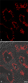
Fixation-dependent appearance of protein PERP in bovine intestinal epithelium as seen after 8 min of fixation with buffer containing 2% formaldehyde, followed by a short washing step and a 4-min treatment with buffer containing 0.1% saponin and then reaction with mAb (26.3.30) to PERP and secondary antibodies (a’ is a shown on a phase contrast background). Note that prominent PERP staining is seen along the lateral membranes and that most of the staining appears in continuous stripes, whereas individual dots and subapical zonula-like rings are rarely present (L lumen). Bar 20 μm
Fig. 8
Fig. 8
Confocal laser-scanning double-label immunofluorescence microscopy of a cryostat section through snap-frozen porcine (a, b) or human (c-c’’) lung tissue, followed by treatment with mAbs (a mAb 26.2.22; b mAb 26.3.30; c-c’’ mAb 8.2.9) against protein PERP (red) and guinea pig pAbs against desmoplakin (green). Note the striking enrichment of PERP-containing structures in a subapical zonula-like region of the pulmonary epithelium, which, in some places, is clearly resolved from the adjacent desmosomes, thus appearing as a red-stained subapical ring-band (many locations in a’, b). While the bronchial cilia (top in a, a’, shown on a phase contrast background) are negative, most of the more basolaterally located cell-cell contact regions show thin but distinct, zonula-like, red-stained PERP-enriched structures (see also human bronchial epithelium in c–c’’). In contrast, desmosomes without any PERP reaction are enriched along the lateral cell-cell contact regions (green). Tissue section samples were solubilized and fixed by a 10-min incubation in acetone and a 5-min treatment with buffer containing 0.2% Triton X-100. Bars 20 μm
Fig. 9
Fig. 9
Confocal laser-scanning double-label immunofluorescence microscopy of an obliquely horizontal cryostat section through snap-frozen bovine bladder epithelium (urothelium) after reaction with murine PERP mAb 26.2.22 (a, red) and rabbit β-catenin pAbs (a’, green). The double-label fluorescence micrograph is presented as a’’, while the double-label picture on a phase contrast background is seen in a’’’. Protein PERP is only seen at the basolateral region of the uppermost cell layer, the so-called umbrella cells, whereas the β-catenin-rich zonulae adhaerentes represent the most apical junction structure in the form of a typical zonula adhaerens (note the long green structures). The more basal layers of much smaller cells are characterized by the presence of only β-catenin-rich (green) cell-cell contacts. Such immunolocalization allows the distinction of an outer, apparently PERP-free, junctional region, a slightly more basal region containing protein PERP and the more basally located cells lacking PERP. Bar 20 μm
Fig. 10
Fig. 10
Higher magnification of a vertical section through the most apical region of umbrella cells in a preparation similar to that in Fig. 9 but stained with mAbs to PERP (red) and rabbit pAbs to β-catenin (green). The urothelium is collapsed so that two surface regions face each other (bracket in a’ residual luminal space). a Double-label immunofluorescence result only. a’ Same area on a phase contrast background. b The double-label appearance of this extended region formed by umbrella cells is again shown on a phase-contrast background in b’. c Higher magnification of b allows the demonstration of the individual cell surface reactions in detail: the most apical reaction is that of β-catenin (green) in the zonulae adhaerentes and is clearly distinguishable from the densely aggregated punctate PERP pattern (red) along the lateral membranes of this cell layer. Bars 20 μm
Fig. 11
Fig. 11
Confocal laser-scanning double-label immunofluorescence microscopy of cryostat sections through frozen bovine heart tissue, prepared with the formaldehyde-saponin procedure (see Fig. 7 and Material and methods) and immunostained with guinea pig pAbs to PERP protein (a’, green) in comparison with murine mAbs (red) to desmoplakin (a, a’’) or β-catenin (b), whereas in c, immunostaining with rabbit pAbs against β-catenin (green) is compared with immunostaining with mouse PERP mAbs (red). Note, in all micrographs, the near-complete co-immunostaining (yellow) of protein PERP with both desmoplakin and β-catenin in the composite junctions of the intercalated disks. Note also, in c, the exclusive β-catenin immunostaining of the zonulae adhaerentes (green) in the arteriole (A) in comparison with the yellow merged immunostaining of the composite junctions (L lumen). Bars 50 μm

Similar articles

Cited by

References

    1. Akat K, Bleck CKE, Lee Y-MA, Haselmann-Weiss U, Kartenbeck J. Characterization of a novel type of adherens junction in meningiomas and the derived cell line HBL-52. Cell Tissue Res. 2008;331:401–412. doi: 10.1007/s00441-007-0512-5. - DOI - PubMed
    1. Attardi LD, Reczek EE, Cosmas C, Demicco EG, McCurrach ME, Lowe SW, Jacks T. PERP, an apoptosis-associated target of p53, is a novel member of the PMP-22/gas3 family. Genes Dev. 2000;14:704–718. - PMC - PubMed
    1. Barth M, Schumacher H, Kuhn C, Akhyari P, Lichtenberg A, Franke WW. Cordial connections: molecular ensembles and structures of adhering junctions connecting interstitial cells of cardiac valves in situ and in cell culture. Cell Tissue Res. 2009;337:63–77. doi: 10.1007/s00441-009-0806-x. - DOI - PubMed
    1. Barth M, Rickelt S, Noffz E, Winter-Simanowski S, Niemann H, Akhyari P, Lichtenberg A, Franke WW. The adhering junctions of valvular interstitial cells: molecular composition in fetal and adult hearts and the comings and goings of plakophilin-2 in situ, in cell culture and upon re-association with scaffolds. Cell Tissue Res. 2012;348:295–307. doi: 10.1007/s00441-011-1315-2. - DOI - PubMed
    1. Beaudry VG, Pathak N, Koster MI, Attardi LD. Differential PERP regulation by TP63 mutants provides insight into AEC pathogenesis. Am J Med Genet. 2009;149A:1952–1957. doi: 10.1002/ajmg.a.32760. - DOI - PMC - PubMed

Publication types

MeSH terms