. 2013 Dec;9(4):687-93.
doi: 10.1007/s11302-013-9365-4. Epub 2013 May 22.

Receptor-independent effects of 2'(3')-O-(4-benzoylbenzoyl)ATP triethylammonium salt on cytosolic pH

Affiliations

Receptor-independent effects of 2'(3')-O-(4-benzoylbenzoyl)ATP triethylammonium salt on cytosolic pH

Juan Pablo Reyes et al. Purinergic Signal. 2013 Dec.

Abstract

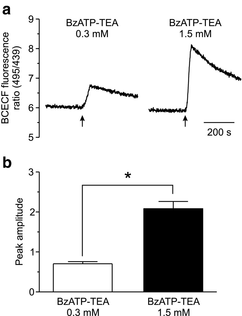
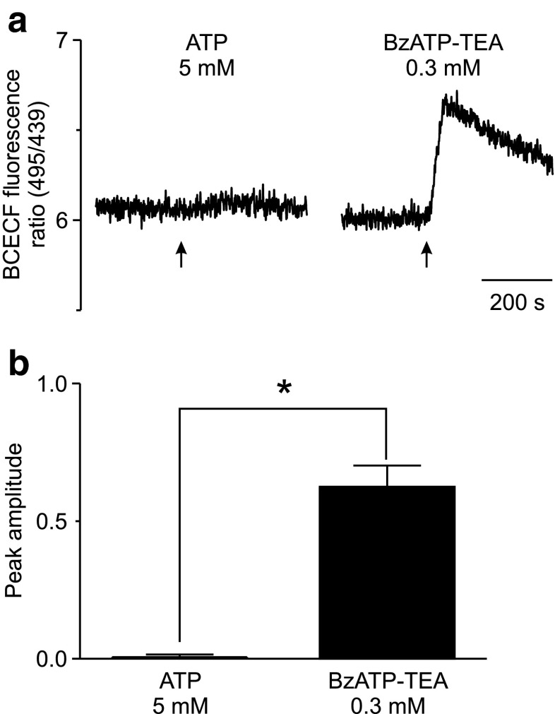
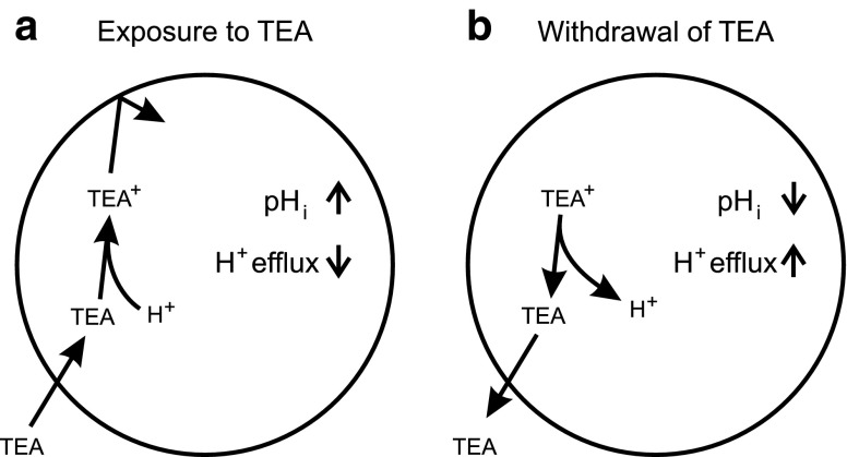
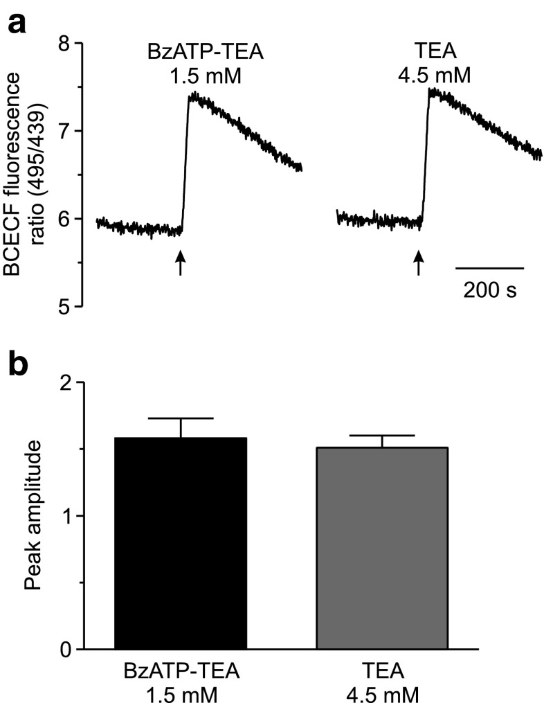
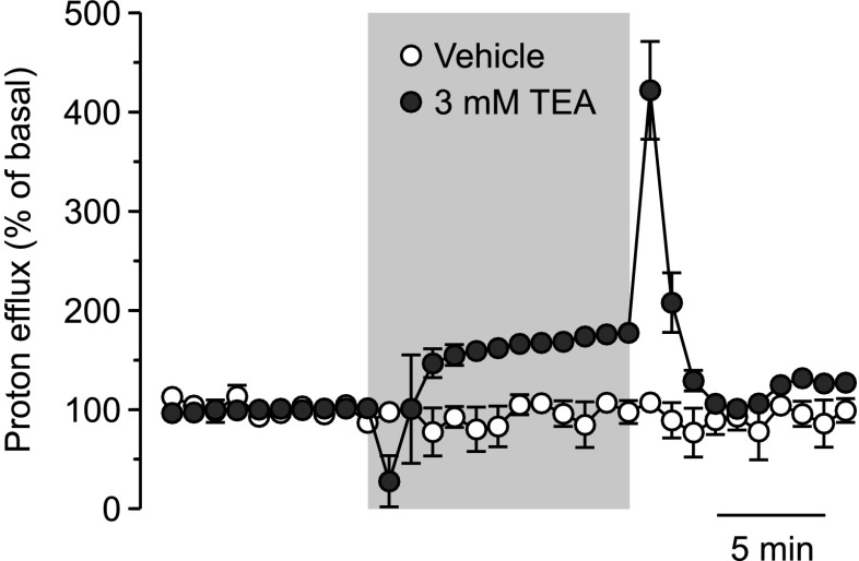
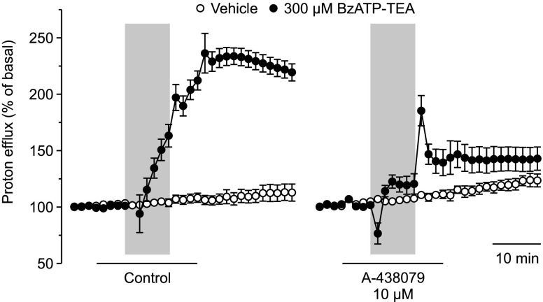
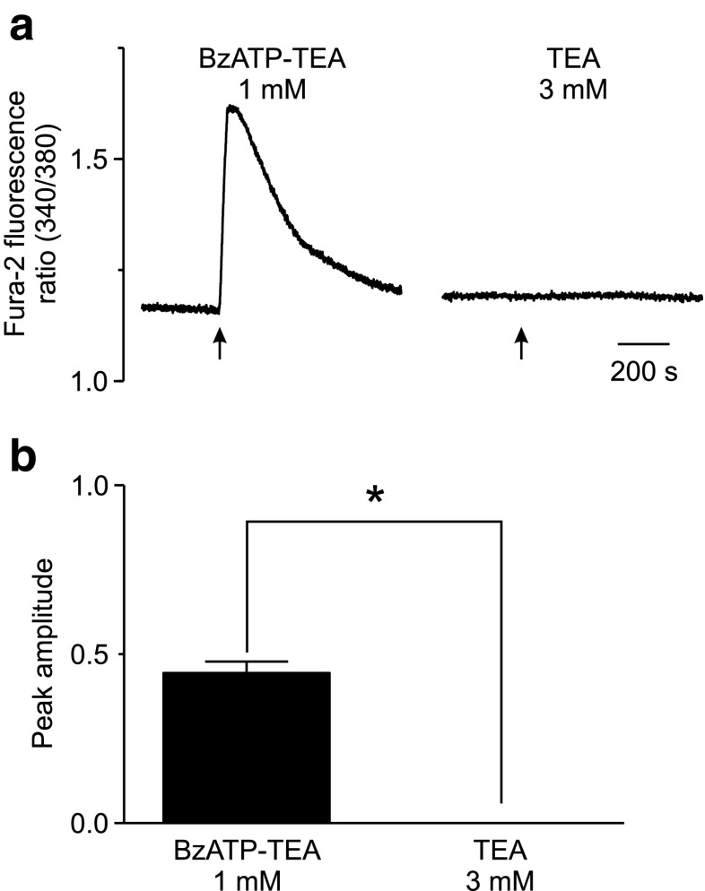
The effect of the relatively potent P2X7 receptor agonist 2'(3')-O-(4-benzoylbenzoyl)adenosine 5'-triphosphate triethylammonium salt (BzATP-TEA) on cytosolic pH (pHi) was studied using MC3T3-E1 osteoblast-like cells, which endogenously express P2X7 receptors. pHi was measured fluorimetrically using the pH-sensitive dye 2',7'-bis(2-carboxyethyl)-5(6)-carboxyfluorescein. BzATP-TEA (0.3-1.5 mM) elicited fast-onset alkalinization responses. In contrast, adenosine 5'-triphosphate disodium salt (5 mM) failed to reproduce the BzATP-TEA-induced responses, indicating a P2 receptor-independent mechanism. We speculated that triethylamine, which is present in solutions of BzATP-TEA, permeates the plasma membrane, and is protonated intracellularly, leading to an increase in pHi. Consistent with this hypothesis, triethylammonium (TEA) chloride mimicked the effects of BzATP-TEA on pHi. Moreover, measurements using a Cytosensor microphysiometer revealed that TEA chloride transiently suppressed proton efflux from cells, whereas washout of TEA transiently enhanced proton efflux. BzATP-TEA also elicited a sustained increase in proton efflux that was blocked specifically by the P2X7 antagonist A-438079. Taken together, we conclude that BzATP-TEA-induced alkalinization is unrelated to P2X7 activation, but is due to the presence of TEA. This effect may confound assessment of the outcomes of P2X7 activation by BzATP-TEA in other systems. Thus, control experiments using TEA chloride are recommended to distinguish between receptor-mediated and nonspecific effects of this widely used agonist. We performed such a control and confirmed that BzATP-TEA, but not TEA chloride, caused the elevation of cytosolic free Ca(2+) in MC3T3-E1 cells, ruling out the possibility that receptor-independent effects on pHi underlie BzATP-TEA-induced Ca(2+) signaling.


Figures

Fig. 1
Fig. 1
BzATP-TEA induces alkalinization of the cytosol. MC3T3-E1 cells were loaded with the pH-sensitive fluorescent dye BCECF and suspended in nominally Na+-free HEPES buffer in a fluorometric cuvette with continuous stirring. Changes in pHi were monitored by fluorescence spectrophotometry, with alternating excitation at 495 and 439 nm and emission at 535 nm. The ratio of emission intensities at 495/439 nm excitation provides a measure of pHi, with increasing values reflecting cytosolic alkalinization. a Where indicated by the arrows, 0.3 or 1.5 mM BzATP-TEA was added to the cuvette. Traces are representative responses. b Changes in pHi were quantified as the peak amplitude of the response above baseline (baseline values were comparable among preparations). *p < 0.05, significant difference between responses to the two BzATP-TEA concentrations. Data are presented as the means ± SEM (n = 5 or 6 independent preparations for 0.3 and 1.5 mM BzATP-TEA, respectively)
Fig. 2
Fig. 2
Cytosolic alkalinization induced by BzATP-TEA is independent of P2X7 receptor activation. MC3T3-E1 cells were loaded with BCECF, suspended in Na+-free HEPES buffer, and changes in pHi were monitored by fluorescence spectrophotometry. a Where indicated by the arrows, ATP disodium salt (5 mM) or BzATP-TEA (0.3 mM) was added to the cuvette. Traces are representative responses. b Changes in pHi were quantified as the peak amplitude of the response above baseline. *p < 0.05, significant difference between responses to 5 mM ATP and 0.3 mM BzATP-TEA. Data are presented as the means ± SEM (n = 4 independent preparations for both ATP and BzATP-TEA)
Fig. 3
Fig. 3
Schematic illustrating permeation and protonation of the weak base triethylamine (TEA). a When in the extracellular fluid, protonated TEA+ is in equilibrium with uncharged TEA, which can permeate the plasma membrane. Once in the cytosol, TEA becomes protonated, increasing pHi. An increase in pHi leads to a decrease in efflux of protons and proton equivalents via Na+/H+ exchange and other pathways. b Upon withdrawal of TEA from the extracellular fluid, uncharged TEA leaves the cell. Protons then dissociate from cytosolic TEA+, decreasing pHi. A decrease in pHi leads to the activation of proton efflux pathways such as Na+/H+ exchange. In both cases, the change in proton efflux is transient, as it occurs only until pHi is restored to its resting level
Fig. 4
Fig. 4
TEA chloride elicits changes in pHi similar to that induced by BzATP-TEA. MC3T3-E1 cells were loaded with BCECF, suspended in Na+-free HEPES buffer, and changes in pHi were monitored by fluorescence spectrophotometry. a Where indicated by the arrows, BzATP-TEA (1.5 mM) or TEA chloride (4.5 mM) was added to the cuvette. Traces are representative responses. b Changes in pHi were quantified as the peak amplitude of the response above baseline. Data are presented as the means ± SEM, p > 0.05 (n = 5 independent preparations for both BzATP-TEA and TEA)
Fig. 5
Fig. 5
TEA chloride elicits transient changes in proton efflux. MC3T3-E1 cells were cultured on porous polycarbonate membranes, and proton efflux was monitored by microphysiometry. Cells were superfused with standard medium, and at 1 min intervals, superfusion was interrupted for 30 s to measure acidification rate. Net efflux of proton equivalents (proton efflux) was calculated from the acidification rate and expressed as a percentage of basal proton efflux. Where indicated by the shaded rectangle, MC3T3-E1 cells were superfused with TEA chloride (3 mM) (closed symbols) or vehicle (open symbols) in standard medium for 12 min. Exposure to TEA promptly induced a transient decrease in proton efflux from 100 to 28 ± 26 % of basal levels. On the other hand, withdrawal of TEA induced a large transient increase in proton efflux to values of 422 ± 49 %. In contrast to the dramatic response to TEA, superfusion with vehicle had no effect. Data are presented as the means ± SEM (n = 11 samples from three independent preparations)
Fig. 6
Fig. 6
BzATP-TEA causes a sustained P2X7-dependent increase in proton efflux. MC3T3-E1 cells were cultured on porous polycarbonate membranes and superfused with standard medium. Superfusion was interrupted for 30 s at 1.5 min intervals to measure acidification rate. Where indicated by the horizontal bar beneath the graph, parallel samples were superfused with solution containing either the P2X7 antagonist A-438079 (10 μM) or control (H2O). After 6 min, cells were stimulated with either BzATP-TEA (300 μM) (closed symbols) or vehicle (open symbols) where indicated by the shaded rectangle in the continued presence of the appropriate medium. In control samples, BzATP-TEA caused a large sustained increase in proton efflux that persisted for at least 60 min. In contrast, no sustained phase was apparent in cultures treated with BzATP-TEA in the presence of A-438079. However, exposure to BzATP-TEA in the presence of A-438079 still induced a transient decrease in proton efflux and withdrawal of BzATP-TEA elicited a large transient increase in proton efflux. Note that the pattern of these changes in proton efflux in the presence of the P2X7 receptor antagonist is similar to that observed in response to TEA chloride alone (compare right panel of Fig. 6 to Fig. 5). Data are presented as the means ± SEM (n = 5–7 samples from three to four independent preparations)
Fig. 7
Fig. 7
BzATP-TEA, but not TEA chloride, induces the elevation of [Ca2+]i. MC3T3-E1 cells were loaded with the Ca2+-sensitive fluorescent dye fura-2 and suspended in Na+-free, Ca2+-containing HEPES buffer in a fluorometric cuvette. Changes in [Ca2+]i were monitored by fluorescence spectrophotometry, with alternating excitation wavelengths of 340 and 380 nm and emission at 510 nm. The ratio of emission intensities at 340/380 nm excitation provides a measure of [Ca2+]i. a Where indicated by the arrows, BzATP-TEA (1 mM) or TEA chloride (3 mM) was added to the cuvette. Traces are representative responses. b Changes in [Ca2+]i were quantified as the peak amplitude of the response above baseline. *p < 0.05, significant difference between responses to BzATP-TEA and TEA chloride. Data are presented as the means ± SEM (n = 5 and 7 independent preparations for BzATP-TEA and TEA chloride, respectively)
Fig. 8
Fig. 8
BzATP elicits a sustained P2X7-dependent elevation of [Ca2+]i. MC3T3-E1 cells were loaded with the Ca2+-sensitive fluorescent dye indo-1 and suspended in Ca2+-containing HEPES buffer in a fluorometric cuvette. Changes in [Ca2+]i were monitored by fluorescence spectrophotometry, with a 355-nm excitation wavelength, and emission recorded at 405 and 485 nm. The ratio of emission intensities at 405/485 nm provides a measure of [Ca2+]i. a BzATP-TEA (300 μM) caused a rapid rise of [Ca2+]i, with an initial peak followed by a sustained phase. The P2X7 antagonist A-438079 (10 μM) specifically suppressed the sustained phase, without affecting the initial transient elevation of [Ca2+]i. Traces are representative responses from four independent preparations. b Changes in [Ca2+]i were quantified as the peak amplitude of the response above baseline. c Changes in [Ca2+]i were also quantified as the amplitude of the sustained phase of the response above baseline, determined at 10 min following the addition of BzATP-TEA. *p < 0.05, significant effect of A-438079. Data are presented as the means ± SEM (n = 4 independent preparations)

Similar articles

Cited by

References

    1. Burnstock G. Physiology and pathophysiology of purinergic neurotransmission. Physiol Rev. 2007;87:659–797. doi: 10.1152/physrev.00043.2006. - DOI - PubMed
    1. North RA. Molecular physiology of P2X receptors. Physiol Rev. 2002;82:1013–1067. - PubMed
    1. Bianchi BR, Lynch KJ, Touma E, Niforatos W, Burgard EC, Alexander KM, Park HS, Yu H, Metzger R, Kowaluk E, Jarvis MF, van Biesen T. Pharmacological characterization of recombinant human and rat P2X receptor subtypes. Eur J Pharmacol. 1999;376:127–138. doi: 10.1016/S0014-2999(99)00350-7. - DOI - PubMed
    1. Jarvis MF, Khakh BS. ATP-gated P2X cation-channels. Neuropharmacology. 2009;56:208–215. doi: 10.1016/j.neuropharm.2008.06.067. - DOI - PubMed
    1. Wildman SS, Unwin RJ, King BF. Extended pharmacological profiles of rat P2Y2 and rat P2Y4 receptors and their sensitivity to extracellular H+ and Zn2+ ions. Br J Pharmacol. 2003;140:1177–1186. doi: 10.1038/sj.bjp.0705544. - DOI - PMC - PubMed

Publication types

MeSH terms

Substances

LinkOut - more resources