. 2013 May 27;201(5):741-57.
doi: 10.1083/jcb.201212094. Epub 2013 May 20.

Diet controls Drosophila follicle stem cell proliferation via Hedgehog sequestration and release

Affiliations

Diet controls Drosophila follicle stem cell proliferation via Hedgehog sequestration and release

Tiffiney R Hartman et al. J Cell Biol. .

Abstract

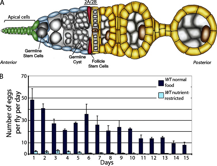
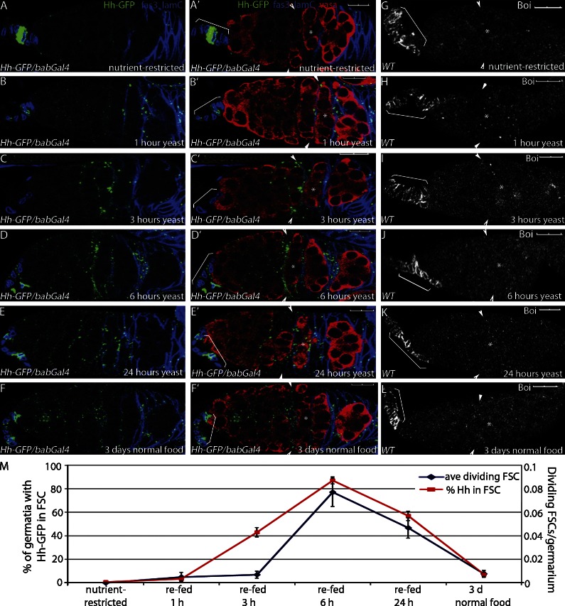
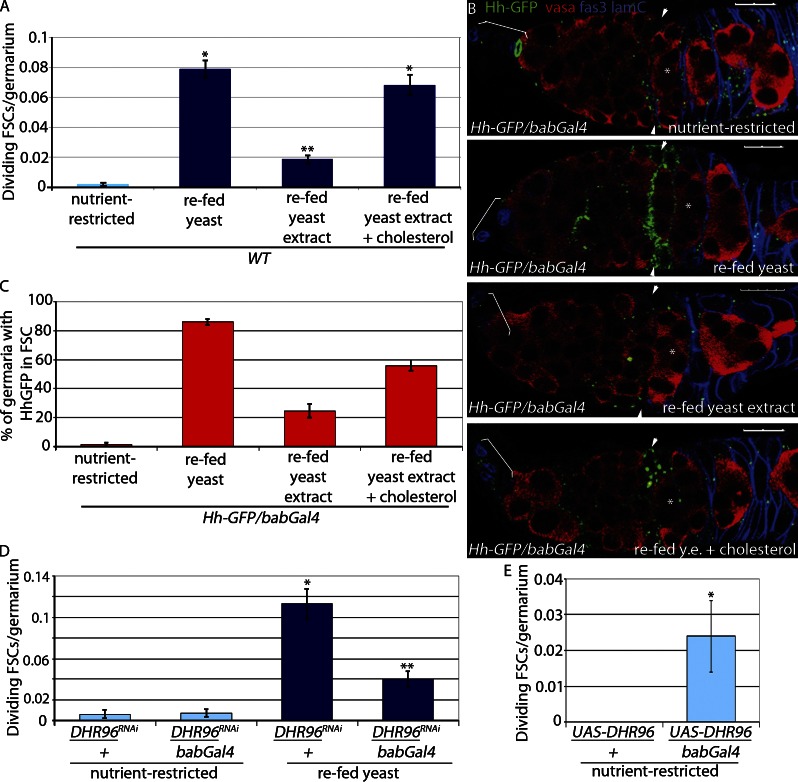
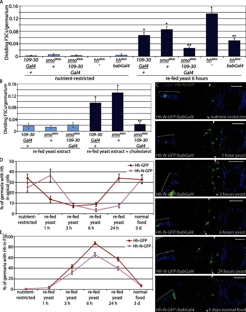
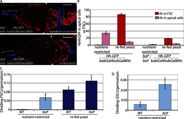
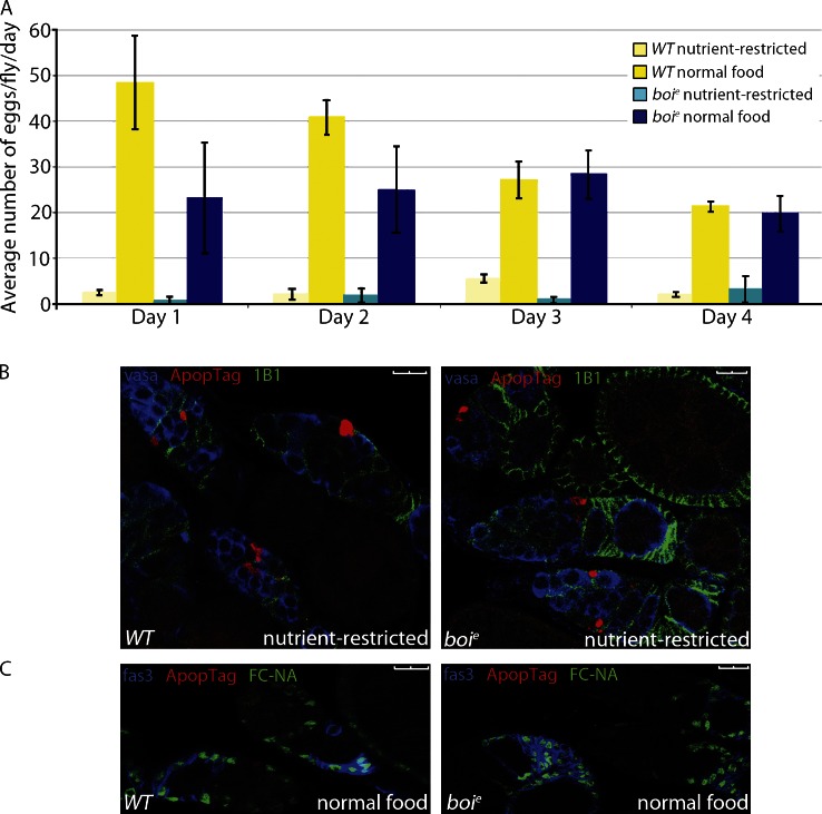
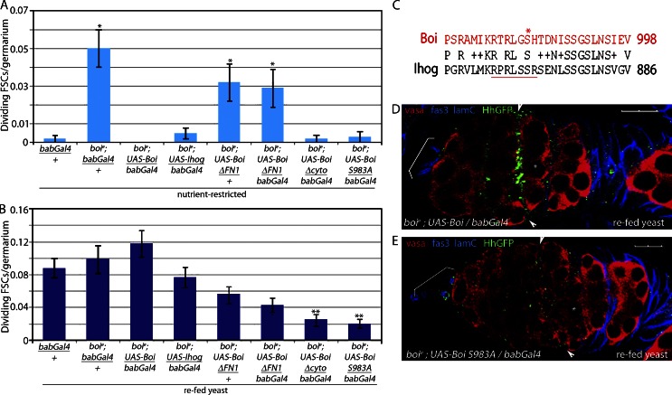
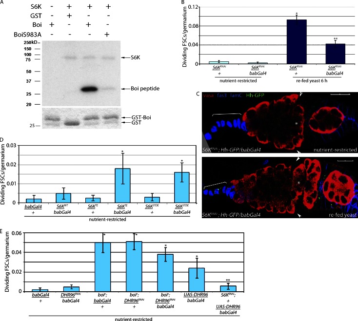
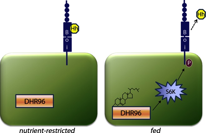
A healthy diet improves adult stem cell function and delays diseases such as cancer, heart disease, and neurodegeneration. Defining molecular mechanisms by which nutrients dictate stem cell behavior is a key step toward understanding the role of diet in tissue homeostasis. In this paper, we elucidate the mechanism by which dietary cholesterol controls epithelial follicle stem cell (FSC) proliferation in the fly ovary. In nutrient-restricted flies, the transmembrane protein Boi sequesters Hedgehog (Hh) ligand at the surface of Hh-producing cells within the ovary, limiting FSC proliferation. Upon feeding, dietary cholesterol stimulates S6 kinase-mediated phosphorylation of the Boi cytoplasmic domain, triggering Hh release and FSC proliferation. This mechanism enables a rapid, tissue-specific response to nutritional changes, tailoring stem cell divisions and egg production to environmental conditions sufficient for progeny survival. If conserved in other systems, this mechanism will likely have important implications for studies on molecular control of stem cell function, in which the benefits of low calorie and low cholesterol diets are beginning to emerge.


Figures

Figure 1.
Figure 1.
Nutrient restriction inhibits egg production in flies. (A) Schematic of early oogenesis. GSCs (gray) contact a cellular niche composed of apical cells (green) found at the anterior of the germarium. FSCs (red) reside three to five cell diameters posterior to apical cells and generate daughter cells (yellow) that form an epithelial layer around 16-cell germline cysts (gray), producing follicles called egg chambers that generate mature eggs in 7 d. The asterisk indicates flattened germline cyst at the region 2A/2B border. (B) Eggs laid/fly/day were scored daily for nutrient-restricted or fed WT flies. Error bars represent standard deviations.
Figure 2.
Figure 2.
Refeeding nutrient-restricted flies stimulates Hh release and FSC proliferation. (A–G) Nutrient-restricted WT flies expressing Hh-GFP in the apical cells (Hh-GFP/bab-Gal4) were refed yeast for the times indicated. (A–F) Follicle cells (Fas3) and apical cells (Lamin C [lamC]) are both labeled in blue to enable mapping of Hh localization. Hh-GFP is also shown. (A′–F′) Same images as A–F with germ cells shown (Vasa). (G–L) Boi localization in apical cells is indicated with brackets in nutrient-restricted (G) and refed (H–L) flies. (A–L) Asterisks mark the flattened germline cyst at the region 2A/2B border. Arrowheads indicate FSCs. Brackets indicate apical cells. Bars, 10 µM. (M) Mean numbers of dividing FSCs and Hh-GFP localization in the FSCs are shown. Error bars represent SEs.
Figure 3.
Figure 3.
Cholesterol triggers Hh release. (A) Nutrient-restricted WT flies were refed for 6 h with yeast or yeast extract (y.e.) ± 0.2 mg/g cholesterol or ethanol vehicle control. Mean numbers of dividing FSCs (PH3+) per germarium are shown. *, P < 0.00001 versus nutrient-restricted WT. **, P < 0.00001 versus WT refed yeast (n = 1,320–2,113; Table 1). (B and C) Nutrient-restricted WT flies expressing Hh-GFP in apical cells (Hh-GFP/bab-Gal4) were refed for 6 h with yeast or yeast extract ± 0.2 mg/g cholesterol or ethanol vehicle control and stained for Hh-GFP. (B) Follicle cells and apical cells are both labeled in blue (Fas3 and Lamin C [lamC], respectively), and germ cells are labeled red (Vasa). Asterisks indicate flattened germline cyst at the region 2A/2B border. Arrowheads indicate FSCs. Brackets indicate apical cells. Bars, 10 µM. (C) The percentage of germaria with Hh-GFP localized to FSCs was scored (n = 85–195). (D) Nutrient-restricted DHR96RNAi/bab-Gal4 and DHR96RNAi/+ flies were refed yeast for 6 h. Mean numbers of dividing FSCs (PH3+) per germarium are shown. *, P < 0.00001 versus nutrient-restricted DHR96 RNAi/+. **, P < 0.00001 versus refed DHR96 RNAi/+ (n = 779–1,194; Table 1). (E) UAS-DHR96/bab-Gal4 and UAS-DHR96/+ flies were nutrient restricted for 3 d. Mean numbers of dividing FSCs (PH3+) per germarium are shown. *, P < 0.005 versus nutrient-restricted UAS-DHR96/+ (n = 184 and 251; Table 1). Error bars represent SEs.
Figure 4.
Figure 4.
Cholesterol-mediated Hh release is sufficient for stem cell proliferation. (A and B) Nutrient-restricted smoRNAi/109-30-Gal4, smoRNAi/+, and 109-30-Gal4/+ flies were refed yeast (A) or yeast extract ± 0.2 mg/g cholesterol (B) for 6 h. Mean numbers of dividing FSCs (PH3+) per germarium are shown. *, P < 0.00001 versus nutrient-restricted 109-30-Gal4/+. **, P < 0.00001 versus smo RNAi/+ refed yeast (n = 366–1,294 [A] and n = 217–514 [B]; Table 1). (A) Nutrient-restricted hhRNAi/bab-Gal4 and hhRNAi/+ flies were refed yeast paste for 6 h. *, P < 0.00001 versus nutrient-restricted bab-Gal4/+. **, P < 0.00001 versus hhRNAi/+ refed yeast (n = 278–699; Table 1). Error bars represent SEs. (C–E) Active Hh-N–GFP is not modified by cholesterol but is retained in the apical cells in nutrient-restricted flies and released in refed flies. Asterisks indicate flattened germline cyst at the region 2A/2B border. Arrowheads indicate FSCs. Brackets indicate apical cells. Bars, 10 µM. Hh-N–GFP is lost from the apical cells (C and D) and accumulates in FSCs (C and E) at similar levels observed with cholesterol-modified Hh-GFP-C. Error bars represent SEs.
Figure 5.
Figure 5.
FSCs proliferate in nutrient-restricted boi mutant flies. (A and B) WT and boie mutant flies expressing Hh-GFP in apical cells (Hh-GFP/bab-Gal4tubGal80ts and boie; Hh-GFP/bab-Gal4tubGal80ts) were nutrient restricted or refed yeast and stained for Hh-GFP. Follicle cells (Fas3) and apical cells (Lamin C [LamC]) are both labeled in blue to enable mapping of Hh-GFP localization, and germ cells are labeled red (Vasa; A). Asterisks indicate flattened germline cyst at the region 2A/2B border. Arrowheads indicate FSCs. Brackets indicate apical cells. Bars, 10 µM. (B) Percentage of germaria with Hh-GFP localized to apical cells or FSCs was quantified; n = 146–654. (C and D) Nutrient-restricted WT and boie mutant flies were refed yeast for 6 h. Mean numbers of dividing FSCs (*, P < 0.00001 vs. nutrient-restricted WT [n = 370–2,113; Table 1]; C) or GSCs (*, P < 0.0007 vs. nutrient-restricted WT [n = 427 and 527]; D) per germarium are shown. Error bars represent SEs.
Figure 6.
Figure 6.
Nutrient restriction blocks egg production in Boi mutant flies. (A) WT and boie mutant flies were nutrient restricted or fed yeast, and eggs laid per fly were scored daily. Error bars represent standard deviations. (B) Germaria from nutrient-restricted WT or boie mutant females were immunostained for follicle cells (1B1), germ cells (Vasa), and apoptosis (ApopTag). (C) WT or boie mutant flies on normal food lacked apoptosis (nuclei [FC-NA]), follicle cells (Fas3), and apoptosis (ApopTag). Bars, 10 µM.
Figure 7.
Figure 7.
Hh release is controlled by phosphorylation of a conserved serine in the Boi cytoplasmic domain. (A and B) boie mutant flies rescued with the indicated form of Boi were nutrient restricted for 3 d (A) and refed yeast for 6 h (B). (A) Forms of Boi with the ability to bind Hh rescued FSC overproliferation in nutrient-restricted boie mutants, consistent with their ability to sequester Hh. *, P < 0.00001 versus nutrient-restricted bab-Gal4/+ (n = 243–753; Table 1). (B) Forms of Boi lacking key regions of the cytoplasmic domain failed to fully rescue FSC proliferation in fed flies, consistent with a failure to release Hh. **, P < 0.00001 versus boie; UAS-boi/bab-Gal4 refed yeast (n = 314–962; Table 1). Error bars represent SEs. (C) Conserved region in the cytoplasmic domains of Boi and Ihog. S983 (asterisk) and the S6K consensus site (RxRxxSx, underlined) are indicated. The alignment was performed using NCBI BLAST (Basic Local Alignment Search Tool). Letters that are identical between two sequences are reported. Those that have positive scores in the scoring matrix are displayed with a plus sign. Gaps and nonpositive scores are blank. (D and E) boie mutant flies expressing HhGFP and WT Boi (boie; HhGFP/+; UAS-Boi/bab-Gal4) accumulate Hh-GFP in FSCs after refeeding (D), whereas flies expressing BoiS983 (boie; HhGFP/+; UAS-boi983A/bab-Gal4) do not accumulate HhGFP in FSCs after refeeding (E). Hh-GFP, follicle cells (Fas3), apical cells (Lamin C [lamC]), and germ cells (Vasa) are shown. Asterisks indicate flattened germline cyst at the region 2A/2B border. Arrowheads indicate FSCs. Brackets indicate apical cells. Bars, 10 µM.
Figure 8.
Figure 8.
Stimulation of FSC proliferation after refeeding is S6K dependent. (A) In the presence of human S6K, BoiS983 is phosphorylated (top, third lane, bottom band). Mutation of S983 to A abrogates phosphorylation (top, forth lane), indicating that S983 is the primary site of phosphorylation. Autophosphorylation of S6K also is observed (top, second to forth lanes, top band). No signal is observed in the absence of S6K (top, first lane) or when GST alone is used as a substrate (top, second lane). (bottom) Coomassie-stained gel showing levels of GST (second lane) or GST-Boi (first, third, and forth lanes) used in the assay. (B) Nutrient-restricted S6KRNAi/bab-Gal4 and S6KRNAi/+ flies were refed yeast for 6 h. Mean numbers of dividing FSCs (PH3+) per germarium are shown. *, P < 0.00001 versus nutrient-restricted S6KRNAi/+. **, P < 0.00001 versus refed S6KRNAi (n = 493–1,037; Table 1). (C) S6KRNAi flies expressing HhGFP in apical cells (S6KRNAi; HhGFP/bab-Gal4) fail to accumulate Hh-GFP in FSCs after refeeding. Hh-GFP, follicle cells (Fas3), apical cells (Lamin C [lamC]), and germ cells (Vasa) are shown. Asterisks indicate flattened germline cyst at the region 2A/2B border. Arrowheads indicate FSCs. Brackets indicate apical cells. Bars, 10 µM. (D) Activated S6K (S6KTE/bab-Gal4, S6KSTDE/bab-Gal4, S6KWT/bab-Gal4, and controls S6KTE/+ and S6KSTDE/+) flies were nutrient restricted for 3 d. Mean numbers of dividing FSCs (PH3+) per germarium are shown. *, P < 0.02 versus nutrient-restricted bab-Gal4/+ (n = 277–1,132; Table 1). (E) boie; DHR96RNAi/bab-Gal4, S6KRNAi/+; UAS-DHR96/bab-Gal4, and control flies were nutrient restricted for 3 d. Mean numbers of dividing FSCs (PH3+) per germarium are shown. *, P < 0.005 versus nutrient-restricted bab-Gal4/+. **, P < 0.03 versus nutrient-restricted UAS-DHR/bab-Gal4 (n = 251–1,014; Table 1). Error bars represent SEs.
Figure 9.
Figure 9.
Cholesterol activation of DHR96 leads to S6K-dependent phosphorylation of BoiS983A, causing release of Hh from apical cells and activation of FSC proliferation. (left) Hh is sequestered by Boi in nutrient-restricted flies. Upon feeding, cholesterol binds to DHR96 and promotes phosphorylation (P) of BoiS983 via S6K activation.

References

    1. Bae G.U., Domené S., Roessler E., Schachter K., Kang J.S., Muenke M., Krauss R.S. 2011. Mutations in CDON, encoding a hedgehog receptor, result in holoprosencephaly and defective interactions with other hedgehog receptors. Am. J. Hum. Genet. 89:231–240 10.1016/j.ajhg.2011.07.001 - DOI - PMC - PubMed
    1. Barcelo H., Stewart M.J. 2002. Altering Drosophila S6 kinase activity is consistent with a role for S6 kinase in growth. Genesis. 34:83–85 10.1002/gene.10132 - DOI - PubMed
    1. Beachy P.A., Hymowitz S.G., Lazarus R.A., Leahy D.J., Siebold C. 2010. Interactions between Hedgehog proteins and their binding partners come into view. Genes Dev. 24:2001–2012 10.1101/gad.1951710 - DOI - PMC - PubMed
    1. Bettoun D.J., Buck D.W., II, Lu J., Khalifa B., Chin W.W., Nagpal S. 2002. A vitamin D receptor-Ser/Thr phosphatase-p70 S6 kinase complex and modulation of its enzymatic activities by the ligand. J. Biol. Chem. 277:24847–24850 10.1074/jbc.C200187200 - DOI - PubMed
    1. Bettoun D.J., Lu J., Khalifa B., Yee Y., Chin W.W., Nagpal S. 2004. Ligand modulates VDR-Ser/Thr protein phosphatase interaction and p70S6 kinase phosphorylation in a cell-context-dependent manner. J. Steroid Biochem. Mol. Biol. 89-90:195–198 10.1016/j.jsbmb.2004.03.087 - DOI - PubMed

Publication types

MeSH terms

LinkOut - more resources