

Longitudinal analyses of early lesions by fluorescence: an observational study
- PMID: 23690351
- PMCID: PMC3706178
- DOI: 10.1177/0022034513490167
Longitudinal analyses of early lesions by fluorescence: an observational study
Abstract
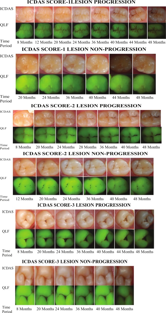
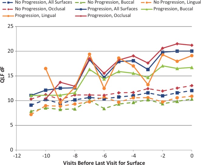
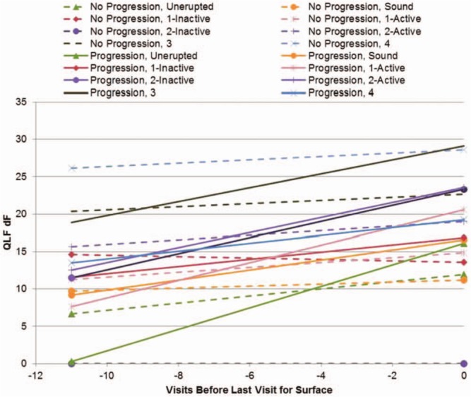
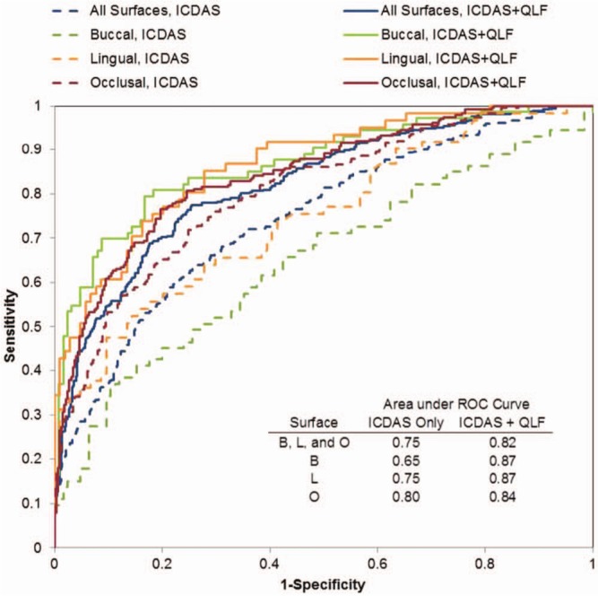
Previous caries experience correlates to future caries risk; thus, early identification of lesions has importance for risk assessment and management. In this study, we aimed to determine if Quantitative Light-induced Fluorescence (QLF) parameters--area (A [mm(2)]), fluorescence loss (F [%]), and Q [% × mm(2)]--obtained by image analyses can predict lesion progression. We secured consent from 565 children (from 5-13 years old) and their parents/guardians and examined them at baseline and regular intervals over 48 months according to the International Caries Detection Assessment System (ICDAS), yearly radiographs, and QLF. QLF images from surfaces with ICDAS 0/1/2/3/4 at baseline that progressed (N = 2,191) to cavitation (ICDAS 5/6) or fillings and surfaces that did not progress to cavitation/fillings (N = 4,141) were analyzed independently for A, F, and Q. Linear mixed-effects models were used to compare means and slopes (changes over time) between surfaces that progressed and those that did not. QLF A, F, and Q increased at a faster rate for surfaces that progressed than for surfaces that did not progress (p = .0001), regardless of type of surface or baseline ICDAS score. AUC for ICDAS ranged from 0.65 to 0.80, but adding QLF information improved AUC (0.82-0.87, p < .0005). We concluded that faster changes in QLF variables can indicate lesion progression toward cavitation and be more clinically relevant than actual QLF values.
Keywords: dental caries; early diagnosis; fluorescence imaging; prospective study; visual examination.
Conflict of interest statement
The authors declare no potential conflicts of interest with respect to the authorship and/or publication of this article.
Figures
References
-
- Ando M, van Der Veen MH, Schemehorn BR, Stookey GK. (2001). Comparative study to quantify demineralized enamel in deciduous and permanent teeth using laser- and light-induced fluorescence techniques. Caries Res 35:464-470. - PubMed
-
- Backer Dirks O. (1966). Posteruptive changes in dental enamel. J Dent Res 45:503-511.
-
- Bader JD, Perrin NA, Maupomé G, Rush WA, Rindal BD. (2008). Exploring the contributions of components of caries risk assessment guidelines. Community Dent Oral Epidemiol 36:357-362. - PubMed
-
- Diniz MB, Boldieri T, Rodrigues JA, Santos-Pinto L, Lussi A, Cordeiro RC. (2012). The performance of conventional and fluorescence-based methods for occlusal caries detection: an in vivo study with histologic validation. J Am Dent Assoc 143:339-350. - PubMed
-
- Ekstrand KR, Martignon S, Ricketts DJ, Qvist V. (2007). Detection and activity assessment of primary coronal caries lesions: a methodologic study. Oper Dent 32:225-235. - PubMed
Publication types
MeSH terms
Grants and funding
LinkOut - more resources
Full Text Sources
Other Literature Sources
Medical
