. 2013 Aug;86(1028):20130115.
doi: 10.1259/bjr.20130115. Epub 2013 May 20.

Optimised z-axis coverage at multidetector-row CT in adults suspected of acute appendicitis

Affiliations

Optimised z-axis coverage at multidetector-row CT in adults suspected of acute appendicitis

N Brassart et al. Br J Radiol. 2013 Aug.

Abstract

Objective: To compare diagnostic performances of two reduced z-axis coverages to full coverage of the abdomen and pelvis for the diagnosis of acute appendicitis and alternative diseases at unenhanced CT.

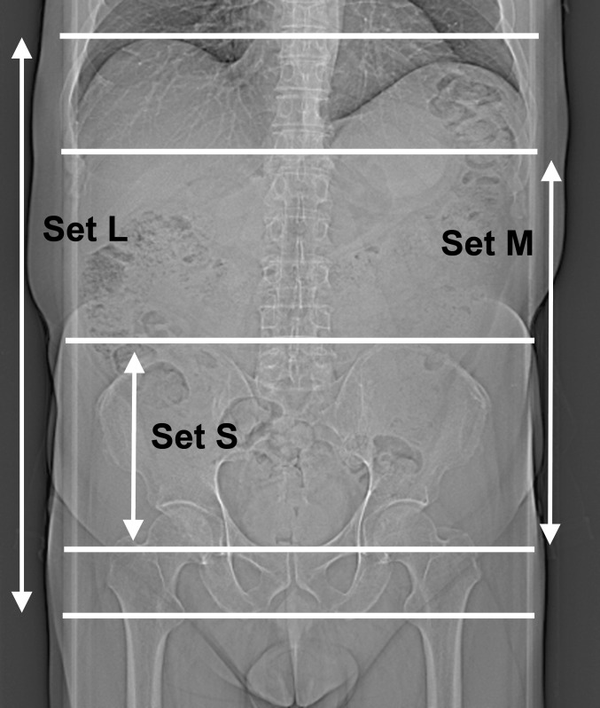
Methods: This study included 152 adults suspected of appendicitis who were enrolled in two ethical committee-approved previous prospective trials. Based on scans covering the entire abdomen and pelvis (set L), two additional sets of images were generated, each with reduced z-axis coverages: (1) from the top of the iliac crests to the pubis (set S) and (2) from the diaphragmatic crus to the pubis (set M). Two readers independently coded the visualisation of the appendix, measured its diameter and proposed a diagnosis (appendicitis or alternative). Final diagnosis was based on surgical findings or clinical follow-up. Fisher exact and McNemar tests and logistic regression were used.

Results: 46 patients had a definite diagnosis of appendicitis and 53 of alternative diseases. The frequency of appendix visualisation was lower for set S than set L for both readers (89% and 84% vs 95% and 91% by Readers A and B, respectively; p=0.021 and 0.022). The probability of giving a correct diagnosis was lower for set S (68%) than set L (78%; odds ratio, 0.611; p=0.008) for both readers, without significant difference between sets L and M (77%, p=0.771); z-axis coverage being reduced by 25% for set M.

Conclusion: Coverage from diaphragmatic crus to pubis, but not focused on pelvis only, can be recommended in adults suspected of appendicitis.

Advances in knowledge: In suspected appendicitis, CT-coverage can be reduced from diaphragmatic crus to pubis.


Figures

Figure 1
Figure 1
Frontal scout view with upper and lower boundaries of each set.
Figure 2
Figure 2
Screen capture showing a 300-mm-thick coronal multiplanar reformated (MPR) image (thick arrowhead) obtained with three-dimensional functionality of the workstation (Syngo® Multimodality Workplace; Siemens Healthcare, Forchheim, Germany) and simulating a frontal scout view. The horizontal line (thin arrow) on the thick MPR image corresponds to the upper boundary of our restricted abdominal coverage and is at the level of the axial image seen in the left lower quadrant (thick arrow).
Figure 3
Figure 3
Unenhanced CT images obtained in an 83-year-old female with definite diagnosis of acute appendicitis. Axial (a) and coronal (b) reformations of a restricted abdominal coverage show enlarged appendix (arrow) containing an appendicolith and periappendiceal fat stranding. The appendix is located above the iliac crest and was not visualised with pelvic focused coverage (set S) by both readers.
Figure 4
Figure 4
Unenhanced axial CT image obtained in a 30-year-old male with definite diagnosis of acute appendicitis showing an enlarged appendix (arrows) and periappendiceal fat stranding. The appendix is located above the iliac crest and was not visualised with pelvic focused coverage (set S) by both readers.
Figure 5
Figure 5
Unenhanced CT images obtained in a 34-year-old female with definite diagnosis of acute pyelonephritis. Axial (a) and sagittal (b) reformations of a restricted abdominal coverage show an enlarged right kidney (arrow), perinephric fat stranding and minimal dilation of the urinary tract consistent with pyelonephritis. Diagnosis was missed with pelvic focused coverage (set S) by both readers.

References

    1. Birnbaum BA, Wilson SR. Appendicitis at the millennium. Radiology 2000;215:337–48 - PubMed
    1. Wise SW, Labuski MR, Kasales CJ, Blebea JS, Meilstrup JW, Holley GP, et al. Comparative assessment of CT and sonographic techniques for appendiceal imaging. AJR Am J Roentgenol 2001;176:933–41 10.2214/ajr.176.4.1760933 - DOI - PubMed
    1. Johnson PT, Horton KM, Mahesh M, Fishman EK. Multidetector computed tomography for suspected appendicitis: multi-institutional survey of 16-MDCT data acquisition protocols and review of pertinent literature. J Comput Assist Tomogr 2006;30:758–64 - PubMed
    1. O'Malley ME, Halpern E, Mueller PR, Gazelle GS. Helical CT protocols for the abdomen and pelvis: a survey. AJR Am J Roentgenol 2000;175:109–13 10.2214/ajr.175.1.1750109 - DOI - PubMed
    1. Liao EA, Quint LE, Goodsitt MM, Francis IR, Khalatbari S, Myles JD. Extra Z-axis coverage at CT imaging resulting in excess radiation dose: frequency, degree, and contributory factors. J Comput Assist Tomogr 2011;35:50–6 10.1097/RCT.0b013e3181f5a652 - DOI - PMC - PubMed