. 2013 Jun 3;210(6):1117-24.
doi: 10.1084/jem.20121588. Epub 2013 May 20.

Retinoic acid expression associates with enhanced IL-22 production by γδ T cells and innate lymphoid cells and attenuation of intestinal inflammation

Affiliations

Retinoic acid expression associates with enhanced IL-22 production by γδ T cells and innate lymphoid cells and attenuation of intestinal inflammation

Lisa A Mielke et al. J Exp Med. .

Abstract

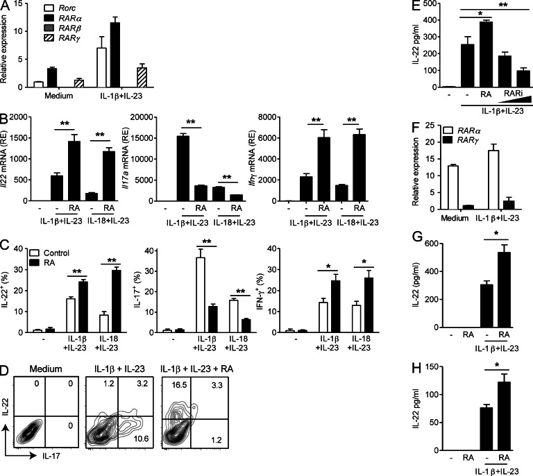
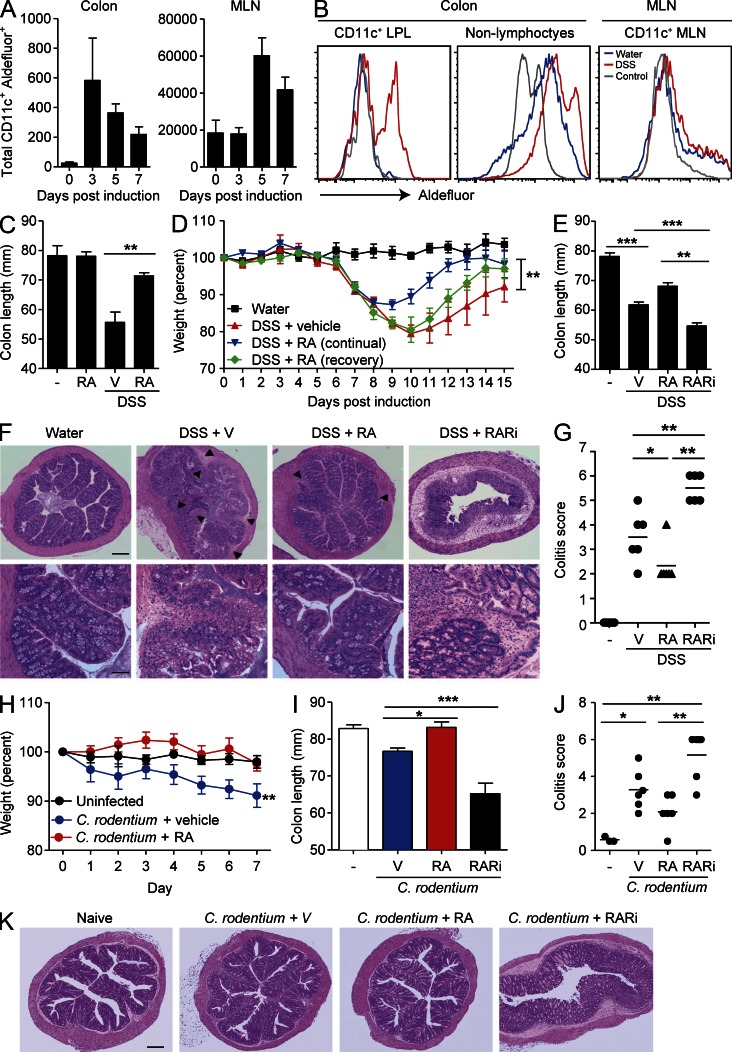
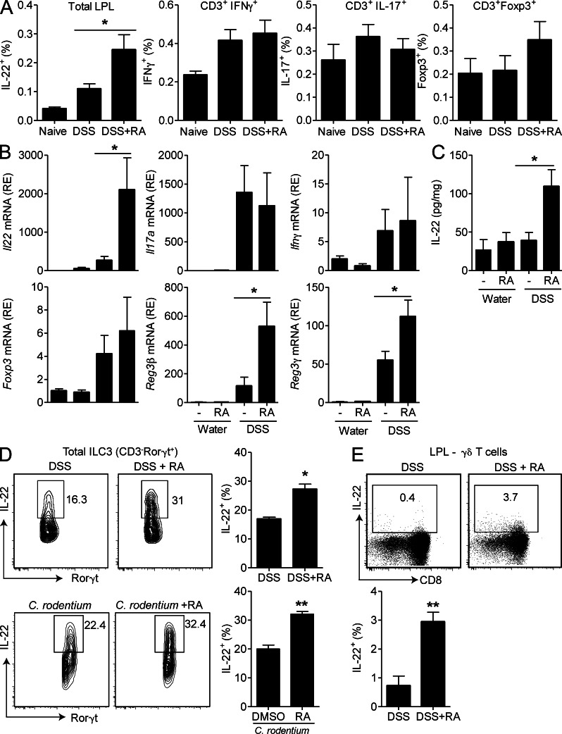
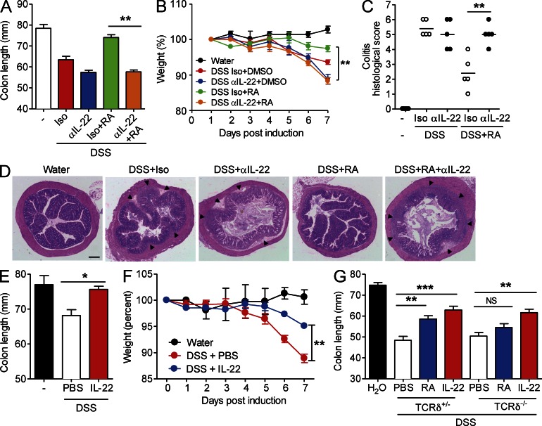
Retinoic acid (RA), a vitamin A metabolite, modulates mucosal T helper cell responses. Here we examined the role of RA in regulating IL-22 production by γδ T cells and innate lymphoid cells in intestinal inflammation. RA significantly enhanced IL-22 production by γδ T cells stimulated in vitro with IL-1β or IL-18 and IL-23. In vivo RA attenuated colon inflammation induced by dextran sodium sulfate treatment or Citrobacter rodentium infection. This was associated with a significant increase in IL-22 secretion by γδ T cells and innate lymphoid cells. In addition, RA treatment enhanced production of the IL-22-responsive antimicrobial peptides Reg3β and Reg3γ in the colon. The attenuating effects of RA on colitis were reversed by treatment with an anti-IL-22 neutralizing antibody, demonstrating that RA mediates protection by enhancing IL-22 production. To define the molecular events involved, we used chromatin immunoprecipitation assays and found that RA promoted binding of RA receptor to the IL-22 promoter in γδ T cells. Our findings provide novel insights into the molecular events controlling IL-22 transcription and suggest that one key outcome of RA signaling may be to shape early intestinal immune responses by promoting IL-22 synthesis by γδ T cells and innate lymphoid cells.


Figures

Figure 1.
Figure 1.
RA enhances IL-22 production by γδ T cells and ILC3. (A) Relative mRNA expression of Rorc, Rarα, , and in purified γδ T cells from LNs, ±IL-1β and IL-23 stimulation for 48 h. (B) Relative mRNA expression (RE) of Il22, Il17a, and Ifnγ in LN γδ T cells stimulated with IL-1β and IL-23 or IL-18 and IL-23 with or without RA. (C) IL-22, IL-17, and IFN-γ production by ICS on purified LN γδ T cells stimulated with IL-1β and IL-23 or IL-18 and IL-23 ± RA or vehicle control for 72 h (mean ± SE). (D) ICS on purified LN γδ T cells stimulated with IL-1β and IL-23 ± RA. (E) IL-22 production by ELISA on purified LN γδ T cells stimulated with IL-1β and IL-23 for 72 h ± 100 nM RA or 0.5 or 5.0 µM RARi (mean ± SD). (F) Relative mRNA expression of Rarα and in FACS-sorted lamina propria NCR+ ILC3 (CD3CD19CD11cNK1.1NKp46+) with and without stimulation with IL-1β and IL-23 for 48 h. (A, B, and F) Results are mean and SD values for triplicate samples. (G and H) IL-22 production detected by ELISA on lamina propria NCR+ ILC3 (G) or γδ T cells (H) stimulated with IL-1β and IL-23 ± RA (mean ± SD). Results are representative of two to four independent experiments (n = 3 for A, B, E, and F; n = 4 for C, G, and H; D is representative of four samples). *, P < 0.05; and **, P < 0.01 versus DMSO control.
Figure 2.
Figure 2.
RA protects against DSS-induced colitis. (A and B) Colon LPLs and MLNs were prepared from naive mice or mice treated with 2% DSS in their drinking water for 3–7 d. Cells were stained with ALDEFLUOR, CD45, and anti-CD11c. (A) Mean ± SEM (n = 5) ALDEFLUOR+CD11c+ cells. (B) Sample FACS plots of CD11c+ cells and non-lymphocytes (CD45 cells) from naive mice (blue) or DSS-treated mice (red) isolated from colon or MLNs. ALDEFLUOR-negative control is shown in gray. (C) Mice were given normal water or 2% DSS in their drinking water for 7 d, and every second day mice were treated i.p. with 200 µg RA or vehicle (V) only. Colon lengths were recorded on day 7 (n = 6). (D) Mice were given water or 2% DSS in their drinking water for 7 d and were then allowed to recover with normal drinking water for a further 7 d. Mice were treated every second day i.p. with 200 µg RA or vehicle from days 1–7 or only in the recovery period (days 7–14). (E) Mice were treated with normal water or 2% DSS in their drinking water for 7 d, and every second day mice were treated i.p. with 200 µg RA, 400 µg RARi, or vehicle. Colon lengths were recorded on day 7 (mean ± SE; n = 6). (F) Sections from the ascending colons were stained with H&E. Areas of inflammatory cell infiltration are shown as arrowheads. (G) Histological scores for inflammatory cell infiltration and tissue disruption. (H–K) Mice were infected with 2 × 10⁹ CFUs of C. rodentium and treated with RA or vehicle, and weight loss was monitored. Weights (H), colon lengths (I), colitis scores (J), and histology on day 7 (K) in mice infected with C. rodentium and treated with RA or RARi. (G and J) Horizontal lines are means. Bars: (F [top] and K) 160 µm; (F, bottom) 45 µm. Results in all panels are mean ± SE (n = 5–6 mice) from two to three independent experiments or representative sections from one of six mice per group. *, P < 0.05; **, P < 0.01; and ***, P < 0.001 versus vehicle.
Figure 3.
Figure 3.
RA enhances IL-22 and antimicrobial peptide expression in the intestine. (A–D) Mice were treated with DSS ± RA, as described in Fig. 2. (A) LPLs were purified and ICS performed for IL-22, IL-17, IFN-γ, and Foxp3; gated on total LPLs or CD3+ LPLs as indicated and pooled data (n = 6). (B and C) Colons were removed on day 7, and relative mRNA expression (RE) of Il22, Il17a, Ifnγ, Foxp3, Reg3β, and Reg3γ was quantified by RT-PCR (B), and IL-22 concentrations in colon homogenates were quantified by ELISA (C; mean ± SEM; n = 6). (D and E) LPLs were purified from the small intestine of RA-treated mice after DSS treatment or C. rodentium infection, and ICS was performed for IL-22 and quantified by flow cytometry. (D) Representative plots of CD3Rorγt+ ILC3; pooled data are shown in the right panel (n = 6). (E) Representative plots of γδ T cells (CD3+γδTCR+), and pooled data are shown in the right panel (n = 6). *, P < 0.05; and **, P < 0.01 versus control. Results are representative of two or three independent experiments.
Figure 4.
Figure 4.
IL-22 mediates the protective effect of RA against DSS-induced colitis. (A–C) Mice were treated with DSS ± RA as described in Fig. 2 and given anti–IL-22 or 500 µg of an isotype control antibody i.p. once on day 0. (A) Colon lengths were recorded on day 7 (mean ± SD; n = 5). (B) Weights of mice were recorded daily (mean ± SD; n = 5). **, P < 0.01. (C) Histological scores for inflammatory cell infiltration and tissue disruption after H&E staining of sections from the ascending colons of mice (n = 5). Horizontal lines are means. **, P < 0.01 for anti–IL-22 + RA versus isotype + RA. (D) Representative sections with areas of inflammatory cell infiltration shown as arrowheads. Bar, 160 µm. (E and F) WT mice were treated with normal water or 2% DSS in their drinking water for 7 d and were treated i.p. with 500 ng rIL-22 or PBS as a control (mice were treated every day for 7 d). (E) Colon lengths were recorded on day 7; mean ± SEM (n = 6); *, P < 0.05. (F) Mice were weighed daily; mean ± SD (n = 6); **, P < 0.01 for rIL-22 versus PBS. (G) TCRδ−/− or litter mate control TCRδ+/− mice were treated with 2% DSS in their drinking water for 7 d and were treated i.p. with RA every second day or treated with rIL-22 daily, and colon lengths were recorded (mean ± SD; n = 5). **, P < 0.01; and ***, P < 0.001.
Figure 5.
Figure 5.
RAR binds the Il22 promoter to enhance Il22 transcription. (A) Putative RARα and RARγ response elements in the mouse Il22 promoter region. The transcription initiation site was designated as 1. (B) A pan-RAR antibody (or control HA antibody) was used to detect binding of RAR to the promoter region of Il22 in LN γδ T cells after stimulation with IL-1β, IL-23, and RA. The predicted RARα-binding site is located −5,495 bp upstream, whereas the predicted adjacent RARγ-binding sites are −1,762/−1,654 bp upstream of the transcription initiation site. RAR binding to the Hoxb3 promoter region is shown as a positive control. Data shown are mean ± SEM (n = 4). **, P < 0.01.

References

    1. Cha H.R., Chang S.Y., Chang J.H., Kim J.O., Yang J.Y., Kim C.H., Kweon M.N. 2010. Downregulation of Th17 cells in the small intestine by disruption of gut flora in the absence of retinoic acid. J. Immunol. 184:6799–6806 10.4049/jimmunol.0902944 - DOI - PubMed
    1. Chen Y., Chou K., Fuchs E., Havran W.L., Boismenu R. 2002. Protection of the intestinal mucosa by intraepithelial gamma delta T cells. Proc. Natl. Acad. Sci. USA. 99:14338–14343 10.1073/pnas.212290499 - DOI - PMC - PubMed
    1. Coombes J.L., Siddiqui K.R., Arancibia-Cárcamo C.V., Hall J., Sun C.M., Belkaid Y., Powrie F. 2007. A functionally specialized population of mucosal CD103+ DCs induces Foxp3+ regulatory T cells via a TGF-β– and retinoic acid–dependent mechanism. J. Exp. Med. 204:1757–1764 10.1084/jem.20070590 - DOI - PMC - PubMed
    1. Denning T.L., Wang Y.C., Patel S.R., Williams I.R., Pulendran B. 2007. Lamina propria macrophages and dendritic cells differentially induce regulatory and interleukin 17-producing T cell responses. Nat. Immunol. 8:1086–1094 10.1038/ni1511 - DOI - PubMed
    1. DePaolo R.W., Abadie V., Tang F., Fehlner-Peach H., Hall J.A., Wang W., Marietta E.V., Kasarda D.D., Waldmann T.A., Murray J.A., et al. 2011. Co-adjuvant effects of retinoic acid and IL-15 induce inflammatory immunity to dietary antigens. Nature. 471:220–224 10.1038/nature09849 - DOI - PMC - PubMed

Publication types

MeSH terms