. 2013 Jul 4;122(1):143-53.
doi: 10.1182/blood-2012-11-459347. Epub 2013 May 20.

Notch1 regulates angio-supportive bone marrow-derived cells in mice: relevance to chemoresistance

Affiliations

Notch1 regulates angio-supportive bone marrow-derived cells in mice: relevance to chemoresistance

Jeanine M L Roodhart et al. Blood. .

Abstract

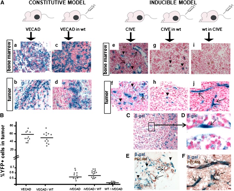
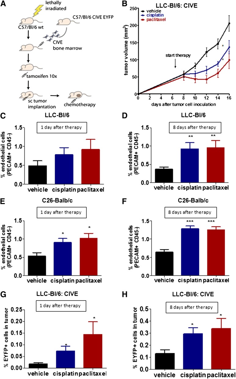
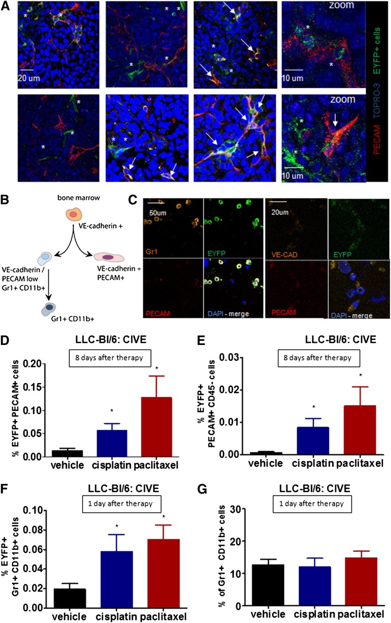
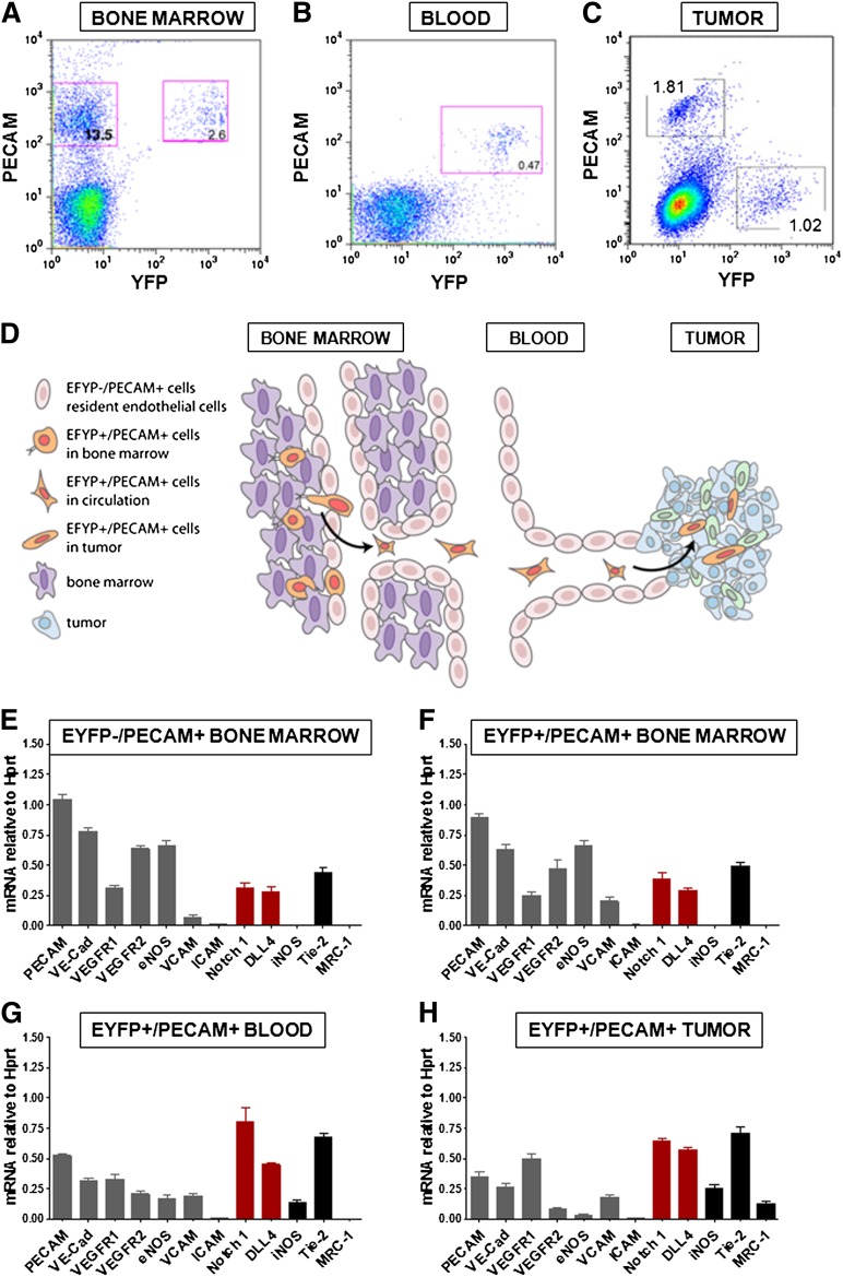
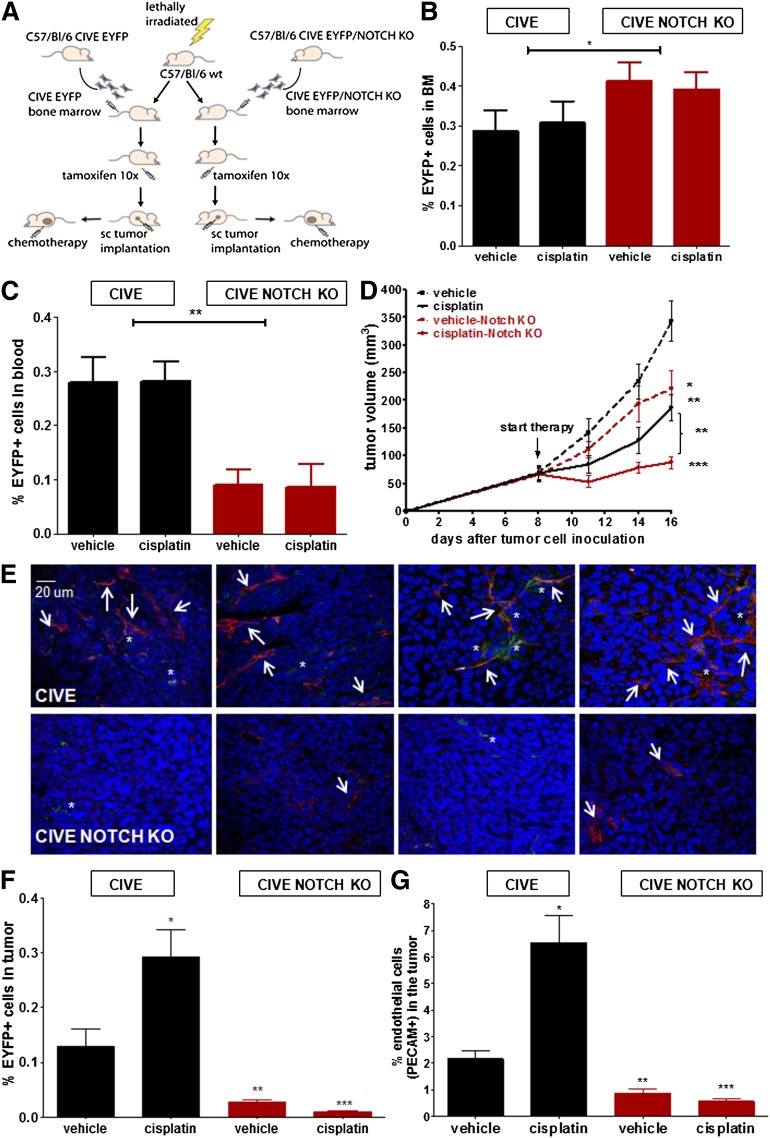
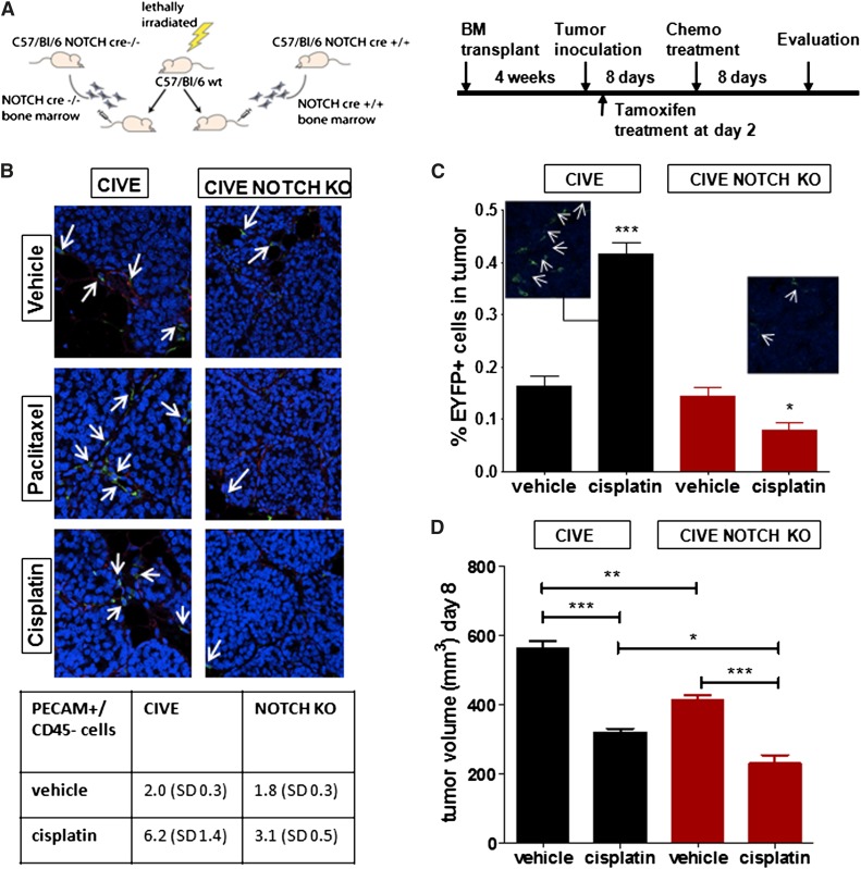
Host responses to chemotherapy can induce resistance mechanisms that facilitate tumor regrowth. To determine the contribution of bone marrow-derived cells (BMDCs), we exposed tumor-bearing mice to chemotherapeutic agents and evaluated the influx and contribution of a genetically traceable subpopulation of BMDCs (vascular endothelial-cadherin-Cre-enhanced yellow fluorescent protein [VE-Cad-Cre-EYFP]). Treatment of tumor-bearing mice with different chemotherapeutics resulted in a three- to 10-fold increase in the influx of VE-Cad-Cre-EYFP. This enhanced influx was accompanied by a significant increase in angiogenesis. Expression profile analysis revealed a progressive change in the EYFP population with loss of endothelial markers and an increase in mononuclear markers. In the tumor, 2 specific populations of VE-Cad-Cre-EYFP BMDCs were identified: Gr1⁺/CD11b⁺ and Tie2high/platelet endothelial cell adhesion moleculelow cells, both located in perivascular areas. A common signature of the EYFP population that exits the bone marrow is an increase in Notch. Inducible inactivation of Notch in the EYFP⁺ BMDCs impaired homing of these BMDCs to the tumor. Importantly, Notch deletion reduced therapy-enhanced angiogenesis, and was associated with an increased antitumor effect of the chemotherapy. These findings revealed the functional significance of a specific population of supportive BMDCs in response to chemotherapeutics and uncovered a new potential strategy to enhance anticancer therapy.


Figures

Figure 1
Figure 1
Contribution of the different VE-cadherin–expressing cells to the tumor vasculature of unperturbed tumors. (A) Immunohistochemical analysis of β-gal expression in bone marrow sections and sections of subcutaneous growing c-neu transformed mammary epithelial tumors in 5 different mouse models is shown: (Aa-b) constitutive VE-cadherin-Cre/R26R mice (VECAD), (Ac-d) bone marrow transplantation of constitutive VECAD mice in lethally irradiated wild-type (WT) C57BL/6, (Ae-f) inducible CIVE mice, (Ag-h) bone marrow transplantation of CIVE mice in lethally irradiated WT C57BL/6 mice, and (Ai-j) bone marrow transplantation of WT C57BL/6 mice into lethally irradiated CIVE mice. (B) The fluorescent versions of the above models with EYFP instead of lacZ expression were used for FACS analysis of subcutaneous growing c-neu transformed mammary epithelial tumors. The graph depicts the percentage of EYFP+ cells in the tumors. (C-F) Shown is the immunohistochemical analysis of β-gal (and CD31 expression for [E] and [F]) in the tumors of the constitutive VE-cadherin transplantation into (C-E) WT C57BL/6 mice and (F) the WT C57BL/6 transplantation into CIVE mice. All images were 20× except for (D) (63×) and (F) (40×).
Figure 2
Figure 2
Chemotherapy-enhanced angiogenesis is associated with an influx of BMDCs. (A) A schematic overview of the mouse model is shown. sc, subcutaneous. (B) The graph shows the tumor growth of LLC cells in C57BL/6 mice either untreated or treated with cisplatin or with paclitaxel. (C-D) The graphs show the percentage of PECAM+/CD45 cells of the total cells in subcutaneous growing LLC cells in BL/6 mice transplanted with CIVE bone marrow, (C) 1 day or (D) 8 days after the start of treatment. (E-F) The graphs show the percentage of endothelial cells in subcutaneous growing C26 cells in BALB/c mice (E) 1 day or (F) 8 days after the start of treatment. (G-H) The graphs show the percentage of EYFP+ cells in LLC tumors in BL/6 mice transplanted with CIVE bone marrow (G) 1 or (H) 8 days after treatment. *P < .05; **P < .01; ***P < .001 compared with the vehicle control.
Figure 3
Figure 3
Influx of 2 subpopulations of VE-cadherin BMDCs. (A) Representative confocal pictures show EYFP+ cells in the LLC tumors (blue: TO-PRO, red: PECAM, green: EYFP). (B) Shown are adult bone marrow cells expressing VE-cadherin from 2 distinct populations: an endotheliallike population expressing PECAM and VE-cadherin and a monocytic population expressing Gr1 and CD11b. (C) Cytospins of FACS sorted BMD-EYFP+ cells stained for Gr1, PECAM, and VE-cadherin are shown. (D-E) The graphs show the percentage of (D) EYFP+/PECAM+ or (E) EYFP+/PECAM+/CD45 cells in subcutaneous growing LLC cells in BL/6 mice transplanted with CIVE bone marrow 8 days after the start of treatment. (F-G) The graphs show the percentage of (F) EYFP+/Gr1+/CD11b+ cells or (G) Gr1+/CD11b+ cells in subcutaneous growing LLC cells in BL/6 mice transplanted with CIVE bone morrow 1 day after the start of treatment. *P < .05 compared with the vehicle control. *, single green EYFP cells; arrowheads, double-positive cells (EYFP+/PECAM+). All images were obtained at 20× except for the zoom images; scale bar sizes are indicated.
Figure 4
Figure 4
Notch is upregulated in VE-cadherin–expressing cells that egressed the bone marrow. (A-C) FACS sorting results are shown for 3 populations of cells from (A) the bone marrow, (B) the blood, and (C) the tumor of the CIVE mice after tamoxifen induction. (D) Shown is a schematic overview of the different stages of the EYFP+ cells. (E-H) From the bone marrow, a population of EYFP/PECAM+ and a population of EYFP+/PECAM+ were sorted. From the blood, the EYFP+/PECAM+ population was sorted. RT-PCR was performed for levels of different proteins in the 4 populations: (E) BM EYFP/PECAM+, (F) BM EYFP+/PECAM+, (G) blood EYFP+/PECAM+, and (H) tumor PECAM+/EYFP+, showing the endothelial markers (gray), Notch1/Dll4 (red), and the macrophage markers (black). eNOS, endothelial nitric oxide synthase; iNOS, inducible nitric oxide synthase; ICAM, intercellular adhesion molecule; mRNA, messenger RNA; VCAM, vascular cell adhesion molecule.
Figure 5
Figure 5
The influx of BMD-VE-cadherin/Notchhigh cells confers chemoresistance and enhanced angiogenesis. (A) Shown is a schematic overview of the transplantation model. Tumor growth occurred in LLC cells in C57BL/6 mice transplanted with either CIVE bone marrow or CIVE–Notch KO bone marrow; mice were either untreated or treated with cisplatin. (B-C) The graphs show a comparison of the EYFP+ cells in (B) the bone marrow (BM) and (C) the blood between the mice transplanted with CIVE or the CIVE–Notch KO bone marrow. (D) The graphs shows a comparison of the tumor growth of LLC cells in C57BL/6 mice transplanted with the CIVE bone marrow vs the CIVE–Notch KO bone marrow, either untreated or treated with cisplatin. (E) Representative confocal pictures show EYFP+ cells in the LLC tumors (blue: DAPI, red: PECAM, green: EYFP) in the mice with the CIVE–Notch KO bone marrow (upper panels) and the mice with the CIVE bone marrow (lower panels). (F) Shown is the contribution of EYFP+ cells to the LLC tumors in BL/6 mice transplanted with CIVE or CIVE–Notch KO bone marrow 8 days after start treatment. (G) The graph shows the percentage of endothelial cells (PECAM+ cells) of the total cells in subcutaneous growing LLC cells in BL/6 mice transplanted with CIVE or CIVE–Notch KO bone marrow 8 days after the start of treatment. *P < .05; **P < .01; ***P < .001 compared with the CIVE vehicle control. The scale bar applies to all images.
Figure 6
Figure 6
Late deletion of Notch, 2 days after tumor cell inoculation, renders the same effect. (A) The schematic overview shows the transplantation model and experimental procedure. (B) Representative confocal pictures show EYFP+ cells in the LLC tumors (red: PECAM, green: EYFP) in the mice with the Notch-Cre−/− bone marrow (left panel) and the mice with the Notch-Cre+/+ bone marrow (right panel) (both treated with the vehicle control, cisplatin, or paclitaxel). In the table (below), mean (standard deviation) PECAM+/CD45 cells per treatment group are shown. (C) The graph shows the contribution of EYFP+ cells to the LLC tumors in BL/6 mice, transplanted, respectively, with the Notch-Cre−/− or the Notch-Cre+/+ bone marrow, shown by FACS analysis and confocal microscopy. (D) The graph shows a comparison of the tumor growth of LLC cells in C57BL/6 mice transplanted with the CIVE bone marrow vs the CIVE–Notch KO bone marrow, either untreated or treated with cisplatin. *P < .05; **P < .01; ***P < .001. Panel B images were obtained with a 20× objective.

References

    1. Shaked Y, Henke E, Roodhart JM, et al. Rapid chemotherapy-induced acute endothelial progenitor cell mobilization: implications for antiangiogenic drugs as chemosensitizing agents. Cancer Cell. 2008;14(3):263–273. - PMC - PubMed
    1. Shaked Y, Ciarrocchi A, Franco M, et al. Therapy-induced acute recruitment of circulating endothelial progenitor cells to tumors. Science. 2006;313(5794):1785–1787. - PubMed
    1. Roodhart JM, Langenberg MH, Vermaat JS, et al. Late release of circulating endothelial cells and endothelial progenitor cells after chemotherapy predicts response and survival in cancer patients. Neoplasia. 2010;12(1):87–94. - PMC - PubMed
    1. Taylor M, Billiot F, Marty V, et al. Reversing resistance to vascular-disrupting agents by blocking late mobilization of circulating endothelial progenitor cells. Cancer Discov. 2012;2(5):434–449. - PubMed
    1. Kioi M, Vogel H, Schultz G, Hoffman RM, Harsh GR, Brown JM. Inhibition of vasculogenesis, but not angiogenesis, prevents the recurrence of glioblastoma after irradiation in mice. J Clin Invest. 2010;120(3):694–705. - PMC - PubMed

Publication types

MeSH terms