. 2013 Aug 1;126(Pt 15):3305-13.
doi: 10.1242/jcs.120246. Epub 2013 May 20.

The iodide-transport-defect-causing mutation R124H: a δ-amino group at position 124 is critical for maturation and trafficking of the Na+/I- symporter

Affiliations

The iodide-transport-defect-causing mutation R124H: a δ-amino group at position 124 is critical for maturation and trafficking of the Na+/I- symporter

Viktoriya Paroder et al. J Cell Sci. .

Abstract

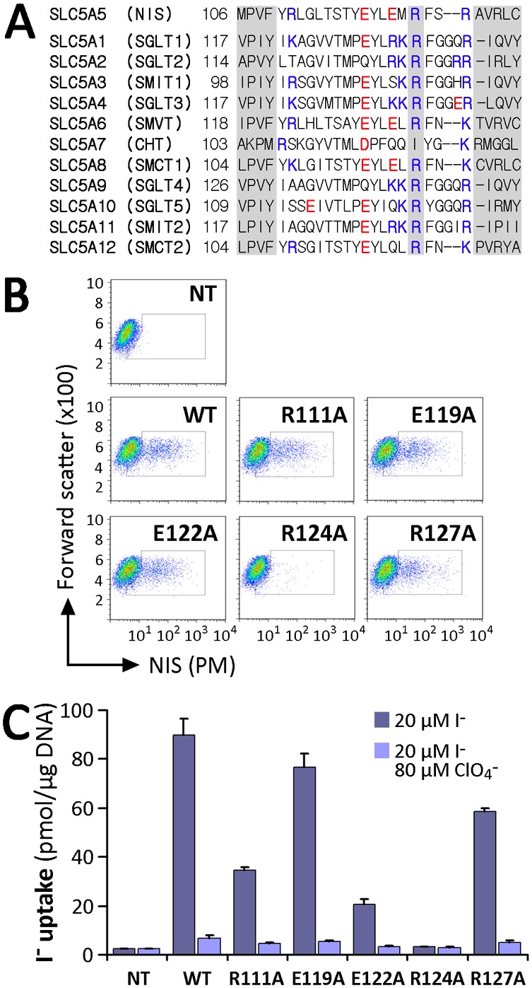
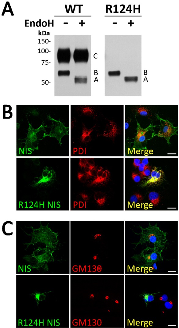
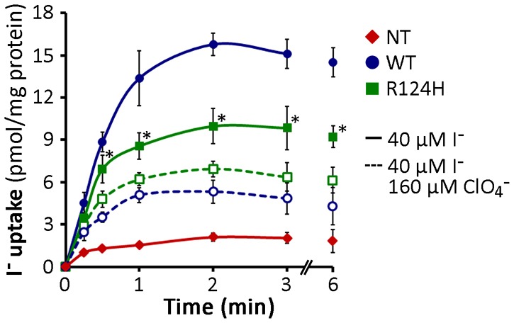
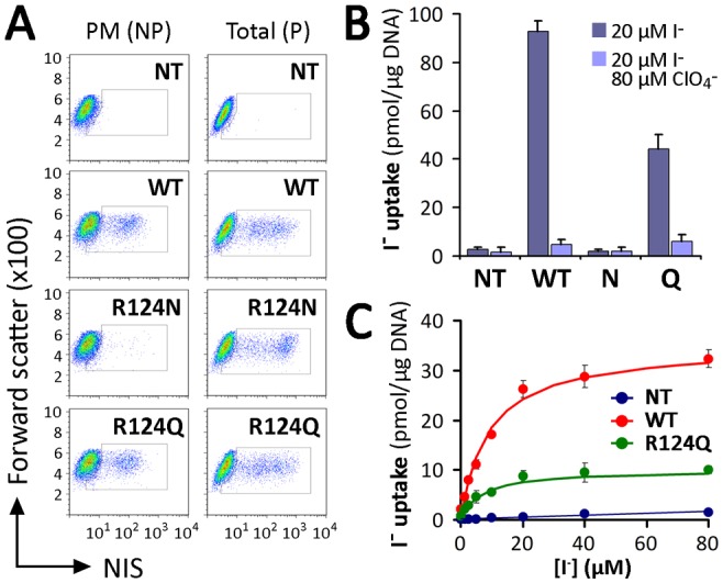
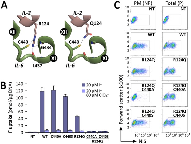
Na(+)/I(-) symporter (NIS)-mediated active accumulation of I(-) in thyrocytes is a key step in the biosynthesis of the iodine-containing thyroid hormones T3 and T4. Several NIS mutants have been identified as a cause of congenital I(-) transport defect (ITD), and their investigation has yielded valuable mechanistic information on NIS. Here we report novel findings derived from the thorough characterization of the ITD-causing mutation R124H, located in the second intracellular loop (IL-2). R124H NIS is incompletely glycosylated and colocalizes with endoplasmic reticulum (ER)-resident protein markers. As a result, R124H NIS is not targeted to the plasma membrane and therefore does not mediate any I(-) transport in transfected COS-7 cells. Strikingly, however, the mutant is intrinsically active, as revealed by its ability to mediate I(-) transport in membrane vesicles. Of all the amino acid substitutions we carried out at position 124 (K, D, E, A, W, N and Q), only Gln restored targeting of NIS to the plasma membrane and NIS activity, suggesting a key structural role for the δ-amino group of R124 in the transporter's maturation and cell surface targeting. Using our NIS homology model based on the structure of the Vibrio parahaemolyticus Na(+)/galactose symporter, we propose an interaction between the δ-amino group of either R or Q124 and the thiol group of C440, located in IL-6. We conclude that the interaction between IL-2 and IL-6 is critical for the local folding required for NIS maturation and plasma membrane trafficking.

Keywords: Congenital iodide transport defect; Impaired intracellular trafficking; Membrane vesicles; Na+/I- symporter (NIS); Plasma membrane targeting.


Figures

Fig. 1.
Fig. 1.
Characterization of expression and activity of R124H Na+/I symporter. (A) Na+/I symporter (NIS) secondary structure model. Cylinders represent the 13 transmembrane segments (TMS), in Roman numerals; branches indicate N-linked glycosylation sites (N225, N489 and N502). The N-terminus faces the extracellular milieu and the C-terminus the cytosol. The iodide transport defect (ITD)-causing NIS mutant R124H is indicated in intracellular loop (IL)-2. IL-6 is also labeled. (B) Steady-state I transport assays in non-transfected (NT), wild-type (WT) or R124H NIS cDNA transiently transfected COS-7 cells. Cells were incubated with 20 µM I in the absence or presence of 80 µM ClO4. The results are expressed in pmol I/µg of DNA ± s.d. Values are representative of at least five different experiments; each experiment was performed in triplicate. (C) Immunofluorescence analysis of permeabilized WT and R124H NIS-transfected COS-7 cells. Immunostaining was performed with anti-human NIS and anti-Na+/K+ ATPase antibodies (Abs), followed by anti-rabbit Alexa 488- and anti-mouse Alexa 555-conjugated Abs. Nuclei were stained with DRAQ5 dye (blue). The overlay of the two images is shown (Merge). Scale bars, 20 µm. (D) Flow cytometry analysis of total and plasma membrane (PM) NIS expression in permeabilized (P) and non-permeabilized (NP) COS-7 cells non-transfected or transfected with WT or R124H NIS. Cells were stained with anti-NIS VJ1 Ab, followed by Alexa-488-conjugated anti-mouse Ab. (E) Immunoblot analysis of cell surface-biotinylated proteins from non-transfected and WT- or R124H NIS-transfected COS-7 cells, performed with anti-human NIS Ab (upper panel). The plasma membrane marker Na+/K+ ATPase was used as a positive control (lower panel). NS, non-specific. (F) Immunoblot of membrane fractions from COS-7 cells transfected with WT or R124H NIS cDNA with anti-human NIS Ab. Letters on the right side of the blot indicate the relative electrophoretic mobilities of the corresponding NIS bands [A: non-glycosylated, B: partially glycosylated, C: fully glycosylated (mature), BB: dimer of the partially glycosylated, and CC: dimer of the mature polypeptide]. α-Tubulin was used as a loading control.
Fig. 2.
Fig. 2.
Analysis of charged residues within IL-2 of NIS. (A) Sequence alignment of IL-2 of SLC5A family members. The R124 position in NIS and corresponding residues in other members are indicated. Positively charged residues are indicated in blue and negatively charged ones in red. Left and right shaded sequences represent parts of TMS III and IV, respectively, according to the crystal structure of vSGLT. (B) Flow cytometry of non-transfected (NT) COS-7 cells or COS-7 cells transfected with WT or IL-2 NIS mutants under non-permeabilized conditions to assess NIS expression at the plasma membrane (PM). Staining was done using anti-NIS VJ1 Ab, followed by Alexa-488-conjugated anti-mouse Ab. (C) Steady-state I transport in WT or R111A, E119A, E122A, R124A and R127A NIS-transfected COS-7 cells. Cells were incubated with 20 µM I in the absence or presence of 80 µM ClO4. Values are expressed in pmol I/µg of DNA ± s.d. and are representative of three different experiments; each experiment was performed in triplicate.
Fig. 3.
Fig. 3.
R124H NIS is incompletely glycosylated and intracellularly retained. (A) Immunoblot of membrane fractions treated with Endo H. Membrane proteins (5 µg) from MDCK-II cells permanently expressing WT or R124H NIS were treated (+) or not (−) with Endo H. Samples were resolved by SDS-PAGE and immunoblotted with polyclonal anti-human NIS Ab. Letters on the right side of the blot indicate the relative electrophoretic mobility of the corresponding NIS polypeptides [A: non-glycosylated, B: partially glycosylated, and C: fully glycosylated (mature)]. The blot shown is representative of two independent experiments. (B,C) Immunofluorescence colocalization experiments. Permeabilized WT or R124H NIS-transfected COS-7 cells were incubated with polyclonal anti-human NIS Ab and either anti-protein disulfide isomerase (PDI) or anti-GM130 Abs, followed by Alexa 488- and Alexa 555-conjugated goat anti-rabbit and anti-mouse Abs. Nuclei stained with DRAQ5 are shown in blue. Overlay images (Merge) are shown. Scale bars, 20 µm.
Fig. 4.
Fig. 4.
R124H NIS is intrinsically active. I uptake in membrane vesicles (MV) prepared from non-transfected (NT) and either WT- or R124H NIS-expressing COS-7 cells. Membrane aliquots (100 µg) were assayed for I uptake at the indicated time points by incubation at room temperature in an equal volume of uptake buffer containing 40 µM I. Incubations were performed in the presence or absence of 160 µM ClO4. The results are expressed as pmol I/mg protein ± s.d.; each experimental point was performed in triplicate. The experiment shown is representative of three independent experiments. *P<0.05 versus same condition in the presence of ClO4 (Student's t-test).
Fig. 5.
Fig. 5.
Substitutions with charged residues at position 124 did not recover NIS plasma membrane targeting. (A) Steady-state I transport in WT and R124K, D, E or W NIS-transfected COS-7 cells. Cells were incubated with 20 µM I in the absence or presence of 80 µM ClO4. The results are expressed in pmol I/µg of DNA ± s.d. Values are representative of at least five different experiments; each experiment was performed in triplicate. (B) Analysis of NIS expression (total) and plasma membrane (PM) targeting by flow cytometry. Non-transfected (NT) or WT and R124K, D, E or W NIS-transfected COS-7 cells were stained under non-permeabilized (NP) or permeabilized (P) conditions with anti-NIS VJ1 Ab, followed by Alexa-488-conjugated anti-mouse Ab.
Fig. 6.
Fig. 6.
Substitution with Gln at position 124 restores NIS plasma membrane targeting. (A) Flow cytometry analysis of NIS expression in non-transfected (NT), WT-, R124N or Q NIS-transfected COS-7 cells. Staining was performed under non-permeabilized (NP) or permeabilized (P) conditions with anti-NIS VJ1 Ab, followed by Alexa-488-conjugated anti-mouse Ab. (B) Steady-state I transport in WT or R124Q and N NIS-transfected COS-7 cells. Cells were incubated with 20 µM I in the absence or presence of 80 µM ClO4. The results are expressed in pmol I/µg of DNA ± s.d. Values are representative of three different experiments; each experiment was performed in triplicate. (C) Initial rates (2 min time points) of I uptake were determined at the indicated concentrations of I. Calculated curves (smooth lines) were generated using the equation v = (Vmax*[I])/(Km+[I]) adjusted to consider background data obtained with non-transfected cells. Data shown are from a representative experiment out of three independent experiments.
Fig. 7.
Fig. 7.
Proposed intramolecular interactions between IL-2 and IL-6. (A) Close-up of NIS homology model based on the structure of vSGLT. C440 interacts with the δ-amino group present in Arg and Gln. R124, but not Q124, interacts with the backbones of G434 and L437. IL-2 and IL-6 are represented in pink and green, respectively, as in Fig. 1A. Potential interactions are indicated by dotted lines. Structural representations were generated using PyMol (Schrödinger, Portland, OR, USA). (B) Steady-state I transport in WT and NIS mutant-transfected COS-7 cells. Cells were incubated with 20 µM I in the absence or presence of 80 µM ClO4. The results are expressed in pmol I/µg of DNA ± s.d. Values are representative of three different experiments; each experiment was performed in triplicate. (C) Analysis of plasma membrane (PM) and total (P) NIS expression by FACS. COS-7 cells transiently expressing WT and mutant NIS constructs were stained under permeabilized (P) or non-permeabilized (NP) conditions with anti-NIS VJ1 Ab, followed by Alexa-488-conjugated anti-mouse Ab.

References

    1. Altorjay A., Dohán O., Szilágyi A., Paroder M., Wapnir I. L., Carrasco N. (2007). Expression of the Na+/I- symporter (NIS) is markedly decreased or absent in gastric cancer and intestinal metaplastic mucosa of Barrett esophagus. BMC Cancer 7, 5 10.1186/1471-2407-7-5 - DOI - PMC - PubMed
    1. Borders C. L., Jr, Broadwater J. A., Bekeny P. A., Salmon J. E., Lee A. S., Eldridge A. M., Pett V. B. (1994). A structural role for arginine in proteins: multiple hydrogen bonds to backbone carbonyl oxygens. Protein Sci. 3, 541–548 10.1002/pro.5560030402 - DOI - PMC - PubMed
    1. Calado J., Loeffler J., Sakallioglu O., Gok F., Lhotta K., Barata J., Rueff J. (2006). Familial renal glucosuria: SLC5A2 mutation analysis and evidence of salt-wasting. Kidney Int. 69, 852–855 10.1038/sj.ki.5000194 - DOI - PubMed
    1. Dai G., Levy O., Carrasco N. (1996). Cloning and characterization of the thyroid iodide transporter. Nature 379, 458–460 10.1038/379458a0 - DOI - PubMed
    1. Danilov S. M., Kalinin S., Chen Z., Vinokour E. I., Nesterovitch A. B., Schwartz D. E., Gribouval O., Gubler M. C., Minshall R. D. (2010). Angiotensin I-converting enzyme Gln1069Arg mutation impairs trafficking to the cell surface resulting in selective denaturation of the C-domain. PLoS ONE 5, e10438 10.1371/journal.pone.0010438 - DOI - PMC - PubMed

Publication types