. 2013 Jun 4;110(23):9505-10.
doi: 10.1073/pnas.1307527110. Epub 2013 May 20.

Ultrastructural analysis of hepatitis C virus particles

Affiliations

Ultrastructural analysis of hepatitis C virus particles

Maria Teresa Catanese et al. Proc Natl Acad Sci U S A. .

Abstract

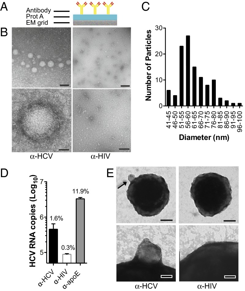
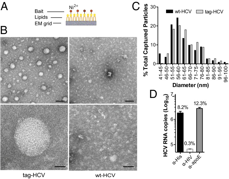
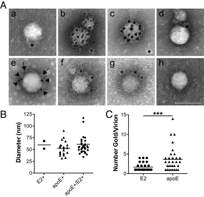
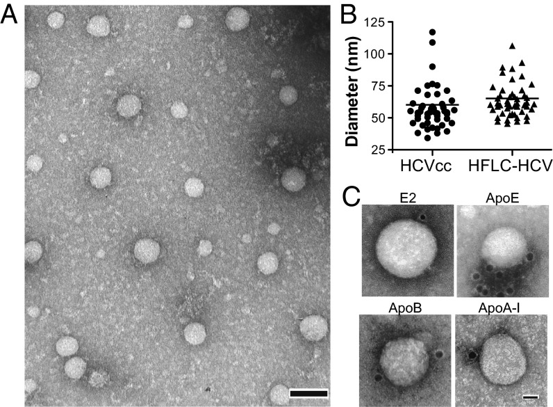
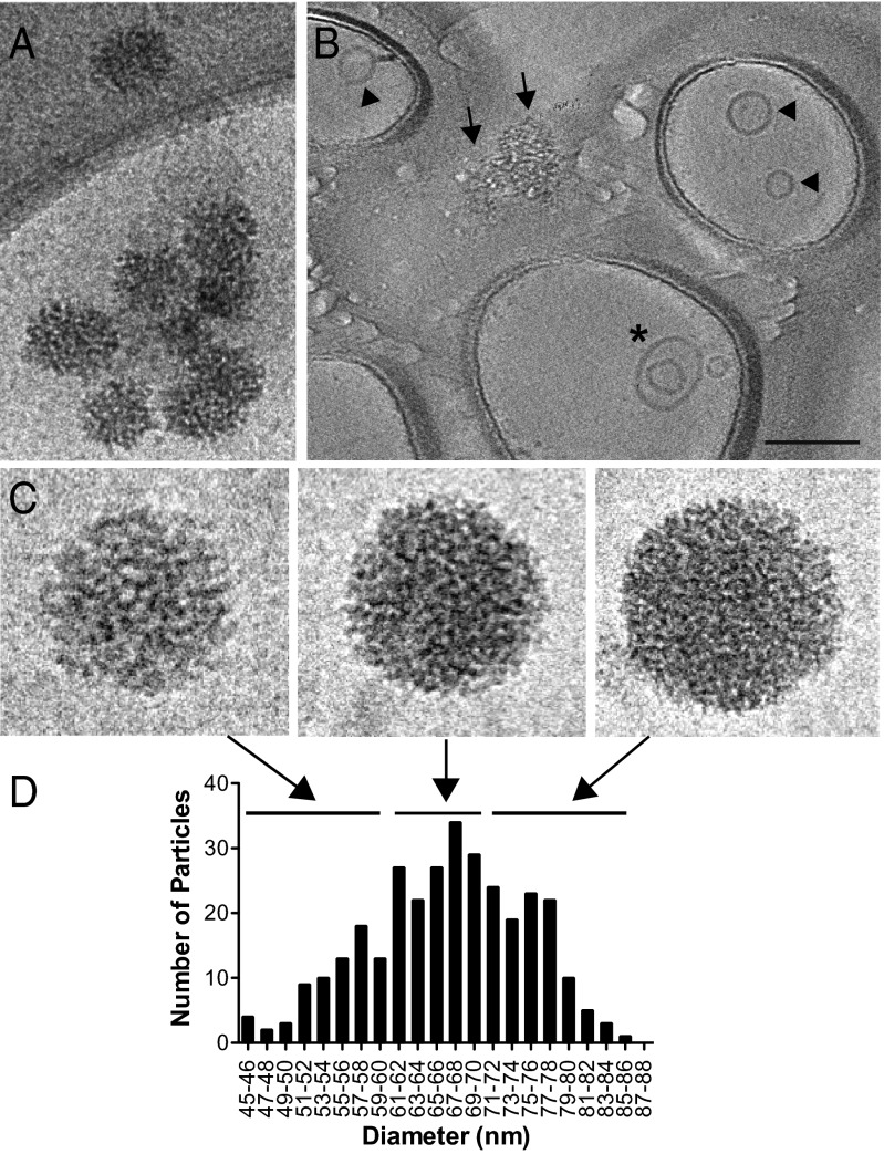
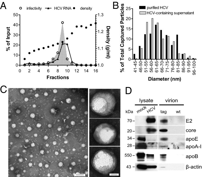
Hepatitis C virus (HCV) is a major cause of chronic liver disease, with an estimated 170 million people infected worldwide. Low yields, poor stability, and inefficient binding to conventional EM grids have posed significant challenges to the purification and structural analysis of HCV. In this report, we generated an infectious HCV genome with an affinity tag fused to the E2 envelope glycoprotein. Using affinity grids, previously described to isolate proteins and macromolecular complexes for single-particle EM, we were able to purify enveloped particles directly from cell culture media. This approach allowed for rapid in situ purification of virions and increased particle density that were instrumental for cryo-EM and cryoelectron tomography (cryo-ET). Moreover, it enabled ultrastructural analysis of virions produced by primary human hepatocytes. HCV appears to be the most structurally irregular member of the Flaviviridae family. Particles are spherical, with spike-like projections, and heterogeneous in size ranging from 40 to 100 nm in diameter. Exosomes, although isolated from unfractionated culture media, were absent in highly infectious, purified virus preparations. Cryo-ET studies provided low-resolution 3D structural information of highly infectious virions. In addition to apolipoprotein (apo)E, HCV particles also incorporate apoB and apoA-I. In general, host apolipoproteins were more readily accessible to antibody labeling than HCV glycoproteins, suggesting either lower abundance or masking by host proteins.

Keywords: enveloped virus; hepacivirus; lipoviral particle; virus assembly; virus structure.


Conflict of interest statement

Conflict of interest statement: This paper discusses hepatitis C virus research and development tools that were developed in academia and licensed to Apath, LLC, a company in which C.M.R. has equity interest.

Figures

Fig. 1.
Fig. 1.
Characterization of HCV virions captured via glycoproteins-specific antibody. (A) Schematic of protein (prot) A EM grids. (B) Representative images of negatively stained HCV virions captured using protein A EM grids coated with α-HCV or α-HIV antibodies. (Scale bar: Upper, 100 nm; Lower, 20 nm.) (C) Size histogram of HCV particles (n = 111; mean = 62 nm, SD = 11 nm). (D) HCV RNA capture assay using protein G beads coupled to α-HCV, α-HIV, or α-apoE antibody and incubated with equal amounts of virus-containing media (2 × 10⁷ viral genome copies). RNA was extracted from each pull-down and HCV genome copy numbers were determined by RT-quantitative PCR. Means and SD from three independent experiments are shown. The percent of input HCV RNA captured by each antibody is indicated. (E) Images of negatively stained HCV virions adsorbed to protein G beads coupled to α-HCV or α-HIV antibody (Scale bars: Upper, 100 nm; Lower, 20 nm.) A bead-bound HCV particle is indicated (arrow).
Fig. 2.
Fig. 2.
Ultrastructural analysis of tag-HCV virions with affinity grids. (A) Schematic of the affinity grid, an EM grid coated with a Ni-NTA lipid monolayer. (B) Representative images of negatively stained HCV virions captured using 2% (vol/vol) Ni-NTA affinity grids. (Scale bars: Upper, 100 nm; Lower, 20 nm.) (C) Comparative analysis of HCV size distribution with different capture methods. The diameter of WT-HCV particles captured on protein A grids via α-E1/E2 antibody (black bars) and tag-HCV bound to affinity grids through the His-Ni interaction (gray bars) was measured and the number of virions in each size group is expressed as percent of total captured particles. (D) The copy number of tag-HCV genomes precipitated by His-, HIV-, or apoE-specific antibodies coupled to protein G beads was measured by RT-quantitative PCR. Means and SDs from three independent experiments are shown and the percent of input HCV RNA captured by each antibody is indicated.
Fig. 3.
Fig. 3.
Viral and host proteins exposed on the envelope of HCV virions. (A) Representative images of tag-HCV particles purified on 2% (vol/vol) Ni-NTA affinity grids and immunolabeled for the (a) viral glycoprotein E2, (b) apoE, (c) apoA-I, (d) apoB, and then (e) double-stained for E2 (arrows, 8 nm gold) and apoE (arrowheads, 18 nm gold), (f) StrepII tag, (g) 6xHis tag, (h) or incubated with secondary antibody only. (Scale bar: 100 nm.) (B) Size distribution of tag-HCV particles immunoreactive for E2 (n = 2, mean = 60 nm, SD = 11 nm), apoE (n = 20, mean = 53 nm, SD = 15 nm), or double-positive for E2 and apoE (n = 38, mean = 61 nm, SD = 22 nm). (C) Number of E2- and apoE-reactive gold particles per virion in apoE+/E2+ group (***P = 0.0003).
Fig. 4.
Fig. 4.
Ultrastructure of HCV purified from primary human hepatocytes cultures. TEM images of virions from HFLC supernatant harvested at day 26 posttransfection and concentrated 10-fold. (A) Tag-HCV particles purified on 2% (vol/vol) Ni-NTA affinity grids. (Scale bar: 100 nm.) (B) Size distribution of apoE+ particles produced in Huh-7.5.1 (HCVcc) or HFLC (HFLC-HCV), captured on affinity grids. (C) TEM images of HFLC-tag-HCV showing immunolabeling with the indicated antibodies. (Scale bar: 20 nm.)
Fig. 5.
Fig. 5.
Cryo-EM analysis of HCVcc virions. (A) Cluster of HCV virions from Huh-7.5.1 cultures purified on 20% (vol/vol) Ni-NTA affinity grids. (B) Area of the grid showing HCV virions on the carbon film (arrows), exosomes (arrowheads), and multivesicular structures (*) inside the holes. (Scale bar: 100 nm.) A reconstructed tomogram of this field is available in Movie S1. (C) Low-dose images of HCV particles at 78,000× magnification, representative of three size classes (50–60, 61–70, and >71 nm). (D) Size distribution of HCV particles (n = 318; mean = 64 nm, SD = 11 nm).
Fig. 6.
Fig. 6.
Biophysical and ultrastructural characterization of purified HCV. (A) High-titer, heparin-purified tag-HCV virions were fractionated according to density on a 10–40% (wt/vol) iodixanol gradient. For each fraction, viral RNA, infectivity, and density were determined and expressed as percent of input. (B) Size distribution of highly infectious tag-HCV particles in fraction 9 (1.13 g/mL) of the buoyant density gradient (black bars; n = 430; mean = 67 nm, SD = 12 nm) is compared with unpurified HCV-containing supernatant (gray bars; n = 317; mean = 64 nm, SD = 11 nm). Data are expressed as percent of total captured particles. (C) Left: Low-magnification TEM image of tag-HCV virions from fraction 9 captured on 2% (vol/vol) Ni-NTA affinity grids. (Scale bar: 100 nm.) Right: Close up views of structures observed in purified preparations with high specific infectivity (Scale bar: 20 nm.) (D) Immunoblot for the indicated proteins in mock- or HCV RNA–electroporated Huh-7.5.1 cells, tag-, and WT-HCV extracellular virions that were heparin- and iodixanol-purified, precipitated with Dynabeads and eluted with 0.9 M imidazole.
Fig. 7.
Fig. 7.
Cryo-ET of purified HCVcc virions. (AD) Two virtual slices through some of the reconstructed purified HCV particles with spikes (arrows), viral envelope (arrowheads), and internal structures (*) indicated. (E and F) Two sections through the 3D tomogram of an HCV particle showing striations in the envelope (arrows) and viral membrane (arrowheads).

References

    1. Lavanchy D. Evolving epidemiology of hepatitis C virus. Clin Microbiol Infect. 2011;17(2):107–115. - PubMed
    1. Delang L, et al. Hepatitis C virus-specific directly acting antiviral drugs. Curr Top Microbiol Immunol. 2013;369:289–320. - PubMed
    1. Moradpour D, Penin F. Hepatitis C virus proteins: From structure to function. Curr Top Microbiol Immunol. 2013;369:113–142. - PubMed
    1. Bartenschlager R, Penin F, Lohmann V, André P. Assembly of infectious hepatitis C virus particles. Trends Microbiol. 2011;19(2):95–103. - PubMed
    1. Lindenbach BD. Virion assembly and release. Curr Top Microbiol Immunol. 2013;369:199–218. - PMC - PubMed

Publication types

Substances

LinkOut - more resources