. 2013 Jun 4;110(23):9535-40.
doi: 10.1073/pnas.1301175110. Epub 2013 May 20.

Brain homogenates from human tauopathies induce tau inclusions in mouse brain

Affiliations

Brain homogenates from human tauopathies induce tau inclusions in mouse brain

Florence Clavaguera et al. Proc Natl Acad Sci U S A. .

Abstract

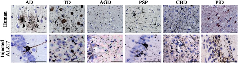
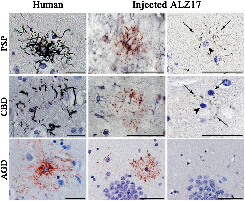
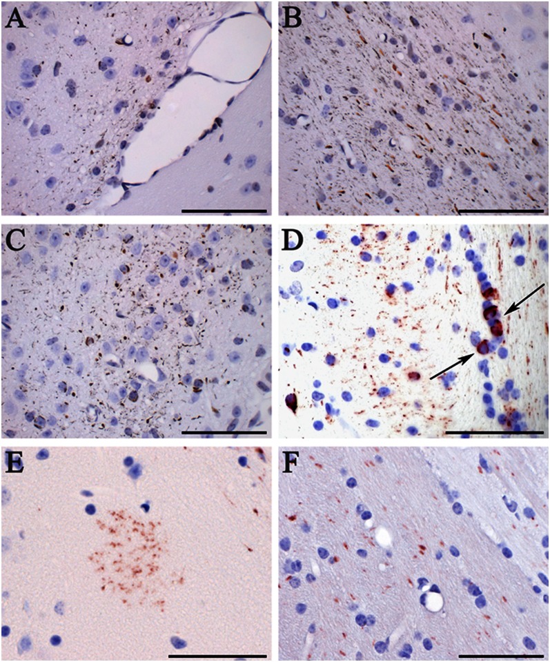
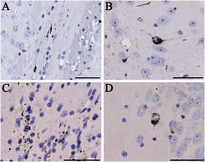
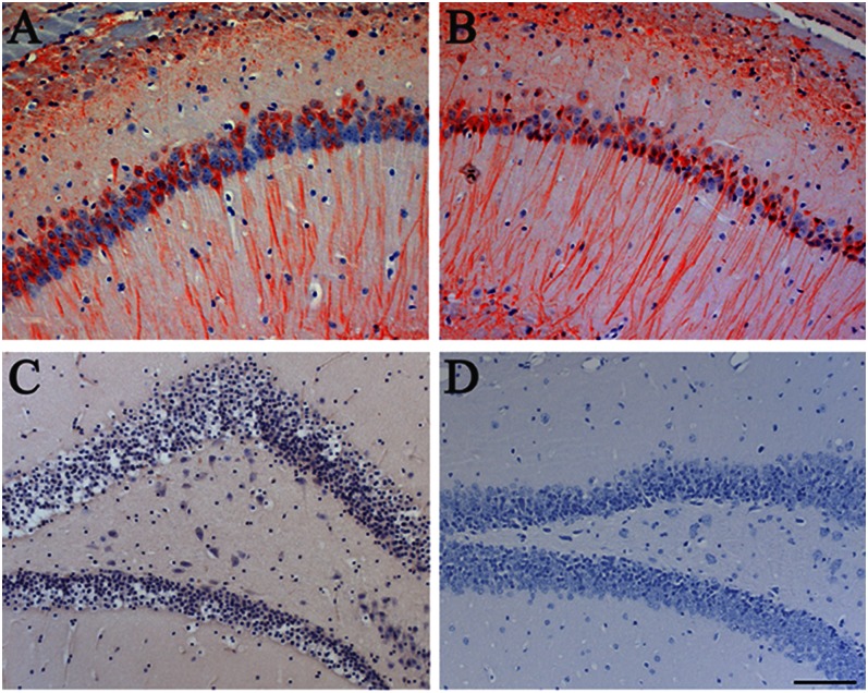
Filamentous inclusions made of hyperphosphorylated tau are characteristic of numerous human neurodegenerative diseases, including Alzheimer's disease, tangle-only dementia, Pick disease, argyrophilic grain disease (AGD), progressive supranuclear palsy, and corticobasal degeneration. In Alzheimer's disease and AGD, it has been shown that filamentous tau appears to spread in a stereotypic manner as the disease progresses. We previously demonstrated that the injection of brain extracts from human mutant P301S tau-expressing transgenic mice into the brains of mice transgenic for wild-type human tau (line ALZ17) resulted in the assembly of wild-type human tau into filaments and the spreading of tau inclusions from the injection sites to anatomically connected brain regions. Here we injected brain extracts from humans who had died with various tauopathies into the hippocampus and cerebral cortex of ALZ17 mice. Argyrophilic tau inclusions formed in all cases and following the injection of the corresponding brain extracts, we recapitulated the hallmark lesions of AGD, PSP and CBD. Similar inclusions also formed after intracerebral injection of brain homogenates from human tauopathies into nontransgenic mice. Moreover, the induced formation of tau aggregates could be propagated between mouse brains. These findings suggest that once tau aggregates have formed in discrete brain areas, they become self-propagating and spread in a prion-like manner.


Conflict of interest statement

The authors declare no conflict of interest.

Figures

Fig. 1.
Fig. 1.
Gallyas-Braak silver-positive neuronal tau inclusions in the hippocampal region of ALZ17 mice 6 mo after the intracerebral injection of brain homogenates from sporadic human tauopathies. (Upper row) Pathological tau hallmark lesions observed in the human tissues used for brain extract preparation. From Left to Right: Gallyas-Braak silver impregnation visualized neurofibrillary tangles (NFTs) and neuropil threads (NTs), as well as dystrophic neurites in the vicinity of plaques in AD; NFTs and NTs in the absence of plaques in TD; argyrophilic grains in AGD; globose NFTs and NTs in PSP and small NFTs and abundant NTs in CBD. Pick bodies were observed in PiD using AT100 immunostaining. (Lower row) Filamentous tau lesions formed in the brain of ALZ17 mice after injection of the corresponding human brain extracts. From Left to Right: NFTs after the injection of AD and TD brain homogenates; argyrophilic grains after the injection of AGD brain homogenate; nerve cell body inclusions and NTs after the injection of PSP homogenate; small numbers of nerve cell body inclusions and numerous NTs after the injection of CBD homogenate; short, thick NTs after the injection of PiD homogenate. (Scale bars, 50 µm.) Sections were counterstained with hematoxylin.
Fig. 2.
Fig. 2.
Induction of astrocytic tau inclusions in ALZ17 mice 12 mo after the intracerebral injection of PSP, CBD, and AGD brain homogenates. (Top row) Gallyas-Braak silver impregnation reveals a tufted astrocyte in the PSP case used for injection (Left). Following the injection of PSP brain homogenate, double labeling with anti-GFAP (dark blue) and AT100 (red) revealed a tufted-like astrocytic tau inclusion (Center) in which aggregated tau was detected by Gallyas-Braak staining in proximal and distal processes (arrows) located around the nucleus (arrowheads) of the tufted-like astrocyte (Right). (Middle row) Gallyas-Braak silver impregnation revealed an astrocytic plaque in the CBD case used for injection (Left). Following the injection of CBD brain homogenate, double labeling with anti-GFAP (dark blue) and AT100 (red) revealed an astrocytic plaque-like lesion (Center) in which silver-positive material was only found in the distal processes (arrows) of the plaque-like lesion after Gallyas-Braak staining (Right). (Bottom row) AT100 immunolabeling shows a tufted-like astrocyte in the AGD case used for injection (Left). Following the injection of AGD brain homogenate, AT100 immunolabeling revealed a tau-positive tufted-like astrocyte (Center) that was negative by Gallyas-Braak silver (Right). (Scale bars, 50 µm.) Sections were counterstained with hematoxylin.
Fig. 3.
Fig. 3.
Induction of tau inclusions in nontransgenic C57BL/6 mice 12 mo after the intracerebral injection of brain homogenates from sporadic human tauopathies. Gallyas-Braak silver impregnation revealed the presence of neuropil threads and coiled bodies in (A) the optic tract following the injection of TD homogenate, (B) the subiculum after the injection of PSP homogenate, and (C) the dorsal thalamus following the injection of AD homogenate. AT100 immunostaining of the hippocampal region showed the presence of (D) coiled bodies (arrows) and (E) a tufted-like astrocyte following the injection of AGD homogenate. (F) MT1 immunostaining detected mouse tau aggregates after the injection of AD brain homogenates. (Scale bars, 50 µm.) Sections were counterstained with hematoxylin.
Fig. 4.
Fig. 4.
Induction of tau inclusions following the intracerebral injection of induced mouse brain homogenates, as detected by Gallyas-Braak silver impregnation. (A) Neuropil threads and coiled bodies in the alveus and (B) neurofibrillary tangle in the subiculum of an ALZ17 mouse 12 mo after the injection of brain homogenate from an ALZ17 mouse that had 18 mo previously been injected with brain homogenate from a mouse transgenic for human mutant P301S tau. (C and D) ALZ17 mouse 12 mo after the injection of brain homogenate from a C57BL/6 mouse that had 18 mo previously been injected with TD brain homogenate. (Scale bars, 50 µm.) Sections were counterstained with hematoxylin.
Fig. 5.
Fig. 5.
Aβ did not induce tau aggregation in ALZ17 mice. The injection of APP23 brain homogenate into the hippocampal formation of ALZ17 mice had no effect on the pretangle tau staining with antibody AT8 at 15 mo after the injection (A), compared with a noninjected ALZ17 mouse (B). (C) Gallyas-Braak silver impregnation failed to detect tau inclusions in the hippocampal formation of an ALZ17 mouse 15 mo after the injection of APP23 brain homogenate. (D) Staining for Aβ with serum NT11 of a tissue section adjacent to that used in C failed to reveal the presence of Aβ deposits. (Scale bar, 100 µm.) Sections were counterstained with hematoxylin.

References

    1. Goedert M, Clavaguera F, Tolnay M. The propagation of prion-like protein inclusions in neurodegenerative diseases. Trends Neurosci. 2010;33(7):317–325. - PubMed
    1. Goedert M, Spillantini MG, Jakes R, Rutherford D, Crowther RA. Multiple isoforms of human microtubule-associated protein tau: Sequences and localization in neurofibrillary tangles of Alzheimer’s disease. Neuron. 1989;3(4):519–526. - PubMed
    1. Götz J, et al. Somatodendritic localization and hyperphosphorylation of tau protein in transgenic mice expressing the longest human brain tau isoform. EMBO J. 1995;14(7):1304–1313. - PMC - PubMed
    1. Goedert M, Spillantini MG, Cairns NJ, Crowther RA. Tau proteins of Alzheimer paired helical filaments: Abnormal phosphorylation of all six brain isoforms. Neuron. 1992;8(1):159–168. - PubMed
    1. Noda K, et al. Quantitative analysis of neurofibrillary pathology in a general population to reappraise neuropathological criteria for senile dementia of the neurofibrillary tangle type (tangle-only dementia): The Hisayama Study. Neuropathology. 2006;26(6):508–518. - PubMed

Publication types