

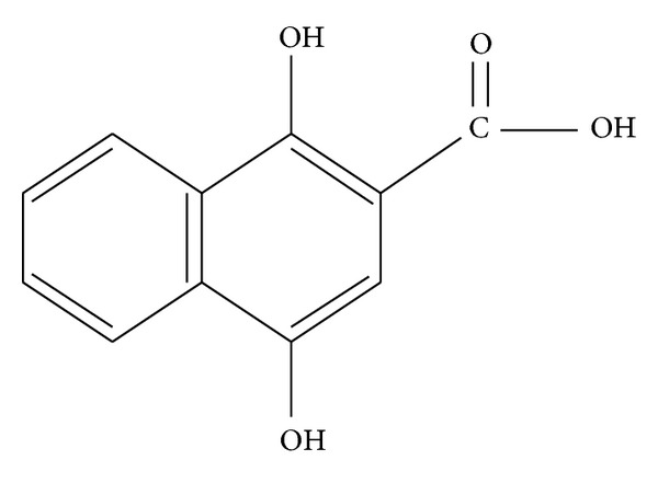
1,4-dihydroxy-2-naphthoic Acid Induces Apoptosis in Human Keratinocyte: Potential Application for Psoriasis Treatment
- PMID: 23690852
- PMCID: PMC3638593
- DOI: 10.1155/2013/792840
1,4-dihydroxy-2-naphthoic Acid Induces Apoptosis in Human Keratinocyte: Potential Application for Psoriasis Treatment
Abstract
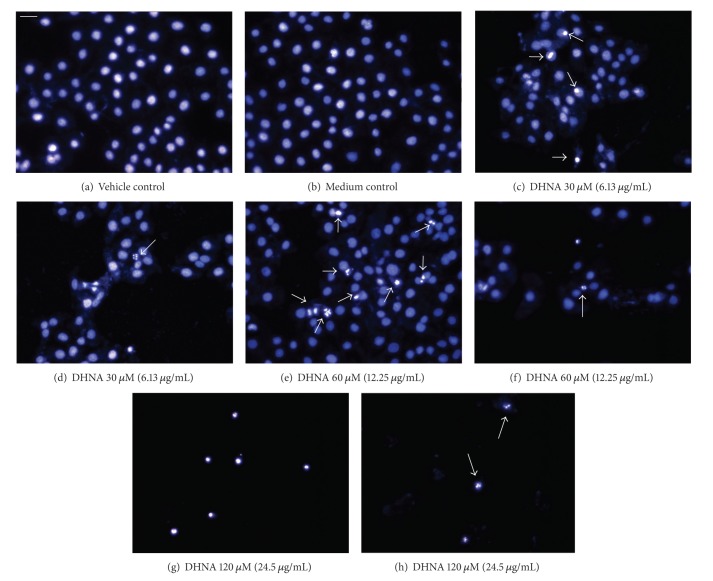
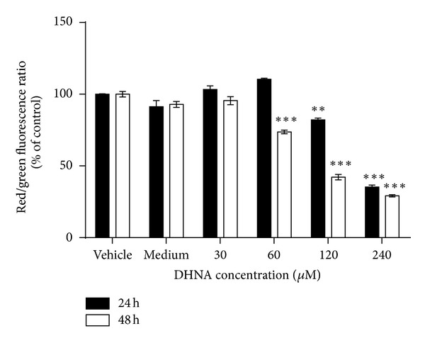
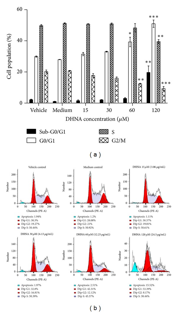
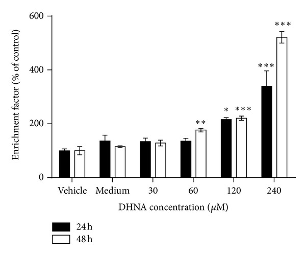
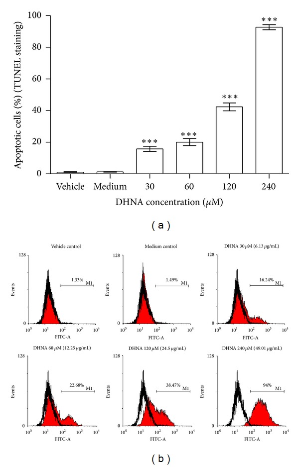
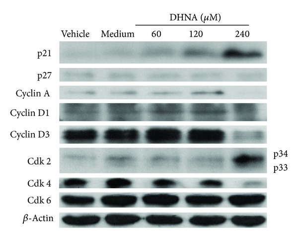
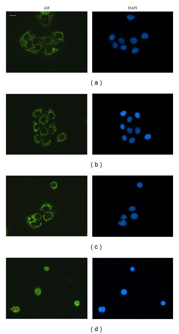
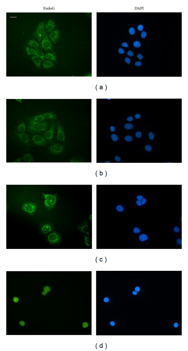
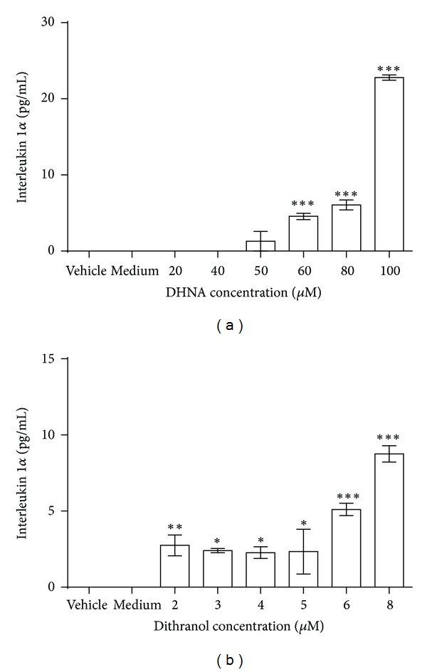
Psoriasis, which affects approximately 1-3% of the population worldwide, is a chronic inflammatory skin disorder characterized by epidermal keratinocytes hyperproliferation, abnormal differentiation, and inflammatory infiltration. Decrease in keratinocyte apoptosis is a specific pathogenic phenomenon in psoriasis. Chinese herbs have been used for the treatment of psoriasis in China showing promising effect in clinical trials. A traditional Chinese medicine has relatively fewer side effects with longer remission time and lower recurrence rate. The extract of Rubia cordifolia L. (EA) was previously found by us to induce HaCaT keratinocytes apoptosis. In this study we identified one of the components in Rubia cordifolia L., the anthraquinone precursor 1,4-dihydroxy-2-naphthoic acid (DHNA), induces HaCaT keratinocytes apoptosis through G0/G1 cell cycle arrest. We have also demonstrated that DHNA acts through both caspase-dependent and caspase-independent pathways. Besides, cytotoxicity and IL-1 α release assays indicate that DHNA causes less irritation problems than dithranol, which is commonly employed to treat psoriasis in many countries. Since DHNA possesses similar apoptotic effects on keratinocytes as dithranol but causes less irritation, DHNA therefore constitutes a promising alternative agent for treating psoriasis. Our studies also provide an insight on the potential of using EA and DHNA, alternatively, as a safe and effective treatment modality for psoriasis.
Figures


References
-
- Griffiths CE, Barker JN. Pathogenesis and clinical features of psoriasis. The Lancet. 2007;370(9583):263–271. - PubMed
-
- Traub M, Marshall K. Psoriasis—pathophysiology, conventional, and alternative approaches to treatment. Alternative Medicine Review. 2007;12(4):319–330. - PubMed
-
- Linden KG, Weinstein GD. Psoriasis: current perspectives with an emphasis on treatment. The American Journal of Medicine. 1999;107(6):595–605. - PubMed
-
- Miller DW, Feldman SR. Cost-effectiveness of moderate-to-severe psoriasis treatment. Expert Opinion on Pharmacotherapy. 2006;7(2):157–167. - PubMed
LinkOut - more resources
Full Text Sources
Other Literature Sources
