. 2013 May 14;8(5):e59269.
doi: 10.1371/journal.pone.0059269. Print 2013.

Analysis of adult neurogenesis: evidence for a prominent "non-neurogenic" DCX-protein pool in rodent brain

Affiliations

Analysis of adult neurogenesis: evidence for a prominent "non-neurogenic" DCX-protein pool in rodent brain

Thomas Kremer et al. PLoS One. .

Abstract

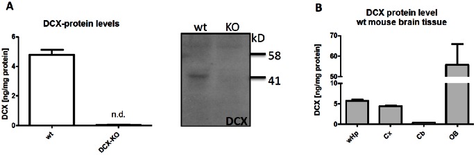
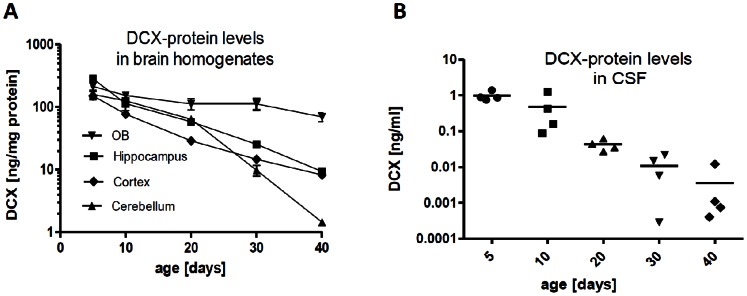
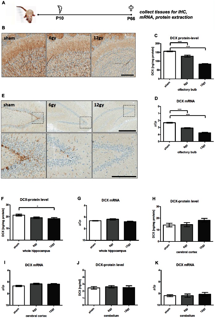
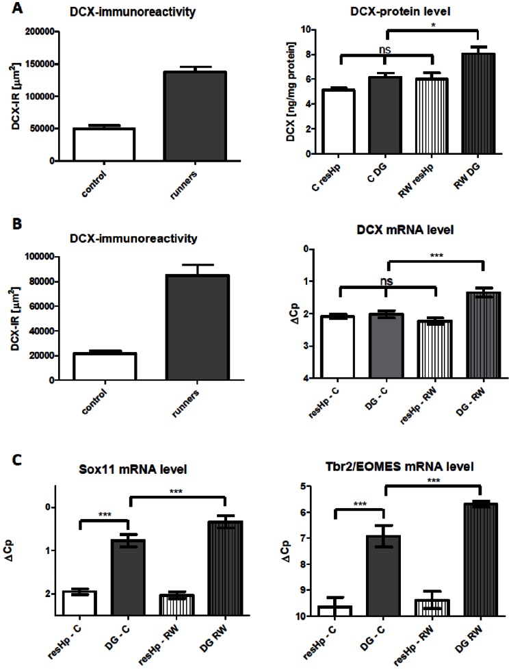
Here, we have developed a highly sensitive immunoassay for Dcx to characterize expression in brain and cerebrospinal fluid (CSF) of rodents. We demonstrate that Dcx is widely expressed during development in various brain regions and as well can be detected in cerebrospinal fluid of rats (up to 30 days postnatal). While Dcx protein level decline in adulthood and were detectable in neurogenic regions of the adult rodent brain, similar levels were also detectable in brain regions expected to bear no neurogenesis including the cerebral cortex and CA1/CA3 enriched hippocampus. We monitored DCX protein levels after paradigms to increase or severely decrease adult hippocampal neurogenesis, namely physical activity and cranial radiation, respectively. In both paradigms, Dcx protein- and mRNA-levels clearly reflected changes in neurogenesis in the hippocampus. However, basal Dcx-levels are unaffected in non-neurogenic regions (e.g. CA1/CA3 enriched hippocampus, cortex). These data suggest that there is a substantial "non-neurogenic" pool of Dcx- protein, whose regulation can be uncoupled from adult neurogenesis suggesting caution for the interpretation of such studies.


Conflict of interest statement

Competing Interests: This study was supported by F. Hoffman-La Roche, the employer of TK, RJ, AH, EB, CC and HM. There are no patents, products in development or marketed products to declare. This does not alter the authors' adherence to all the PLOS ONE policies on sharing data and materials, as detailed online in the guide for authors.

Figures

Figure 1
Figure 1. Murine Dcx-protein expression using a Dcx-specific immunoassay.
A, Left, Dcx is detected in whole brain homogenates from adult C57BL/6 wt and Dcx-KO mice using a sandwich immunoassay. No signal can be observed in Dcx-KO mice (N = 3). Right, Representative Dcx immunoblot. Dcx can be detected in hippocampal tissue of adult wildtype but not Dcx KO mice. B, expression levels of Dcx-protein in various mouse brain regions (wHp: whole hippocampus, Cx: cortex, Cb: Cerebellum, OB: olfactory bulb, N = 6).
Figure 2
Figure 2. Dcx protein-levels in rat brain homogenates and CSF during development.
Olfactory bulb, whole hippocampus, pieces of cerebral cortex and cerebellum, and CSF were analyzed from rats at different developmental stages (postnatal day 5 to 40). A, Dcx-protein levels in rat brain tissue homogenates during postnatal development. B, Dcx-protein levels in CSF during postnatal development (N = 4).
Figure 3
Figure 3. Irradiation-induced ablation of neurogenesis.
A, schematic diagram of experimental procedures. Female wistar rats received a high (12 gy) or low (6 gy) irradiation dose or were sham-treated at P10 (N = 10 per group). Mice were sacrificed 7 weeks after treatment. A subset of brains were processed for immunohistochemistry (12 gy: N = 2, 6 gy: N = 3, sham: N = 3). Residual brains were split into hemispheres and dissected for mRNA and protein analysis (N = 6/group). B, Representative images of Dcx-IR in the olfactory bulb of formalin-fixed paraffin embedded (FFPE) sections. Left-to-right: A high density of dendritic Dcx-IR is observed in the olfactory bulb granular layer sham-irradiated animals. Dendritic labeling is reduced with low-irradiation and virtually absent in animals after 12 gy-irradiation. Scale bar: 200 µm. C, bar graphs of Dcx-protein-levels in the olfactory bulb. A dose-dependent decrease in Dcx-protein levels is observed in irradiated animals vs sham-controls. D, bar graphs of Dcx-mRNA-levels in the olfactory bulb. A dose-dependent decrease in Dcx-mRNA levels is observed in irradiated animals vs sham-controls. E, Representative images Dcx-IR in the dentate gyrus of FFPE sections. Upper panel: overview of Dcx-IR in the dentate gyrus. Left-to-right: Dcx-IR is restricted to cells in the dentate gyrus SGZ with dendrites spanning into the granular and molecular layer. Lower panel: higher magnification of the SGZ. F, bar graphs of Dcx-protein-levels in the hippocampus. A slight dose-dependent decrease in DCX-protein levels is observed in irradiated animals. G, bar graphs of Dcx-mRNA-levels in the hippocampus. DCX-mRNA levels do not change significantly between sham and irradiated groups. H, bar graphs of Dcx-protein-levels in the cerebral cortex. I, bar graphs Dcx-mRNA-levels in cerebral cortex. DCX-mRNA levels do not change significantly between sham and irradiated groups. J, bar graphs of Dcx-protein-levels in the cerebellum. K, bar graphs of Dcx-mRNA-levels in cerebellum. Dcx-mRNA levels do not change significantly between sham and irradiated groups. Dunnett’s Multiple Comparisons Test.
Figure 4
Figure 4. 14-day voluntary running wheel experiment.
In two separate experiments, adult mice with or without access to a running wheel were sacrificed after 2 weeks. The right hemisphere was dissected for either protein or mRNA-analysis while the left hemisphere was used to confirm exercise-induced increase in Dcx-IR via immunohistochemistry. A, Dcx-IR was quantified by calculating the total Dcx-IR area in µm2 for four different sections within the dorsal hippocampus (left). Bar graph of hippocampal Dcx-protein-levels in DG and resHp (right). N = 12/group. B, Dcx-IR was quantified by calculating the total Dcx-IR area in µm2 for four different sections within the dorsal hippocampus (left). Bar graph of hippocampal Dcx-mRNA-level in DG and resHp (right). N = 15/group. C, Bar graph mRNA-level in DG and resHp for Sox11 and Tbr2/EOMES. N = 15/group. Bonferroni’s Multiple Comparisons Test.

References

    1. Eriksson PS, Perfilieva E, Bjork-Eriksson T, Alborn AM, Nordborg C, et al. (1998) Neurogenesis in the adult human hippocampus. Nature medicine 4: 1313–1317. - PubMed
    1. Kuhn HG, Dickinson-Anson H, Gage FH (1996) Neurogenesis in the dentate gyrus of the adult rat: age-related decrease of neuronal progenitor proliferation. The Journal of neuroscience : the official journal of the Society for Neuroscience 16: 2027–2033. - PMC - PubMed
    1. Knoth R, Singec I, Ditter M, Pantazis G, Capetian P, et al. (2010) Murine features of neurogenesis in the human hippocampus across the lifespan from 0 to 100 years. PloS one 5: e8809. - PMC - PubMed
    1. Wolf SA, Melnik A, Kempermann G (2011) Physical exercise increases adult neurogenesis and telomerase activity, and improves behavioral deficits in a mouse model of schizophrenia. Brain, behavior, and immunity 25: 971–980. - PubMed
    1. Danzer SC (2012) Depression, stress, epilepsy and adult neurogenesis. Experimental neurology 233: 22–32. - PMC - PubMed

Publication types

MeSH terms